Original Article
Increased expression of Sestrin2 in human
and experimental heart failure
Menglong Wang*, Jianfang Liu*, Juanjuan Qin, Menglin Liu, Ying Feng, Lei Shi, Wenhui Yuan, Jing Ye, Jun Wan
Department of Cardiology, Cardiovascular Research Institute, Hubei Key Laboratory of Cardiology, Renmin Hospi-tal of Wuhan University, Wuhan, PR China. *Equal contributors.
Received February 16, 2016; Accepted June 13, 2016; Epub August 1, 2016; Published August 15, 2016
Abstract: Background: Sestrins (Sesns) are originally identified as critical antioxidant proteins and involved in com-plex regulation of cell viability in response to diverse stress conditions. However, the exact expression and func-tion of Sesns in heart failure (HF) remains unclear. Methods and results: Sesns expression was detected in heart samples from end-stage HF patients and unmatched donors by real-time polymerase chain reaction (RT-PCR), Western Blotting and Immunofluorescence. Mice myocardial ischemia/ reperfusion (I/R) and myocardial infarc-tion (MI) models and doxorubicin induced rat HF model were established to confirm the heart expression of Sesns. Sesn2 was significantly increased, whereas, no alteration for Sesn1 or Sesn3 in failing hearts when comparing with control hearts. Unexpectedly, the mRNA expression of Sesn2 in ischemic cardiomyopathy was higher than that in dilated cardiomyopathy. Immunofluorescence staining revealed all Sesns were expressed in non-cardiomyocytes in human heart. Furthermore, Sesn2 was co-expressed with vimentin, a marker for fibroblasts. Correlation analysis demonstrated Sesn2 levels were significantly correlated with expression levels of natriuretic peptide B (NPPB) and connective tissue growth factor (CTGF), markers for severity of HF and cardiac fibrosis. Meanwhile, the increased Sesn2 was validated in mice I/R and MI models, and also in doxorubicin induced rat HF model. Conclusions: Sesn2, which is expressed in cardiac fibroblasts, could be a potential biomarker to reflect the severity of HF and might play an important role during cardiac remodeling.
Keywords: Heart failure, Sestrin, oxidative stress, fibrosis, biomarker
Introduction
Redox signaling plays important role not only in physiological processes, but also in pathologi-cal conditions in heart. At low concentrations, ROS modulate excitation-contraction coupling, cell proliferation and differentiation through changes in cellular signalling and gene expres-sion [1-3]. However, overproduction of ROS may result adverse cardiac remodeling, leading to further worsening of cardiac function [1]. It is accepted that oxidative stress is at least one main cause of heart failure (HF) [4]. Target- ing oxidative stress is becoming an important strategy to fight HF.
Sestrins (Sesns) are highly conserved proteins originally identified as critical antioxidant with sulfinic acid reductase activity, which is neces-sary for the recycling of peroxiredoxin [5-7]. Mammals express three Sesns (Sesn1, Sesn2,
and Sesn3), while most invertebrate express only one Sesn. The Sesn family proteins can be induced by a variety of environmental stresses, including DNA damage, oxidative stress, and hypoxia [6, 8]. Once induced, Sesns can protect cells against oxidative stress [9-11].
Materials and methods
Human heart tissues
Human heart tissues were collected from transplanted hearts with end-stage heart fail-ure, without diabetes mellitus. All samples were obtained from identical myocardial loci of left-ventricular to extremely ensure optimal uniformity. Control hearts were procured from organ donators who died of traffic accidents with no cardiac disease history. These hearts had originally been intended for transplan- tation, but failed to get suitable matching re- cipients. The hearts were immediately dissec-tion into small pordissec-tions, snap-frozen in liquid nitrogen, and then maintained at -80°C until using. Heart samples used for pathological study were fixed in 10% phosphate-buffered formalin and embedded in paraffin.
Fully informed consent was obtained from all patients or family members before partici- pating in the study. This study protocol was approved by the Ethics Committee of Renmin Hospital of Wuhan University.
Mice myocardial ischemia/reperfusion and myocardial infarction models
Male C57BL/6 (8-10 weeks old) mice were pur-chased from Hunan SJA Laboratory Animal Co., Ltd. Pentobarbital sodium was used to
anes-myocardial infarction (MI) model, and the tho-racic cavity was closed after occlusion of the LCA. The hearts were harvested at 1 week and 4 weeks after operation. Sham-operated animals underwent the same surgical proce-dure without LCA occlusion.
Doxorubicin induced rat heart failure model
Male SD (6-8 weeks old) rats were purchased from Hunan SJA Laboratory Animal Co., Ltd. and were randomly divided into two groups. The doxorubicin (Dox) group received Dox (2.5 mg/kg/week) through intraperitoneal inject- ion for 6 weeks. The control group received same volume saline as the same way. Two weeks after the last injection, the rats were sacrificed and heart samples were collected for research.
Quantitative real-time polymerase chain reac-tion
[image:2.612.91.377.82.310.2]Total RNA was extracted from heart tissue with TRIzol reagent (Roche, Indiana) as the manual instructed, and the concentration was assessed by Nanodrop 2000. A total of 2 μg RNA was used to synthesis cDNA using Transcriptor First Stand cDNA Synthesis Kit (Roche). Quantitative real-time PCR (qRT-PCR) was performed with the LightCycler 480 SYBR Green I Master (Roche) using the LightCycler 480 real-time PCR system according to the
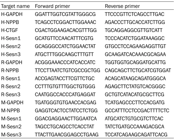
Table 1. Primer pairs used in the present study
Target name Forward primer Reverse primer
H-GAPDH GGATTTGGTCGTATTGGGCG TTCCCGTTCTCAGCCTTGAC
H-NPPB TCAGCCTCGGACTTGGAAAC AGACCCTTGCACCATCTTGG
H-CTGF CGACTGGAAGACACGTTTGG TGCAGGAGGCGTTGTCATT
H-Sesn1 GCATGTTCCAACATTTCGTG TCCCACATCTGGATAAAGGC
H-Sesn2 GCAGGGCCATCTGGAACTAT GTGCCTCCAGAAGAGGTTGT
H-Sesn3 ATGCTTTGGCAAGCTTTGTT GCAAGATCACAAACGCAGAA
R-GAPDH ACGGGAAACCCATCACCATC TGGTGGTGCAGGATGCATTG
R-NPPB TTCCTTAATCTGTCGCCGCTGG CAGCAGCTTCTGCATCGTGGAT
R-Sesn1 ACCGAGTACCTTCGTTCTGC ACAGCATAAGCAGATGGGCA
R-Sesn2 CCTTTGTGTTTGGCTGTGGG AGAGCTTCTATGTCACGGGC
R-Sesn3 CAATGGCCACCCATGAGGAT GCTGTCATACATGCGCTTCG
M-GAPDH TGATGGGTGTGAACCACGAG TCATGAGCCCTTCCACGATG
M-NPPB GAGGTCACTCCTATCCTCTGG GCCATTTCCTCCGACTTTTCTC
M-Sesn1 GGACGAGGAACTTGGAATCA ATGCATCTGTGCGTCTTCAC
M-Sesn2 TAGCCTGCAGCCTCACCTAT TATCTGATGCCAAAGACGCA
M-Sesn3 TTACTTGAACGGAGCCTGAAG TCCATCAGAAGCAGATTCACG
manufacturer’s instructions (Roche). The prim-er pairs wprim-ere designed online using Primprim-er- Primer-Blast and were shown in Table 1. The PCR con-ditions used were as follows: initial denatur-ation at 95°C for 10 minutes, followed by 40 cycles of 95°C for 10 seconds (denaturation), 60°C for 10 seconds (annealing), and 72°C for 20 seconds (extension). The relative expres-sion levels of mRNAs were normalized to the reference gene GAPDH. All reactions were con-ducted in triplicates and the data was calculat-ed using the 2-ΔΔCT method.
Western blot
Western blot was used to examine the protein expression of specific targets. Total proteins were extracted from heart tissues using RIPA lysis buffer (720 μL of RIPA, 20 μL of PMSF, 100 μL of complete protease inhibitor cock- tail, 100 μL of Phosstop, 50 μL of NaF, and 10 μL of Na3VO4 in 1 mL of lysis buffer). The protein concentration was determined using a BCA Protein Assay Kit (Thermo Fisher Scienti- fic, USA). 50 μg protein samples were separat-ed by sodium dodecyl sulphate polyacryla- mide gel electrophoresis and then transferr- ed to a PVDF membrane (Millipore, USA) that was blocked with 5% skim milk in Tris-buffer- ed saline for 60 min at room temperature. The membrane was incubated overnight at 4°C with primary antibodies anti-sesn1 (1:1000, Santa Cruz Biotechnology, USA), anti-sesn2 (1: 1000, Proteintech, China), anti-sesn3 (1:1000, Abcam, USA) and anti-Nrf2 (1:1000, Abcam, USA). Then the membrane was incubated with a secondary antibody. The blots were detect- ed using a Bio-Rad imaging system. Specific protein expression levels were normalized to GAPDH protein.
Histological analysis
Paraffin-embedded hearts were cut transver- sely into 5-μm sections. Picrosirius red (PSR)
and subsequently incubated overnight at 4°C with the primary antibodies. The sections were then washed with PBS and incubated with the appropriate secondary antibodies for 1 h at 37°C. The secondary antibodies used were goat anti-rabbit IgG Alexa Fluor 568 conju- gate (Invitrogen, CA) and anti-mouse IgG Alexa Fluor 568 conjugate (Invitrogen, CA). The nu- clei were stained with 4,6-diamidino-2-pheny- lindole (DAPI). Images were all obtained at 400× magnification with a fluorescence micro-scope (Olympus Dx51) and DP2-BSW software (Version 2.2), and the images were analyzed with Image-Pro Plus (Version 6.0) in a blinded manner.
Statistical analysis
Data were presented as mean ± SD. The SP- SS18.0 was used for statistical analysis. Mul- tiple-group comparison was performed by one-way analysis of variance. Nonparametric test was applied to assess the enumeration data. Spearman’ correlation was used to assess the relation of Sesn2 mRNA levels with heart fail-ure maker and myocardial fibrosis marker. Significance was assumed as P<0.05.
Results
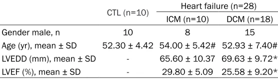
Clinical characteristics
[image:3.612.91.369.86.165.2]The clinical characteristics for all patients were shown in Table 2. Briefly, heart samples from 10 patients with ischemic cardiomyopathy (ICM) and 18 patients with dilated cardiomyop-athy (DCM) were included as the failing hearts in this study. 10 donor hearts were included as control (CTL). Of the 10 ICM patients, 8 were males and the average age were 54.00 ± 5.42 years. Among DCM patients, 15 were males and the average age were 52.93 ± 7.40 years. There was no difference for age between HF
Table 2. Characteristics for heart failure patients
CTL (n=10) Heart failure (n=28)
ICM (n=10) DCM (n=18)
Gender male, n 10 8 15
Age (yr), mean ± SD 52.30 ± 4.42 54.00 ± 5.42# 52.93 ± 7.40#
LVEDD (mm), mean ± SD - 65.60 ± 10.37 69.63 ± 9.72*
LVEF (%), mean ± SD - 29.80 ± 5.09 25.58 ± 9.20*
CTL: Control hearts; ICM: Ischemic cardiomyopathy; DCM: Dilated cardiomyopathy; LVEDD: Left ventricular end diastolic diameter; LVEF: Left ventricular ejection frac-tion. #P>0.05 vs. CTL; *P>0.05 vs. ICM.
groups and CTL. These failing hearts remained severely decreased left ventricular function (ejection fraction: 29.80 ± 5.09 and 25.58 ± 9.20, respectively for ICM and DCM) as well as significantly enlarged left ventricular cavity size (left ventricular end-diastolic dimension: 65.60 ± 10.37 and 69.63 ± 9.72, respective- ly for ICM and DCM).
Expression of Sesns in human hearts
Comparing with control hearts, there was no difference for mRNA levels of Sesn1 or Sesn3 in failing hearts (Figure 1A). However, a significant increase of Sesn2 in failing hearts was found (Figure 1A). And unexpectedly, the expression of Sesn2 was higher in ICM than
increased in failing myocardium, including in ICM and DCM.
Expression of Sens2 and its correlation with severity of heart failure and myocardial fibrosis
Natriuretic peptide B (NPPB), a marker for heart failure, was up-regulated in failing hearts. To clarify the relationship between Sesn2 levels and severity of heart failure, Spearman’ corre-lation was applied. The levels of Sesn2 were positively associated with that of NPPB (Figure 4A). PSR staining showed a relatively disperse distribution of collagen fibers in control hearts and an excessive accumulation of collagen in extracellular matrix of failing hearts (Figure 4B). Connective tissue growth factor (CTGF), Figure 1. The mRNA expression of Sesns in control and HF patients. A:
Com-pare of mRNA levels between heart failure patients and controls. B: ComCom-pare of mRNA levels between ICM and DCM patients. #P<0.05 vs. CTL; *P<0.05 vs. DCM.
Figure 2. The protein expression of Sesns in control and HF patients. A: Rep-resentative bands showing the expression of Sesns in heart tissues. B-D: Quantitative analysis of Sesn1, Sesn2 and Sesn3 protein levels. **P<0.01 vs. CTL; n. s, no significance.
that in DCM (Figure 1B). Analysis of the protein levels by Western blot confirmed that Sesn2 protein was signi- ficantly higher in the cardiac tissues of heart failure pa- tients (Figure 2A-D). However, for the etiology-specific differ-ence in Sesn2 protein levels, there was just an up-trend in ICM than that in DCM (P= 0.054).
To further clarify the heart expression of Sesns, we per-formed immunofluorescence staining. As shown in Figure 3A-C, control hearts express- ed all three members of Se- sns. Meanwhile, Sesns were all expressed in the non-car-diomyocytes. To clarify the cell types that express Se- sn2, we performed double immunofluorescence staining for Sesn2 and vimentin, a well-known marker for fibro-blasts. The results revealed Sesn2 was localized in fibro-blasts (Figure 3D).
which can regulate fibrillar collagen gene tran-scription in the heart and often up-regulated in failing hearts, was also found significantly cor-related with Sesn2 (Figure 4C).
(Figure 5A and 5B). Meanwhile, Sesn2 levels were significantly higher in MI hearts after 4 weeks than that in 1 week (**P<0.01 vs. MI 1 w). Dox induced HF model is one model used to
Figure 3. Immunofluorescence staining of Sesns in human hearts. A-C: Immunofluores-cence staining showing Sesn1, Sesn2 and Sesn3 all expressed in human hearts. D: Co-staining for Sesn2 (red) and vimentin (green) showing the localization of Sesn2 in human hearts.
Figure 4. Sesn2 positively correlated with the severity of heart dysfunction and cardiac fibrosis in human hearts. A: Correlation between mRNA levels of Sesn2 and NPPB in human hearts (r=0.542, P<0.01). B: PSR staining showed a relatively disperse distribution of collagen fibers in CTL and an ex-cessive accumulation of collagen in extracellular matrix of ICM and DCM (col-lagen was stained with red). C: Correlation between mRNA levels of Sesn2 and CTGF in human hearts (r=0.561, P<0.01). PSR, picrosirius red.
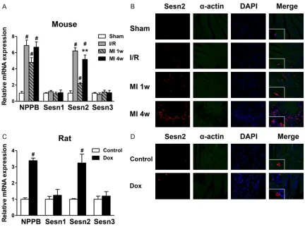
Increased heart expression of Sesn2 in animal HF mod-els
To further investigate the expression of Sesn2 in HF, we assessed the expression of Sesns in mice I/R, MI mod-els and in doxorubicin in- duced rat HF model using RT-PCR and immunofluores-cent staining. After construc-tion of the models, NPPB was significantly increased in all three models (Figure 5A
[image:5.612.92.514.70.589.2]simulation human DCM process [13]. As shown in Figure 5C and 5D, increased expression of Sesn2 was found in rat failing heart, but no change for Sesn1 and Sesn3.
Discussion
In the present study, we investigated the expression and function of Sesns in human and experimental failing hearts. We discovered that Sesn1, Sesn2 and Sesn3 were all expressed in non-cardiomyocytes in human heart, and Sesn2 was up-regulated in failing hearts, including in ICM and DCM. Furthermore, Sesn2 levels were significantly correlated with expres-sion levels of NPPB and CTGF, makers for sever-ity of heart failure and cardiac fibrosis. The up-regulation of Sesn2 was validated in mouse I/R and MI models, and also in doxorubicin induced rat heart failure model.
[image:6.612.92.524.72.394.2]dependent ROS generation, thus to prevent fibrotic injury in diabetes [20]. Also, studies in mouse models of chronic obstructive pulmo-nary disease revealed that inactivation of Se- sn2 could induce TGF-β signaling and PDGFR β signaling [21, 22], two well-known pathways participated in fibrosis. All these studies indi-cated an important function of Sesn2 in re- gulating cell apoptosis and fibrosis.
More importantly, heart protective function of Sesns has been reported. Sesn played a significant role in maintaining basal cardiac integrity as Sesn null mutants and Sesn defi-cient hearts display irregularity of heartbeat, cardiac dilation, ultimately leading to decre- ased cardiac function [12]. Also, Alex Morri- son et al. demonstrated increased myocardial infarct size after I/R challenge in Sesn2 knock-out mice when compared with wild type mice [23]. Our results firstly illustrated the expres-sion patterns of Sesns in human normal and failing hearts. Besides, increased expression of Sesn2 was revealed and the positive corre- lation between Sesn2 levels and cardiac fibro-sis was illustrated. Similar to the increased antioxidant enzyme catalase in human end-stage HF [15], the up-regulation of Sesn2 in our study might be a compensatory reaction in response to cardiac stress. However, the increase might not enough to maintaining nor-mal cardiac function. Furthermore, our results of increased Sesn2 in mice I/R model was con-sistent with the result of Alex Morrison et al [23], who reported increased Sesn2 in wild type mice after 5 min ischemia.
Study limitations
First, human heart samples in this study are not large enough and hearts from organ dona-tors who died of traffic accidents are not real- ly normal. More heart samples should be col-lected to identify the increased expression of Sesn2 in failing hearts. Second, although Sesn2 was found up-regulated in human fail- ing hearts, and its levels were significantly correlated with heart dysfunction and cardiac fibrosis, these results cannot provide a causal relationship between the increased Sesn2 and the progress of HF. Intervention studies should be applied to investigate the function of Sesn2 during HF.
Conclusions
Our findings indicate that Sesns family mem-bers are expressed in human and rodent hearts and Sesn2, which is expressed in fibroblasts, might participate in the development and main-tenance of cardiac fibrosis during HF. Further investigation should be done to investigate the exact mechanisms of Sesn2 during cardiac remodeling.
Acknowledgements
This work was supported by National Natural Science Foundation of China (No. 81170208), and the Natural Science Foundation of Hubei province, China (No. 2007ABA208).
Disclosure of conflict of interest
None.
Address correspondence to: Dr. Jun Wan, Depart- ment of Cardiology, Cardiovascular Research In- stitute, Hubei Key Laboratory of Cardiology, Ren- min Hospital of Wuhan University, 238 Jiefang Road, Wuhan 430060, Hubei, PR China. Tel: 86-27-88041911-86603; Fax: 86-27-88042292; E-mail: [email protected]
References
[1] Burgoyne JR, Mongue-Din H, Eaton P, Shah AM. Redox signaling in cardiac physiology and pathology. Circ Res 2012; 111: 1091-1106. [2] Munzel T, Gori T, Keaney JJ, Maack C, Daiber A.
Pathophysiological role of oxidative stress in systolic and diastolic heart failure and its ther-apeutic implications. Eur Heart J 2015; 36 2555-2564.
[3] Zhang M, Perino A, Ghigo A, Hirsch E, Shah AM. NADPH oxidases in heart failure: poachers or gamekeepers? Antioxid Redox Signal 2013; 18: 1024-1041.
[4] Hafstad AD, Nabeebaccus AA, Shah AM. Novel aspects of ROS signalling in heart failure. Basic Res Cardiol 2013; 108: 359-369. [5] Budanov AV, Sablina AA, Feinstein E, Koonin
EV, Chumakov PM. Regeneration of peroxire-doxins by p53-regulated sestrins, homologs of bacterial AhpD. Science 2004; 304: 596-600. [6] Budanov AV, Shoshani T, Faerman A, Zelin E,
[7] Velasco-Miguel S, Buckbinder L, Jean P, Gel- bert L, Talbott R, Laidlaw J, Seizinger B, Kley N. PA26, a novel target of the p53 tumor sup-pressor and member of the GADD family of DNA damage and growth arrest inducible genes. Oncogene 1999; 18: 127-137.
[8] Budanov AV, Karin M. p53 target genes ses-trin1 and sestrin2 connect genotoxic stress and mTOR signaling. Cell 2008; 134: 451-460. [9] Ro SH, Nam M, Jang I, Park HW, Park H,
Semple IA, Kim M, Kim JS, Park H, Einat P, Damari G, Golikov M, Feinstein E, Lee JH. Sestrin2 inhibits uncoupling protein 1 expres-sion through suppressing reactive oxygen spe-cies. Proc Natl Acad Sci U S A 2014; 111: 7849-7854.
[10] Yang Y, Cuevas S, Yang S, Villar VA, Escano C, Asico L, Yu P, Jiang X, Weinman EJ, Armando I, Jose PA. Sestrin2 decreases renal oxida- tive stress, lowers blood pressure, and medi-ates dopamine D2 receptor-induced inhibition of reactive oxygen species production. Hyper- tension 2014; 64: 825-832.
[11] Yang YL, Loh KS, Liou BY, Chu IH, Kuo CJ, Chen HD, Chen CS. SESN-1 is a positive regu- lator of lifespan in Caenorhabditis elegans. Exp Gerontol 2013; 48: 371-379.
[12] Lee JH, Budanov AV, Park EJ, Birse R, Kim TE, Perkins GA, Ocorr K, Ellisman MH, Bodmer R, Bier E, Karin M. Sestrin as a feedback inhibitor of TOR that prevents age-related pathologies. Science 2010; 327: 1223-1228.
[13] Leontyev S, Schlegel F, Spath C, Schmiedel R, Nichtitz M, Boldt A, Rubsamen R, Salameh A, Kostelka M, Mohr FW, Dhein S. Transplantation of engineered heart tissue as a biological diac assist device for treatment of dilated car-diomyopathy. Eur J Heart Fail 2013; 15: 23-35. [14] Heymes C, Bendall JK, Ratajczak P, Cave AC,
Samuel JL, Hasenfuss G, Shah AM. Increased myocardial NADPH oxidase activity in human heart failure. J Am Coll Cardiol 2003; 41: 2164-2171.
[15] Dieterich S, Bieligk U, Beulich K, Hasenfuss G, Prestle J. Gene expression of antioxidative en-zymes in the human heart: increased expres-sion of catalase in the end-stage failing heart. Circulation 2000; 101: 33-39.
[16] Park HW, Park H, Ro SH, Jang I, Semple IA, Kim DN, Kim M, Nam M, Zhang D, Yin L, Lee JH. Hepatoprotective role of Sestrin2 against chronic ER stress. Nat Commun 2014; 5: 4233-4243.
[17] Hagenbuchner J, Kuznetsov A, Hermann M, Hausott B, Obexer P, Ausserlechner MJ. FOX- O3-induced reactive oxygen species are re- gulated by BCL2L11 (Bim) and SESN3. J Cell Sci 2012; 125: 1191-1203.
[18] Nogueira V, Park Y, Chen CC, Xu PZ, Chen ML, Tonic I, Unterman T, Hay N. Akt determines rep-licative senescence and oxidative or oncogen-ic premature senescence and sensitizes cells to oxidative apoptosis. Cancer Cell 2008; 14: 458-470.
[19] Ben-Sahra I, Dirat B, Laurent K, Puissant A, Auberger P, Budanov A, Tanti JF, Bost F. Sestrin2 integrates Akt and mTOR signaling to protect cells against energetic stress-induced death. Cell Death Differ 2013; 20: 611-619. [20] Eid AA, Lee DY, Roman LJ, Khazim K, Gorin Y.
Sestrin 2 and AMPK connect hyperglycemia to Nox4-dependent endothelial nitric oxide syn-thase uncoupling and matrix protein expres-sion. Mol Cell Biol 2013; 33: 3439-3460. [21] Heidler J, Fysikopoulos A, Wempe F, Seimetz
M, Bangsow T, Tomasovic A, Veit F, Scheibe S, Pichl A, Weisel F, Lloyd KC, Jaksch P, Klepetko W, Weissmann N, von Melchner H. Sestrin-2, a repressor of PDGFRbeta signal-ling, promotes cigarette-smoke-induced pul-monary emphysema in mice and is upregu- lated in individuals with COPD. Dis Model Mech 2013; 6: 1378-1387.
[22] Wempe F, De-Zolt S, Koli K, Bangsow T, Parajuli N, Dumitrascu R, Sterner-Kock A, Weissmann N, Keski-Oja J, von Melchner H. Inactivation of sestrin 2 induces TGF-beta signaling and partially rescues pulmonary emphysema in a mouse model of COPD. Dis Model Mech 2010; 3: 246-253.