ORIGINAL RESEARCH
Extent of Microstructural White Matter Injury in
Postconcussive Syndrome Correlates with
Impaired Cognitive Reaction Time: A 3T Diffusion
Tensor Imaging Study of Mild Traumatic Brain
Injury
S.N. Niogi P. Mukherjee J. Ghajar C. Johnson R.A. Kolster R. Sarkar H. Lee M. Meeker R.D. Zimmerman G.T. Manley B.D. McCandliss
BACKGROUND AND PURPOSE: Diffusion tensor imaging (DTI) may be a useful index of microstructural changes implicated in diffuse axonal injury (DAI) linked to persistent postconcussive symptoms, especially in mild traumatic brain injury (TBI), for which conventional MR imaging techniques may lack sensitivity. We hypothesized that for mild TBI, DTI measures of DAI would correlate with impairments in reaction time, whereas the number of focal lesions on conventional 3T MR imaging would not.
MATERIALS AND METHODS: Thirty-four adult patients with mild TBI with persistent symptoms were assessed for DAI by quantifying traumatic microhemorrhages detected on a conventional set of T2*-weighted gradient-echo images and by DTI measures of fractional anisotropy (FA) within a set of a priori regions of interest. FA values 2.5 SDs below the region average, based on a group of 26 healthy control adults, were coded as exhibiting DAI.
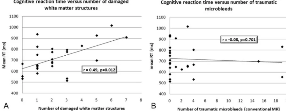
RESULTS: DTI measures revealed several predominant regions of damage including the anterior corona radiata (41% of the patients), uncinate fasciculus (29%), genu of the corpus callosum (21%), inferior longitudinal fasciculus (21%), and cingulum bundle (18%). The number of damaged white matter structures as quantified by DTI was significantly correlated with mean reaction time on a simple cognitive task (r⫽0.49,P⫽.012). In contradistinction, the number of traumatic microhemorrhages was uncorrelated with reaction time (r⫽ ⫺0.08,P⫽.71).
CONCLUSION:Microstructural white matter lesions detected by DTI correlate with persistent cognitive deficits in mild TBI, even in populations in which conventional measures do not. DTI measures may thus contribute additional diagnostic information related to DAI.
T
raumatic brain injury (TBI) is the leading cause of death and disability in young people, with 1.4 million annually reported cases in the United States and an estimated 57 million people worldwide hospitalized with 1 or more TBIs.1Further-more, approximately 80% of the hospital-reported patients with TBI are categorized as having mild TBI on the basis of a Glasgow Coma Scale score between 13 and 15. Although those patients with mild TBI with normal CT findings and no traumatic amnesia usually have complete resolution of post-traumatic symptoms within 1 month, approximately 30% of patients with mild TBI with posttraumatic amnesia have per-sistent posttraumatic symptoms, and a significant number at 1-year postinjury have decreased functional outcome.2,3
Structural imaging studies of acute TBI demonstrate that MR imaging is more sensitive than CT in the number of trau-matic lesions visualized.4However, the relationship between
focal structural lesions detected by conventional MR imaging and long-term patient outcome is controversial.3,5-7 Never-theless, patients with TBI with posttraumatic symptoms often have cognitive impairment, and their cognitive function is a major predictor of poor outcome.8-12In particular, attention, working memory, cognitive manipulation of temporal infor-mation, and processing speed are vulnerable.13,14Sequelae of
TBI cause significant disability, which compelled the National Institutes of Health (NIH) to declare mild TBI as a major public health problem.15
Although conventional MR imaging techniques can readily visualize posttraumatic focal structural lesions, they fail to ad-equately detect diffuse axonal injury (DAI), the key mecha-nism of damage following TBI.16DAI results from unequal
rotational or acceleration/deceleration forces that cause mul-tifocal lesions in white matter due to a shear-strain deforma-tion.17-19DAI is primarily responsible for transient deficits in
cognitive performance in domains such as processing speed, working memory, and attention.20,21More recent studies
sug-gest that DAI causes persistent postconcussive symptoms in executive function and memory dysfunction.8,22-25
MR diffusion tensor imaging (DTI) may be used to better assess DAI. In DTI, the characteristics of water diffusion in the brain are used to assess microstructural integrity of white mat-ter pathways.26In white matter, water diffuses more readily
along the orientation of axonal fibers than across the fibers due
Received October 5, 2007; accepted after revision December 4.
From the Department of Psychiatry (S.N.N., R.A.K., B.D.M.), Sackler Institute, Weill Medical College of Cornell University, New York, NY; Department of Radiology (P.M.), University of California, San Francisco, San Francisco, Calif; Brain Trauma Foundation (J.G., R.A.K., R.S.), New York, NY; Departments of Neurological Surgery (J.G.) and Radiology (C.J., R.D.Z.), Weill Medical College of Cornell University, New York, NY; Department of Neurological Surgery (H.L., M.M., G.T.M.), University of California, San Francisco, Calif.
S.N. Niogi and P. Mukherjee contributed equally to this work.
This work was supported by a collaborative grant from the James S. McDonnell Foundation to the Brain Trauma Foundation with lead investigators of the Cognitive Neurobiological Research Consortium from Weill Cornell Medical College, University of California San Francisco, and University of California Berkeley.
Please address correspondence to Bruce D. McCandliss, MD, Sackler Institute, Department of Psychiatry, Weill Medical College of Cornell University, 525 E. 68th St, New York, NY 10065; e-mail: [email protected]
Indicates article with supplemental on-line tables
DOI 10.3174/ajnr.A0970
FUNCTIONAL
ORIGINAL
to hindrance from structural elements such as the axolemma and the myelin sheath. One can calculate the apparent diffu-sion coefficient (ADC), which is a rotationally invariant mea-sure of the magnitude of diffusion. The degree of directional-ity of diffusion is termed “anisotropy.” This is the variation in the eigenvalues of the diffusion tensor.27Fractional anisotropy
(FA), a normalized measure of anisotropy, has been shown to be sensitive to microstructural changes in white matter integ-rity.28,29Such measurements quantify the extent of damage following TBI24,30-32and are more sensitive than conventional
MR imaging to axonal injury in a mouse model of TBI.33
In a group of patients with mild TBI with persistent postconcussive symptoms, we tested the hypothesis that the extent of microstructural white matter injury on DTI would account for deficits in cognitive reaction time, whereas the number of focal lesions on conventional MR imaging would not. The purpose of this study was to determine the predom-inant areas of damage in mild TBI and whether the spatial extent of white matter injury on DTI can be used as an effective biomarker for global cognitive outcome.
Methods
Participants
The group with mild TBI consisted of 34 patients (18 male, 16 female) who had Glasgow Coma Scale scores of 13–15 at the time of injury, loss of consciousness, and posttraumatic amnesia. All subjects with mild TBI were examined at least 1 month postinjury (range, 1– 65 months) and had at least 1 persistent postconcussive symptom deter-mined from a self-completed head injury symptom checklist survey. The subset of postconcussive symptoms in the head injury symptom checklist included headaches, fatigue, dizziness, irritability (lack of patience), anxiety or depression, difficulty sleeping, personality
changes, or apathy.34,35Exclusion criteria included any prior history
of TBI and any history of neurologic or psychiatric illness including drug or alcohol abuse. The average age was 37.4 years (range, 16 – 61 years). Control subjects included 26 healthy volunteers (19 male, 7 female) with an average age of 28.3 (range, 17–58 years). Written informed consent was obtained from all subjects in accordance with NIH guidelines and as approved by protocols reviewed by the re-search ethics committees of each participating institution.
MR Imaging and DTI Acquisition and Analysis
MR imaging was performed in accordance with protocols approved by the Internal Review Board of Weill Cornell Medical College or the Committee on Human Research at University of California, San Francisco before testing. MR imaging was acquired on a 3T Excite scanner (GE Healthcare, Milwaukee, Wis) equipped with an 8-chan-nel phased-array head coil. DTI was performed with a multisection
single-shot spin-echo echo-planar pulse sequence (TE⫽63 ms, TR⫽
14 seconds, NEX⫽1) by using 55 diffusion-encoding directions
iso-tropically distributed over the surface of a sphere with electrostatic
repulsion, acquired atb⫽1000 s/mm2, 1 acquisition withb⫽0
s/mm2, 72 interleaved sections of 1.8-mm thickness, each with no gap
between sections, with a 128⫻128 matrix that was zero-filled during
reconstruction to 256⫻256, and an FOV of 230 mm. The total
acquisition time was 13.07 minutes. Images were postprocessed off-line by using DTIstudio software (Johns Hopkins University,
Balti-more, Md)36to obtain FA maps, ADC maps, and directionally
en-coded color FA maps. We acquired the following conventional 3T MR
imaging sequences: 1) axial 3D inversion recovery fast-spoiled
gradi-ent T1-weighted images (TE⫽1.5 ms, TR⫽6.3 ms, TI⫽400 ms, flip
angle⫽15°) with a 230-mm FOV, one hundred fifty-six 1.0-mm
contiguous partitions at a 256⫻256 matrix; 2) axial T2-weighted
fluid-attenuated inversion recovery (FLAIR) images (TE⫽126 ms,
TR⫽10 seconds, TI⫽2200 ms) with a 220-mm FOV, forty-seven to
forty-eight 3.0-mm contiguous sections at a 256⫻256 matrix; and 3)
axial magnetization-prepared gradient echo (MPGR) T2*-weighted
images (TE⫽15 ms, TR⫽500 ms, flip angle⫽20°) with a 220⫻170
mm FOV, and forty-seven to forty-eight 3.0-mm contiguous sections
at a 256⫻192 matrix. Conventional MR images were interpreted by
attending neuroradiologists certified by the American Board of Radiology.
To avoid several confounds associated with spatial normalization of white matter tracts, we adopted a region-of-interest approach to
test specific structures throughout the brain selected in ana priori
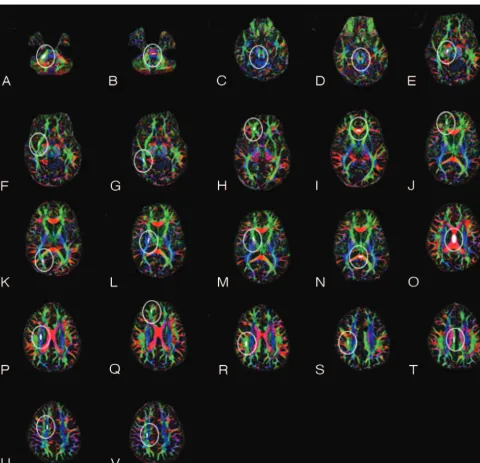
fashion. Each region of interest was placed in an anatomically identi-fiable white matter tract on the directionally encoded color FA images (Fig 1). The region-of-interest procedure consisted of using standard ellipse-shaped regions of interest. Selection and analysis were imple-mented with software written in Interactive Data Language, Version 6.0 (ITT Visual Information Solutions, Boulder, Colo). Ellipses were prescribed on axial directionally encoded anisotropy maps at the cen-ter and within the boundaries of each structure in normal-appearing white matter, as assessed from the conventional MR imaging se-quences. The sizes and dimensions of ellipses were kept constant for each tract across subjects. Subsequently, the anatomic accuracy of the region-of-interest placement was validated by a board-certified neu-roradiologist. Intrarater reliability was assessed from the coefficient of variation of FA from repeat region-of-interest measurements in 5 healthy subjects. The selected structures, following the nomenclature
of Mori et al,37are displayed and listed in Fig 1.
Mean and SD of FA values for each region of interest were re-corded. In patients with TBI, a white matter structure with an FA value reduced by more than 2.5 SDs below the mean for that region of interest in the control subjects was considered damaged. The poste-rior and anteposte-rior centrum semiovales were combined into 1 measure such that the centrum semiovale was defined as damaged if the FA was below the threshold in either region of interest. Similarly, the poste-rior and anteposte-rior infeposte-rior longitudinal fasciculi were combined into 1 measure. For paired structures, an FA value below the 2.5-SD thresh-old in either hemisphere designated the tract as damaged, regardless of whether the FA fell below the threshold in 1 or both hemispheres. The number of damaged white matter structures measured on the DTI scan and the number of traumatic microhemorrhages detected on the T2*-weighted MPGR scan by a board-certified neuroradiolo-gist were tallied and correlated with the global measure of cognitive performance described below.
Cognitive Assessment
The Attention Network Task38provided quantitative assessment of
and a general measure of cognitive reaction time was evaluated by calculating the mean reaction time across the various conditions.
Statistics
Significance was demonstrated by nonparametric statistical tests. The
Spearmanstatistic was used to test for significance of correlations
be-tween FA and mean reaction time as well as bebe-tween the number of
self-reported symptoms and MR imaging findings. The Spearman
sta-tistic was also used to test for any correlations between age and FA in the regions of interest measured in the control and patient groups. In this, a Bonferroni correction was used to correct for multiple comparisons.
Mann-WhitneyUtests were used to test statistical significance when
comparing the mild TBI cohort and the healthy control group.
Results
Conventional MR imaging findings were normal in all 26 con-trol subjects. Demographic data of the patients, including age, sex, Glasgow Coma Scale score, head injury symptom check-list, time since injury, and conventional MR imaging findings are displayed in On-line Table 1. The coefficient of variation for intraoperator reliability was⬍3% for all regions of interest measured. Although a significant difference of age was present between the TBI and control groups (Mann WhitneyUtest⫽ 248.0,P⫽.004), no significant correlations were found be-tween FA and age in any of the regions of interest in either the control or patient groups. On the basis of the MR imaging examination excluding DTI, 11 patients with TBI had normal Fig 1.Region-of-interest placement for DTI analysis. Shown are corresponding regions of interest for the right hemisphere. The solid ellipse within the white outline indicates the location and size of the region of interest.A, Middle cerebral peduncle.B, Pontine crossing tract.C, Superior cerebellar peduncle.D, Decussation of the superior cerebellar peduncle.E, Cerebral peduncle.F, Anterior inferior longitudinal fasciculi.G, Posterior inferior longitudinal fasciculi.H, Uncinate fasciculus.I, Genu of the corpus callosum.J, Forceps minor.K, Forceps major.
[image:3.594.55.536.40.503.2]findings and 11 patients with TBI had traumatic microhem-orrhages on 3T MPGR T2*-weighted imaging. The remaining 12 patients with TBI had either nonspecific white matter hy-perintensities evident on T2-weighted FLAIR images or evi-dence of chronic hemorrhagic contusions. There was no cor-relation between the number of self-reported symptoms (head injury symptom checklist) and MR imaging findings (r ⫽ 0.14,P⫽.46).
For the DTI analysis, a white matter tract was considered to show microstructural injury in a patient with TBI if the FA was reduced by more than 2.5 SDs below the control mean. Fol-lowing this definition, On-line Table 2 summarizes the struc-tures damaged in the patients with mild TBI on the basis of DTI. The most frequently damaged tracts were the anterior corona radiata in 14 of 34 patients and the uncinate fasciculus in 10 of 34 patients. The genu of the corpus callosum was frequently damaged (7/34), as well as the cingulum bundle (7/34) and the inferior longitudinal fasciculi (6/34). The 5 most frequently injured white matter tracts were all located in frontal and temporal lobes, corresponding to the rostral por-tions of the anterior and middle fossae.
Almost one third of the patients with mild TBI had normal findings on conventional 3T MR images of the brain (subjects 4, 8, 13, 18, 19, 21–24, 27, and 32 in On-line Table 1), yet 10 of these 11 patients had at least 1 structure with FA values in the range defined as having microstructural injury.
DTI results were significantly correlated with cognitive re-action time, as measured by the mean rere-action time score in the Attention Network Task. Figure 2Ashows the correlation between mean reaction time and the number of white matter structures with microstructural injury detected by DTI (r⫽ 0.49,P⫽.012). In contradistinction, Fig 2Bshows that the number of traumatic microhemorrhages detected by T2* MPGR did not correlate with mean reaction time (r⫽ ⫺0.08, P⫽.70).
Discussion
Conventional MR imaging and CT often underestimate the extent of axonal injury following a TBI. Patient reports of postconcussion syndrome may be dismissed in the context of
normal findings on CT or conventional MR imaging. Even when focal structural lesions such as traumatic microhemor-rhages are identified by routine clinical neuroimaging, their prognostic value for patient outcome is uncertain.6,7,39In this
3T DTI study, we showed variable extent of microstructural injury in normal-appearing white matter in a cohort of 34 patients with isolated mild TBI. This study also demonstrated that 10 of 11 patients with mild TBI with no abnormalities on conventional 3T MR imaging had evidence of microstructural white matter injury on DTI. Most important, the extent of microstructural white matter injury on DTI correlated with impaired cognitive reaction time, unlike the number of trau-matic microhemorrhages detected on conventional MR imaging.
Evidence of DAI in Mild TBI
Despite the high incidence of TBI in the population, there are relatively few studies of subjects with TBI with postconcussive symptoms.9 There are rare cases in which neuropathologic
information can be acquired from human postmortem studies typically involving patients who die from other causes soon after their injury. For example, Blumbergs et al40reported 5
patients with mild TBI who died of other causes, with DAI based on postmortem histopathology. Bigler9reported that a
patient with mild TBI had working memory deficits and, at autopsy, was noted to have hemosiderin-laden macrophages in the perivascular space and macrophages in the white matter particularly in the frontal lobe. A gross macroscopic inspec-tion of the brain at autopsy was unrevealing, indicating the need for techniques that can probe microscopic structural changes.9
These studies and others indicate axonal shear injury to be the primary mechanism of damage in mild TBI.17,19Most in-teresting, in a mouse model of TBI, reduced anisotropy on DTI correlated with the attenuation of axons stained with amyloid precursor protein, suggesting that DTI is indeed sen-sitive to the microstructural effects of traumatic axonal inju-ry.33This mouse study showed that conventional MR imaging
was not as sensitive as DTI to axonal injury, consistent with the results of our study in human patients with TBIs. This pattern Fig 2.A, Correlation of the number of damaged white matter tracts and speed of processing. The correlation is statistically significant (P⫽.012) withr⫽0.49.B, In contradistinction, the corresponding analysis, by using conventional MR imaging of the number of traumatic microhemorrhages correlated with speed of processing, is not statistically significant (r⫽ ⫺0.08,
[image:4.594.65.521.47.224.2]of conventional MR imaging underdiagnosing the extent of damage has been documented in previous studies of patients with TBI with mild-to-severe head injury.41,42Although the
exact mechanisms that cause changes in DTI measures such as FA are not fully understood, it is generally accepted that loss of the microstructural integrity of white matter reduces FA val-ues.30Disruption of the parallel organization of fiber tracts,
loss of myelination, and increased permeability between inter-nal and exterinter-nal axointer-nal environments are plausible conse-quences of DAI.17,43,44
DAI and Pattern of DTI Lesions
To study the pattern of microstructural axonal injury, we de-fined a white matter pathway to be damaged if the FA value was reduced to values⬍2.5 SDs below the mean FA of the corresponding structure in the control group. Although DTI can change significantly when broad age ranges are consid-ered,28we did not match the age of the control group (used to
derive an objective criterion indicating damage in FA within each region of interest) to the age of the patient group overall, or to each patient specifically. Thus, this criterion might change with age in a systematic fashion, which could, in turn, impact the data reported. The highest rate of FA changes, how-ever, is in the early years of life. A recent study shows that, when using a region-of-interest analysis, only FA of the cen-trum semiovale shows a correlation with age in a young adult group.45 Furthermore, no significant correlations existed in
any of our regions of interest measured between age and FA in either the healthy group or the control group, suggesting that criterion estimates may be fairly stable over the age range in this study. Given the relative stability of FA in adults and the conservative threshold chosen to define a lesion, it is unlikely that age had a significant impact on the categorization of pa-tient results.
With the 2.5-SD threshold, in a healthy population, an FA measure under this threshold would occur⬍0.62% of the time. That means that only approximately 4 false-positives would be expected in the multiple comparisons of On-line Table 2 if the patients with TBI did not differ from the control sample. However, as seen in On-line Table 2, there are many more locations throughout the brain where reduced FA, fall-ing beyond this criterion for damage, occurs after a mild TBI. Given the conservative nature of this threshold, it is likely that smaller or minor white matter lesions would not be detected by this statistical test. Nevertheless, the data clearly show that frontal and temporal lobe fibers, especially the anterior corona radiata and uncinate fasciculus, tend to be selectively dam-aged. This pattern of damage is consistent with a rotational shearing mechanism that would cause the greatest damage to tracts that are the farthest from the axis of rotation at the neck. Other common areas of damage include the genu of the cor-pus callosum, cingulum bundle, and inferior longitudinal fas-ciculi, which are also rostrally located.
Methodology of DTI Measurements
The DTI sequence used in this study contained 55 diffusion-encoding directions. There has been debate in the literature about the optimal number and orientational distribution of diffusion-encoding directions; however, the emerging con-sensus is that diffusion tensor estimation is more robust with
data acquired from many diffusion-encoding directions rather than repeated scans of the minimal (ie, 6) number of directions.46,47The rationale for sampling more directions is
that this reduces the orientational dependence and increases the accuracy and precision of diffusion tensor parameters such as FA, mean diffusivity, and eigenvalues and eigenvectors. In other words, measurement errors will not be as dependent on relative orientation of the measured diffusion tensor com-pared with the set of diffusion gradient directions. According to 1 Monte Carlo computer simulation study,46 at least 20
unique directions are necessary for a robust estimation of an-isotropy, whereas at least 30 directions are required for a ro-bust estimation of tensor orientation (ie, the primary eigen-vector) and mean diffusivity. This has been validated in real data from the human brain, which show that there is less bias and greater precision in FA measurements made with greater numbers of diffusion-encoding directions.47
Although region-of-interest analysis is the most commonly used method for DTI research and has many merits, there are limitations to its use in the study of TBI. Region-of-interest analysis only lends itself to sample a finite number of regions, despite the fact that axonal injury is often diffuse. The rater for this study placed regions of interest only on normal-appearing white matter, avoiding areas that appeared damaged on con-ventional MR imaging. Hence, it is likely that the extent of damage determined from this region-of-interest analysis of FA maps is underestimated.
Another method for analyzing DTI is a voxel-based ap-proach. Salmond et al25used this technique in a study of 16 chronic TBI survivors, reporting FA changes in a number of white matter tracts that revealed a heterogeneous distribution of damage among the cohort. Although voxel-based analysis is operator-independent, fast, and suited for group analyses, there are concerns as to the accuracy of the final measurement due to errors that may arise from the required spatial normal-ization and coregistration processes. The accuracy of the nor-malization may be compromised by the presence of focal le-sions and atrophy in some patients, abnormalities that are heterogeneous in their spatial extent and distribution across subjects. In our study, we chose to use a region-of-interest technique to avoid confounds from such normalization and coregistration errors. An added advantage of the region-of-interest approach is that it is appropriate for individual anal-ysis, unlike voxel-based techniques that have yet to establish their validity in single-subject analysis.
Correlation of DTI with Cognitive Deficits
Our finding of a lack of correlation between conventional MR imaging results and patient outcome in TBI is consistent with recent studies. Scheid et al7defined DAI in 66 patients with
impairments, especially memory and executive function. However, there was no correlation between the number of microhemorrhages and global cognitive performance. A 1T MR imaging study of 80 patients with acute mild TBI demon-strated no correlation of conventional MR imaging lesions with neurocognitive tests of memory, learning, attention, and executive function at long-term follow-up.39
There is mounting evidence that individual differences in microstructural white matter integrity account for variation in a wide range of cognitive skills. For example, groups have shown significant correlations of frontoparietal white matter FA values to memory performance and of frontostriatal FA measurements to cognitive control.48-52However, there is a dearth of prior work relating DTI to neurocognitive function in TBI. Two prior DTI studies of TBI showed that reduced white matter FA values are correlated with the Glasgow Coma Score at presentation24,53and with the modified Rankin Scale score at hospital discharge.24A prior DTI investigation has
evaluated neurocognitive status, finding that ADC is associ-ated with impaired learning and working memory in patients with chronic TBI and that there are significant bilateral de-creases in anisotropy in major white matter tracts in the tem-poral, frontal, parietal, and occipital lobes.25
In a more recent DTI study of TBI, Kraus et al54examined
a group of 20 patients with mild TBI and demonstrated that these subjects had reduced FA in the corticospinal tract (which corresponds to our superior corona radiata, posterior limb of the internal capsule, and cerebral peduncle), sagittal stratum (which corresponds to our forceps major), and superior lon-gitudinal fasciculus. In their study, a 1-SD threshold (in con-trast to the 2.5-SD threshold used in our study) below the control mean FA was used to indicate reduced anisotropy. Using this criterion, Kraus et al found an average of 5.9 regions of interest with reduced FA in their subjects with mild TBI, but, most important, also reported that control subjects had, on average, 3.6 regions of interest with reduced FA. Thus, with such a criterion, it is difficult to assess whether FA reduced by 1 SD is due to normal interindividual variability in white mat-ter integrity or is truly a result of DAI. In contradistinction, with a 2.5-SD threshold, none of our control subjects had re-duced FA, suggesting that FA values below this are likely pathologic in nature. Kraus et al also found that the number of structures with reduced FA correlated with executive, atten-tional, and memory measures. This is consistent with our finding that the extent of posttraumatic microstructural ax-onal injury on DTI correlates with reduced cognitive reaction time, suggesting that DTI can reveal a biologic substrate for cognitive impairments in TBI.
Anterior corona radiata fibers pass through the Brodmann area 32, which consists of the dorsal anterior cingulate. Addi-tionally, uncinate fasciculus fibers pass through the Brod-mann area 11, which is part of the frontal cortex, covering the medial portion of the frontal lobe and connecting to the tem-poral lobe at Brodmann areas 20, 28, 34, and 36. DTI research in schizophrenia indicates that FA reduction in this pathway corresponds to poor verbal and visual memory perfor-mance.55,56The relative frequency of injury to the anterior corona radiata and the uncinate fasciculus, as well as to the cingulum and the genu of the corpus callosum, may explain the impairment of cognitive reaction time found in this study
of mild TBI. Future studies will more closely examine the neu-ronal circuitry involved in cognitive domains commonly im-paired following TBI to predict cognitive deficits on the basis of physical injury. This research will also investigate whether relationships of normal interindividual variation of cognitive function and white matter integrity are also evident in the extended range of function and integrity that exist in patients with TBI. Prospective longitudinal assessment of patients with TBI from the acute phase through the chronic phase is needed to understand the relationship between the evolution of mi-crostructural white matter injury and the recovery of function after TBI, which is underway at the Cognitive Neurobiological Research Consortium in Traumatic Brain Injury.
Conclusion
We find that the extent of microstructural white matter injury on DTI in patients with mild TBI with postconcussive syn-drome is associated with poorer reaction time in a simple cog-nitive task, whereas the number of traumatic microhemor-rhages on conventional 3T MR imaging is not. This study provides preliminary evidence that DTI may serve as a micro-structural imaging biomarker for long-term neurocognitive impairments in TBI.
References
1. Langlois JA, Rutland-Brown W, Wald MM.The epidemiology and impact of traumatic brain injury: a brief overview. J Head Trauma Rehabil
2006;21:375–78
2. Dikmen S, McLean A Jr, Temkin NR, et al.Neuropsychologic outcome at one-month postinjury.Arch Phys Med Rehabil1986;67:507–13
3. van der Naalt J, Hew JM, van Zomeren AH, et al.Computed tomography and magnetic resonance imaging in mild to moderate head injury: early and late imaging related to outcome.Ann Neurol1999;46:70 –78
4. Mittl RL, Grossman RI, Hiehle JF, et al.Prevalence of MR evidence of diffuse axonal injury in patients with mild head injury and normal head CT findings.
AJNR Am J Neuroradiol1994;15:1583– 89
5. Hammoud DA, Wasserman BA.Diffuse axonal injuries: pathophysiology and imaging.Neuroimaging Clin N Am2002;12:205–16
6. Scheid R, Preul C, Gruber O, et al.Diffuse axonal injury associated with chronic traumatic brain injury: evidence from T2*-weighted gradient-echo imaging at 3T.AJNR Am J Neuroradiol2003;24:1049 –56
7. Scheid R, Walther K, Guthke T, et al.Cognitive sequelae of diffuse axonal injury.Arch Neurol2006;63:418 –24
8. Himanen L, Portin R, Isoniemi H, et al.Cognitive functions in relation to MRI findings 30 years after traumatic brain injury.Brain Inj2005;19:93–100 9. Bigler ED.Neuropsychological results and neuropathological findings at
au-topsy in a case of mild traumatic brain injury.J Int Neuropsychol Soc
2004;10:794 – 806
10. Arcia E, Gualtieri CT.Neurobehavioural performance of adults with closed-head injury, adults with attention deficit, and controls. Brain Inj
1994;8:395– 404
11. Hellawell DJ, Taylor RT, Pentland B.Cognitive and psychosocial outcome fol-lowing moderate or severe traumatic brain injury.Brain Inj1999;13:489 –504 12. Kaplan CP, Corrigan JD.The relationship between cognition and functional independence in adults with traumatic brain injury.Arch Phys Med Rehabil
1994;75:643– 47
13. Levin HS.Memory deficit after closed head injury.J Clin Exp Neuropsychol
1990;12:129 –53
14. Stuss DT, Ely P, Hugenholtz H, et al.Subtle neuropsychological deficits in patients with good recovery after closed head injury. Neurosurgery
1985;17:41– 47
15. Ragnarsson KT.Results of the NIH consensus conference on “rehabilitation of persons with traumatic brain injury.”Restor Neurol Neurosci2002;20:103– 08 16. Medana IM, Esiri MM.Axonal damage: a key predictor of outcome in human
CNS diseases.Brain2003;126:515–30
17. Povlishock JT, Katz DI.Update of neuropathology and neurological recovery after traumatic brain injury.J Head Trauma Rehabil2005;20:76 –94 18. Smith DH, Meaney DF, Shull WH.Diffuse axonal injury in head trauma.
J Head Trauma Rehabil2003;18:307–16
20. Wallesch CW, Curio N, Galazky I, et al.The neuropsychology of blunt head injury in the early postacute stage: effects of focal lesions and diffuse axonal injury.J Neurotrauma2001;18:11–20
21. Wallesch CW, Curio N, Kutz S, et al.Outcome after mild-to-moderate blunt head injury: effects of focal lesions and diffuse axonal injury.Brain Inj
2001;15:401–12
22. Fork M, Bartels C, Ebert AD, et al.Neuropsychological sequelae of diffuse traumatic brain injury.Brain Inj2005;19:101– 08
23. Himanen L, Portin R, Isoniemi H, et al.Longitudinal cognitive changes in traumatic brain injury: a 30-year follow-up study.Neurology2006;66:187–92 24. Huisman TA, Schwamm LH, Schaefer PW, et al.Diffusion tensor imaging as potential biomarker of white matter injury in diffuse axonal injury.AJNR Am J Neuroradiol2004;25:370 –76
25. Salmond CH, Menon DK, Chatfield DA, et al.Diffusion tensor imaging in chronic head injury survivors: correlations with learning and memory indi-ces.Neuroimage2006;29:117–24
26. Basser PJ, Pierpaoli C.Microstructural and physiological features of tissues eluci-dated by quantitative-diffusion-tensor MRI.J Magn Reson B1996;111:209 –19 27. Ulug AM, van Zijl PC.Orientation-independent diffusion imaging without
tensor diagonalization: anisotropy definitions based on physical attributes of the diffusion ellipsoid.J Magn Reson Imaging1999;9:804 –13
28. Mukherjee P, McKinstry RC.Diffusion tensor imaging and tractography of human brain development.Neuroimaging Clin N Am2006;16:19 – 43, vii 29. Watts R, Liston C, Niogi S, et al.Fiber tracking using magnetic resonance
diffusion tensor imaging and its applications to human brain development.
Ment Retard Dev Disabil Res Rev2003;9:168 –77
30. Arfanakis K, Haughton VM, Carew JD, et al.Diffusion tensor MR imaging in diffuse axonal injury.AJNR Am J Neuroradiol2002;23:794 – 802
31. Huisman TA.Diffusion-weighted imaging: basic concepts and application in cerebral stroke and head trauma.Eur Radiol2003;13:2283–97
32. Lee ZI, Byun WM, Jang SH, et al.Diffusion tensor magnetic resonance imaging of microstructural abnormalities in children with brain injury.Am J Phys Med Rehabil2003;82:556 –59
33. Mac Donald CL, Dikranian K, Song SK, et al.Detection of traumatic axonal injury with diffusion tensor imaging in a mouse model of traumatic brain injury.Exp Neurol2007;205:116 –31. Epub 2007 Feb 12
34. Kay T, Harrington DE, Adams R, et al.Definition of mild traumatic brain injury.J Head Trauma Rehabil1993;8:86 – 87
35. McLean A Jr, Dikmen S, Temkin N, et al.Psychosocial functioning at 1 month after head injury.Neurosurgery1984;14:393–99
36. Jiang H, van Zijl PC, Kim J, et al.DTIStudio: resource program for diffusion tensor computation and fiber bundle tracking.Comput Methods Programs Biomed2006;81:106 –16. Epub 2006 Jan 18
37. Mori S, Wakana S, Van Zijl PCM. MRI Atlas of Human White Matter.
Amsterdam: Elsevier; 2005
38. Fan J, McCandliss BD, Fossella J, et al.The activation of attentional networks.
Neuroimage2005;26:471–79
39. Hughes DG, Jackson A, Mason DL, et al.Abnormalities on magnetic resonance
imaging seen acutely following mild traumatic brain injury: correlation with neu-ropsychological tests and delayed recovery.Neuroradiology2004;46:550 –58 40. Blumbergs PC, Scott G, Manavis J, et al.Staining of amyloid precursor protein
to study axonal damage in mild head injury.Lancet1994;344:1055–56 41. Anderson CV, Wood DM, Bigler ED, et al.Lesion volume, injury severity, and
thalamic integrity following head injury.J Neurotrauma1996;13:59 – 65 42. Gale SD, Johnson SC, Bigler ED, et al.Trauma-induced degenerative changes
in brain injury: a morphometric analysis of three patients with preinjury and postinjury MR scans.J Neurotrauma1995;12:151–58
43. Inglese M, Makani S, Johnson G, et al.Diffuse axonal injury in mild traumatic brain injury: a diffusion tensor imaging study.J Neurosurg2005;103:298 –303 44. Povlishock JT, Erb DE, Astruc J.Axonal response to traumatic brain injury: reactive axonal change, deafferentation, and neuroplasticity.J Neurotrauma
1992;9(suppl 1):S189 –200
45. Snook L, Plewes C, Beaulieu C.Voxel based versus region of interest analysis in diffusion tensor imaging of neurodevelopment.Neuroimage2007;34:243–52. Epub 2006 Oct 27
46. Jones DK.The effect of gradient sampling schemes on measures derived from diffusion tensor MRI: a Monte Carlo study.Magn Reson Med2004;51:807–15 47. Landman BA, Farrell JA, Jones CK, et al.Effects of diffusion weighting schemes on the reproducibility of DTI-derived fractional anisotropy, mean diffusivity, and principal eigenvector measurements at 1.5T.Neuroimage2007;36:1123–38 48. Liston C, Watts R, Tottenham N, et al.Frontostriatal microstructure
modu-lates efficient recruitment of cognitive control.Cereb Cortex2006;16:553– 60 49. Nagy Z, Westerberg H, Klingberg T.Maturation of white matter is associated
with the development of cognitive functions during childhood.J Cogn Neuro-sci2004;16:1227–33
50. Niogi SN, McCandliss BD.Left lateralized white matter microstructure ac-counts for individual differences in reading ability and disability. Neuropsy-chologia2006;44:2178 – 88. Epub 2006 Mar 9
51. Olesen PJ, Nagy Z, Westerberg H, et al.Combined analysis of DTI and fMRI data reveals a joint maturation of white and grey matter in a fronto-parietal network.Brain Res Cogn Brain Res2003;18:48 –57
52. Tuch DS, Salat DH, Wisco JJ, et al.Choice reaction time performance corre-lates with diffusion anisotropy in white matter pathways supporting visuo-spatial attention.Proc Natl Acad Sci U S A2005;102:12212–17. Epub 2005 Aug 15
53. Benson RR, Meda SA, Vasudevan S, et al.Global white matter analysis of dif-fusion tensor images is predictive of injury severity in traumatic brain injury.
J Neurotrauma2007;24:446 –59
54. Kraus MF, Susmaras T, Caughlin BP, et al.White matter integrity and cogni-tion in chronic traumatic brain injury: a diffusion tensor imaging study.Brain
2007;130:2508 –19
55. Nakamura M, McCarley RW, Kubicki M, et al.Fronto-temporal disconnectiv-ity in schizotypal personaldisconnectiv-ity disorder: a diffusion tensor imaging study.Biol Psychiatry2005;58:468 –78. Epub 2005 Jun 22