PREVALENCE OF ROOT MORPHOLOGY USING CONE BEAM
COMPUTED TOMOGRAPHY; A RETROSPECTIVE STUDY.
Dr. Nimmi Singh1, Dr. Devika Singh*2, Dr. Samar Ali Faraz3 and Dr. Mamta Sharma4
1
Reader, Department of Oral Medicine & Radiology. Buddha Institute of Dental Sciences &
Hospital, Patna.
2
Senior Resident, Indira Ghandhi Institute of Medical Sciences, Patna.
3
MDS, Department of Oral Medicine & Radiology.
4
MDS, Department of Oral Medicine & Radiology. Jammu Government College, Jammu.
ABSTRACT
Aim- To evaluate the root and canal morphology of maxillary first and
second premolar by Cone Beam Computed Tomography (CBCT) -A
retrospective study. Material and method- The following study was
conducted at CBCT centre where CBCT scans of patients who were
undergoing various dental treatment were evaluated. The scans were
evaluated to study the number of canals and their morphology in
maxillary first and second premolar. Result- Out of 120 maxillary first
premolar 54 (45%) had one root, 61 (51%) had two root and 5 (4.1%)
had three root. Out of 135 maxillary second premolar 110(81.4%) had
one root, 20 (14.8%) had two root and 5 (3.7%) had three root. Conclusion- No significant
correlation was found between root number and tooth position in both first and second
maxillary premolar (P=0.35 & P=0.41 respectively for first and second maxillary premolar.
KEYWORDS: Cone Beam Computed Tomography, maxillary premolar.
INTRODUCTION
Traditional radiography is limited in its ability to give reliable information on the number and
morphology of root canals. The application of cone-beam computed tomography (CBCT)
provides a non-invasive three-dimensional confirmatory diagnosis as a complement to
conventional radiography[1], A thorough knowledge of root canal morphology is essential for successful endodontic treatment. As a group, the mandibular premolars are among the most
difficult teeth to treat endodontically, because they have a high incidence of multiple roots or
Volume 7, Issue 9, 1635-1640. Research Article ISSN 2277– 7105
Article Received on 18 March 2018,
Revised on 08 April 2018, Accepted on 28 April 2018
DOI: 10.20959/wjpr20189-12193
*Corresponding Author
Dr. Devika Singh
canals.[2] Cone beam computed tomography (CBCT) can provide dentists with high-quality 3-dimensional images of dental structures because of its high spatial resolution.[3]
This study was conducted to evaluate the root canal morphology of maxillary first and second
premolars in our population by using cone-beam computed tomography (CBCT).
MATERIALS AND METHODS
The present study was conducted at CBCT centre Patna, India. Images were taken from
Patient attending for CBCT scanning for dental treatment. CBCT images were obtained by
I-CAT 17-19 Platinum machine. 8×8 FOV with voxel size .125 (resolution) with exposure time
of 7 sec, KV 120, mA 5. were used. CBCT images of 120 scans for maxillary first premolar
and 135 for maxillary second premolar were visualized in 3 orthogonal planes i.e axial,
coronal and sagittal sections. All the images were scroll from pulp chamber region upto
apical end of root of each tooth.[4] Root number, canal configuration, (Vertussi's configuration), Number of apical foramen per root. Number of roots in axial plane (Pecora et
al) were recorded.
The inclusion criteria included images who currently taken, tooth with no periapical
pathology, no obturated tooth, tooth undergoing any treatment. Informed consent was
obtained from all patients prior to scan Details of all parameters of scanned images were
recorded. Statistical analysis was done. Regarding interexaminar agreement, the Cohen
Kappa value was determine for first and second premolar.
RESULTS
Table shows the frequency distribution of the number of roots according to tooth position.
Out of 120 maxillary first premolar 54 (45%) had one root, 61 (51%) had two root and 5
(4.1%) had three root. Out of 135 maxillary second premolar 110(81.4%) had one root, 20
(14.8%) had two root and 5 (3.7%) had three root. No significant correlation was found
between root numberand tooth position in both first and second maxillary premolar (P=0.35
Tooth position One root (%) Two root (%) Three root (%) Total
Maxillary first premolar
Right 21 (46%) 22 (49%) 2 (5%) 45
Left 33 (44%) 39 (52%) 3 (4%) 75
Total 54 (45%) 61 (51%) 5 (4.1%) 120
Maxillary second premolar
Right 43 (78%) 10 (18%) 2 (4%) 55
Left 67 (84%) 10 (13%) 3 (3%) 80
Total 110 (81.4%) 20 (14.8%) 5 (3.7%) 135
Maxillary First premolar
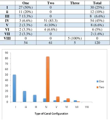
Table shows prevalence of different root canal type. The most prevalent root canal type
among maxillary first premolar was type IV (n=54, 45%) followed by type I (n=30, 25%) and
type II (n=12, 10%). Among Single rooted first premolar type I found in 27 (50%) patients
and type II found in 11 (20%) patients. Most two rooted first premolar exhibited a type IV
canal configuration (n=51, 83.3%).
One Two Three Total
I 27 (50%) 0 30 (25%)
ii 11 (20%) 0 12 (10%)
III 7 13.3%) 0 8 (6.6%)
IV 3 (6.6%) 51 (83.3) 54 (45%)
V 2 (3.3%) 6 (10%) 8 (6.6%)
VI 2 (3.3%) 4 (6.6%) 6 (5%)
VII 2 (3.3%) 0 2 (1.6%)
VIII 0 0 5 (100%) 0
[image:3.595.122.477.351.739.2]Maxillary Second premolar
Table shows prevalence of different root canal type. The most prevalent root canal type
among maxillary second premolar was type I (n=54, 40%) followed by type IV (n=38,
28.3%) and type II (n=20, 15%). Among Single rooted second premolar type I found in 52
(47%) patients and type II found in 34 (31%) patients. Most two rooted second premolar
exhibited a type IV canal configuration (n=10, 50%).
One Two Total
I 52 (47%) 7 (33.3%) 54 (40%)
II 34 (31%) 0 20 (15%)
III 10 (9.5%) 0 7 (5%)
IV 6 (5.6%) 10 (50%) 38 (28.3%)
V 4 (3.3%) 2 (10%) 9 (6.6%)
VI 0 1 (5%) 5(3.3%)
VII 4 (3.3%) 0 2 (1.6%)
VIII 0 0 5 (100%) 0
110 20 5 135
Regarding interexaminar agreement, the Cohen Kappa value was determine for first and
second premolar whiich was found to be 0.919 and 0902 respectively.
DISCUSSION
In the present study, the number of canals and their morphology were evaluated using CBCT
I-CAT machine.
The study was conducted using scans of patients who were had visited Cbct centre seeking
[image:4.595.100.496.207.577.2]CBCT films were retrieved from the database and differents sections, i.e, axial, coronal and
sagittal sections were evaluated to study the number of pulp canals and their morphology in
maxillary first and second premolar. Thus the subjects were not exposed to any radiations.
A total number of 255 scans of maxillary first and second premolar were evaluated.
Out of 120 maxillary first premolar 54 (45%) had one root, 61 (51%) had two root and 5
(4.1%) had three root. Out of 135 maxillary second premolar 110(81.4%) had one root, 20
(14.8%) had two root and 5 (3.7%) had three root.
The most prevalent root canal type among maxillary first premolar was type IV (n=54, 45%)
followed by type I (n=30, 25%) and type II (n=12, 10%). Among Single rooted first premolar
type I found in 27 (50%) patients and type II found in 11 (20%) patients. Most two rooted
first premolar exhibited a type IV canal configuration (n=51, 83.3%).
The most prevalent root canal type among maxillary second premolar was type I (n=54, 40%)
followed by type IV (n=38, 28.3%) and type II (n=20, 15%). Among Single rooted second
premolar type I found in 52 (47%) patients and type II found in 34 (31%) patients. Most two
rooted second premolar exhibited a type IV canal configuration (n=10, 50%).[6]
No significant correlation was found between root numberand tooth position in both first and
second maxillary premolar (P=0.35 & P=0.41 respectively for first and second maxillary
premolar). Abella F et al conducted a similar study in 2015 where 804 images of first and
second premolar were evaluated and number of roots, root canal configuration (Vertucci's
classification), number of root canals, and number of apical foramina per root and used the
χ(2) test to analyze the correlation between root number and tooth position. No statistical
correlation was evident between root number and gender and tooth position.
CONCLUSION
Since this study was conducted on a small group, a more extensive study needs to be
conducted in order to be able to evaluate the morphology of root canal for the population of
Bihar In vivo CBCT analysis is a noninvasive and clinically effective tool for examining root
REFRENCES
1. Xuan Yu†, Cone-beam computed tomography study of root and canal morphology of mandibular premolars in a western Chinese population BMC Medical Imaging, 2012; 12:
18.
2. Torabinejad M, Walton RE: Principles and Practice of Endodontics. 2009, Saunders, St.
Louis, 216-218. 4.
3. Jérôme Michetti, Validation of Cone Beam Computed Tomography as a Tool to Explore
Root Canal Anatomy DOI: https://doi.org/10.1016/j.joen.2010.03.029
4. Sezer Demirbuga et al, Use of Cone Beam Computed Tomography to evaluate root and
canal morphology of mandibular first and secong molars in turkish individuals. Med Oral
Patol Oral Cir Bucal., 2013 Jul; 18(4): e737- e744.
5. Frances Abella et al Cone Beam Computed Tomography analysis of root canal
morphology first and second premolars in spanish population. JOE, August 2015; 41: 8.
6. Tian Y.‐Y, Guo B, Zhang R, Yu X, Wang H, Hu T, Dummer PMH. Root and canal
morphology of maxillary first premolars in a Chinese subpopulation evaluated using
cone‐beam computed tomography. International Endodontic Journal., 2012; 45: