Diaphragmatic Dysfunction in Paediatrics
Avaliação Ecográfica da Disfunção Diafragmática
Induzida pelo Ventilador em Idade Pediátrica
Maria Teresa DIONÍSIO1, Armanda REBELO2, Carla PINTO1,2, Leonor CARVALHO1, José Farela NEVES1 Acta Med Port 2019 Jul–Aug;32(7–8):520–528 ▪ https://doi.org/10.20344/amp.10830
1. Serviço de Cuidados Intensivos Pediátricos. Hospital Pediátrico de Coimbra. Centro Hospitalar e Universitário de Coimbra. Coimbra. Portugal. 2. Clínica Universitária de Pediatria. Faculdade de Medicina. Universidade de Coimbra. Coimbra. Portugal.
Autor correspondente: Maria Teresa Dionísio. [email protected]
Recebido: 23 de maio de 2018 – Aceite: 31 de janeiro de 2019 | Copyright © Ordem dos Médicos 2019 ABSTRACT
Introduction: Invasive mechanical ventilation contributes to ventilator-induced diaphragmatic dysfunction, delaying extubation and increasing mortality in adults. Despite the possibility of having a higher impact in paediatrics, this dysfunction is not routinely monitored. Diaphragm ultrasound has been proposed as a safe and non-invasive technique for this purpose. The aim of this study was to describe the evolution of diaphragmatic morphology and functional measurements by ultrasound in ventilated children.
Material and Methods: Prospective exploratory study. Children admitted to Paediatric Intensive Care Unit requiring mechanical ven-tilation > 48 hours were included. The diaphragmatic thickness, excursion and the thickening fraction were assessed by ultrasound.
Results: Seventeen cases were included, with a median age of 42 months. Ten were male, seven had comorbidities and three in seventeen had malnutrition at admission. The median time under mechanical ventilation was seven days. The median of the initial and minimum diaphragmatic thickness was 2.3 mm and 1.9 mm, respectively, with a median decrease in thickness of 13% under pressure-regulated volume control. Diaphragmatic atrophy was observed in 14/17 cases. Differences in the median thickness variation were found between patients with sepsis and without (0.70 vs 0.25 mm; p = 0.019). During pressure support ventilation there was a
tendency to increase diaphragmatic thickness and excursion. Extubation failure occurred for diaphragmatic thickening fraction ≤ 35%.
Discussion: Under pressure-regulated volume control there was a tendency for a decrease in diaphragmatic thickness. In the pre-ex-tubation stage under pressure support, there was a tendency for it to increase. These results suggest that, by titrating ventilation using physiological levels of inspiratory effort, we can reduce the diaphragmatic morphological changes associated with ventilation.
Conclusion: The early recognition of diaphragmatic changes may encourage a targeted approach, namely titration of ventilation, in order to reduce ventilator-induced diaphragmatic dysfunction and its clinical repercussions.
Keywords: Child; Diaphragm/ultrasonography; Respiration, Artificial/adverse effects; Ultrasonography
RESUMO
Introdução: A ventilação mecânica invasiva condiciona disfunção diafragmática, atrasando a extubação e aumentando a mortalidade
em adultos. Em pediatria, apesar de eventualmente mais relevante, essa disfunção não é sistematicamente avaliada. A ecografia dia
-fragmática tem sido proposta como uma técnica não invasiva e segura para esse fim. O objetivo deste estudo foi descrever a evolução dos índices ecográficos de morfologia e função diafragmáticas em crianças ventiladas.
Material e Métodos: Estudo exploratório, prospetivo. Foram incluídas crianças admitidas num Serviço de Cuidados Intensivos
Pediá-tricos sob ventilação mecânica invasiva > 48 horas e realizadas medições ecográficas de espessura, excursão e fração de espessa -mento diafragmáticas.
Resultados: Foram incluídos 17 casos. Mediana de idades: 42 meses. Eram do género masculino 10/17, tinham comorbilidades 7/17 e manifestavam desnutrição na admissão 3/17 casos. Mediana do tempo sob ventilação invasiva: sete dias. Medianas das espes-suras diafragmáticas inicial e mínima: 2,3 e 1,9 mm, respetivamente, tendo-se observado uma diminuição mediana da espessura de
13% sob volume controlado regulado por pressão. Observou-se atrofia diafragmática em 14/17 casos. Verificaram-se diferenças na
mediana da variação da espessura entre os grupos com e sem sépsis (0,70 vs 0,25 mm; p = 0,019). Durante a ventilação em pressão de suporte, observou-se uma tendência para aumento da espessura e excursão diafragmáticas. Ocorreu falência de extubação para
fração de espessamento ≤ 35%.
Discussão: Sob volume controlado regulado por pressão verificou-se tendência para diminuição da espessura diafragmática. Sob pressão de suporte, verificou-se uma tendência para o seu aumento. Estes resultados sugerem que, titulando a ventilação, podemos
reduzir as alterações morfológicas diafragmáticas associadas à ventilação.
Conclusão: O reconhecimento precoce de alterações diafragmáticas poderá fomentar uma abordagem dirigida, de forma a limitar a disfunção diafragmática induzida pelo ventilador e suas repercussões.
Palavras-chave: Criança; Diafragma/ultrassonografia; Respiração Artificial/efeitos adversos; Ultrassonografia
INTRODUCTION
Invasive mechanical ventilation (IMV) is widely used in intensive care units.1,2
Even though it is crucial to the management of critically ill patients, deleterious effects on respiratory muscles have been shown by multiple studies, even with a short-term use.1–4 These are mainly reflected in myofibrillar and mi -tochondrial changes, leading to sarcomere disruption and
intracellular lipid accumulation.3,5,6 There is a decrease in diaphragm thickness, the main respiratory muscle, leading to atrophy and progressive loss in diaphragm contractile function. This is known as ventilator-induced diaphragmatic dysfunction (VIDD).1
ARTIGO ORIGINAL the development of sepsis, the use of corticosteroids,
aminoglycoside antibiotics and neuromuscular blocking agents, as well as the patient’s nutritional status.2,3,6,7
The definition of VIDD in critically ill adult patients is a relatively recent concept, even though its frequency and relevance has been widely described in different publica-tions.1,2,6 A high incidence of diaphragmatic dysfunction has been found, with an interference on the outcomes, delaying extubation, extending the length of stay, increasing mortal-ity5 and leading to poorer outcomes in the paediatric pop-ulation, in whom accessory muscles are more fragile and susceptible to fatigue, involving a more difficult approach to diaphragmatic dysfunction.8
However, despite its relevance, diaphragmatic function is not systematically monitored in intensive care units, sug-gesting that VIDD could be underdiagnosed.2,3
Invasive and troublesome bedside procedures are in-volved in current gold standard methods for the evaluation of diaphragm function in adults, including the measurement of trans-diaphragmatic pressure with oesophageal and gas-tric balloon or magnetic phrenic nerve stimulation.2,3
Therefore, diaphragm ultrasound imaging has emerged as a promising technique for the evaluation of diaphragm morphology and contractile activity.2,9 It is a safe, non-in-vasive, painless and radiation-free technique which has proved to be cost-effective and easy-to-use in patients re-ceiving IMV.1,2,4,5 Diaphragm thickness, excursion and thick-ening fraction (DTf) are the major ultrasound parameters,3
allowing for the identification of dysfunction / paralysis, the evaluation of change in diaphragm thickness in ventilated patients and prediction of extubation outcome.2
Even though ultrasound criteria have already been pub-lished for the evaluation of diaphragm function in adults,2 studies in the paediatric population are still scarce and the prevalence of VIDD in mechanically ventilated paediatric patients is still unknown, in addition to the contribution of other related factors and the impact of dysfunction on clin-ical outcome.
This study was primarily aimed at describing the pro-gression of ultrasound diaphragmatic morphological and functional indices – thickness, thickening fraction and ex-cursion — in mechanically ventilated paediatric patients admitted to the intensive care unit. The assessment of the impact of ventilation modes on ultrasound indices was the secondary aim of the study.
MATERIAL AND METHODS
This was an exploratory observational study with pro-spective data based on diaphragm ultrasound imaging in paediatric patients admitted to the Paediatric Intensive Care Unit [Serviço de Cuidados Intensivos Pediátricos (CIPE)] of the Hospital Pediátrico de Coimbra (HP).
The study was approved by the ethics committee of the institution, according to the Helsinki declaration. A written informed consent has been obtained from each patient’s le-gal representative prior to medical examination.
Study sample
All paediatric patients admitted to the CIPE between 1 Jun 2017 and 31 Mar 2018 (ten-month study period) and meeting the following criteria were included in the study: 1) patients aged between one month and 18 years; 2) patients in need for IMV for more than 48 hours.
These were the exclusion criteria: 1) patients in need for IMV for at least 12 hours within three months prior to the study; 2) neuromuscular disorder or brain stem injury; 3) congenital thoracic malformation; 4) subphrenic abscess; 5) thoracic surgery within the six months prior to the study; 6) chest drain at the site of examination; 7) pneumothorax; 8) post liver transplant.
A total of 329 paediatric patients were admitted to the CIPE during the study period (101 were aged under one month, 85 were not receiving IMV and 109 had received IMV for less than 48 hours, in addition to two patients who had been in need for IMV for over 12 hours within the pre-vious three months and two patients who presented with neuromuscular dysfunction and five who were admitted for liver transplant were excluded from the study, such as two patients who presented with neuromuscular dysfunction and five who were recovering from liver transplant were ex -cluded from the study). Eight patients were not assessed due to technical drawbacks and a total of 17 patients were included in the study.
Patient characteristics and clinical outcomes
Data were based on the analysis of the patient’s clinical records, by using FileMaker Pro-6® and B-ICU.Care® data-base of the CIPE of the HP.
The following variables were obtained and analysed: patient’s age (months), gender, admitting diagnosis, co-morbidities, body weight (kg), height/length (cm), therapy (sedoanalgesia, neuromuscular blockade, aminoglycoside antibiotics and corticosteroids) and presence of sepsis. Data regarding duration of IMV (hours/days), as well as re-garding ventilation parameters and modes, Paediatric Index of Mortality 3 (PIM3)10 and mortality were also obtained.
Daily doses in mg/kg of each drug used during IMV were obtained. All prescribed corticosteroids were converted to equivalent doses of prednisolone, according to the conver-sion formulae described by Martindale: The Complete Drug Reference.11
Body mass index (BMI) and BMI-for-age percentile (P) were obtained according to the World Health Organization reference curves. The presence of malnutrition was defined at BMI ≤ the third percentile (P3).
Extubation failure was defined as the need for re-intu -bation within 48 hours and ventilator-free days were calcu-lated and defined as the number of days from successful weaning from mechanical ventilation (invasive or non-inva-sive) up to day 28 after study inclusion.
Ultrasound assessment
ARTIGO ORIGINAL two-dimensional (B-mode) and M-mode imaging.Three measurements of the ultrasound parameters —
thickness, excursion and thickening fraction - were obtained in each evaluation— and the average value of the three measurements was considered as the final value, in order to establish its reproducibility. Only measurements made on the right hemi-diaphragm were obtained.12
Ultrasound parameters were assessed considering the different phases of invasive mechanical ventilation. Pres-sure regulated volume control (PRVC) mode is usually used in paediatric patients, which is a mode of ventilation that combines volume and pressure control ventilation. Pres-sure support ventilation (PSV) or spontaneous breathing through an endotracheal tube (ETT) are predominantly used in pre-extubation, aimed at maintaining the patient’s inspiratory effort and subsequent inspiratory muscle train-ing.
Considering that the ultrasound evaluation of diaphragm thickness does not depend on maintaining the patient’s in-spiratory effort, its evaluation was carried out in every phase of the ventilation process, while the first measurement was obtained within the first 24 hours of IMV and subsequently on a daily basis over a 24 ± 8 hour period, up to the day of extubation or day 28 of intubation, or in case of transfer to another hospital or death.
On the contrary, the preservation of the patient’s inspira-tory effort is required for an accurate ultrasound evaluation of diaphragmatic excursion and thickening fraction. There-fore, these were only evaluated on a daily basis in patients in pre-extubation, during PSV or spontaneous breathing through an endotracheal tube.
In the presence of extubation failure, in need for a new ventilation cycle, a daily evaluation of these ultrasound pa-rameters was carried out in pre-extubation, while each cy-cle was considered as an independent evaluation moment. All ultrasound imaging evaluations were carried out by the same operator, a paediatric cardiologist, sub-special-ist in paediatric intensive care, with adequate training and qualifications.
a) Diaphragm thickness
All evaluations were carried out in B-mode according to a standardised technique6 by using a 10 – 5 MHz transduc-er linear ultrasound probe. Patients wtransduc-ere lying supine in a 30 – 45º position while the probe was positioned perpen-dicular to the rib surface between the eighth and the tenth intercostal spaces, between midaxillary and right anterior axillary lines, for the acquisition of a bi-dimensional coronal image of the zone of apposition.
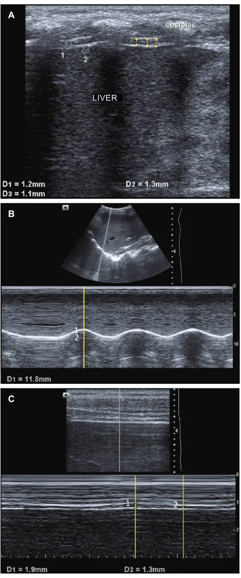
The diaphragm was visualised at a 2-3.5 cm depth, showing a hypo-echoic muscular layer amongst two echo-genic layers, one above and one below, corresponding to the diaphragmatic pleura and the parietal peritoneum, re-spectively (Fig. 1A).
Diaphragm thickness was defined as the vertical dis -tance in millimetres between the midpoint of the diaphrag-matic pleura and the midpoint of the parietal peritoneum,
measured on the most perpendicular axis to the longitudinal plane.1,6
Airway pressure and flow real-time charts were ana -lysed in each evaluation and three consecutive measure-ments were obtained during the expiration within the same cycle.
Change in diaphragm thickness from baseline to nadir was obtained for each patient. The presence of diaphragm atrophy was considered in this study at ≥ 10% decrease in thickness from baseline.5
b) Diaphragmatic excursion
The measurement of diaphragmatic excursion was obtained by use of a 6 – 2 MHz convex probe positioned perpendicular to the subcostal area, between midclavicular and right anterior axillary lines.
The diaphragm was visualised in B-mode showing a hyper-echoic line produced by the diaphragmatic pleura ad-herent to the muscle, while diaphragmatic excursion was assessed in M-mode, corresponding to the vertical distance in millimetres of the diaphragmatic line between maximum inspiration (peak of the curve) and maximum expiration (base of the curve) (Fig. 1B).
Whenever two or more measurements of this parameter were available, the change in diaphragmatic excursion was obtained, corresponding to the difference between peak and baseline values.
c) Diaphragmatic thickening fraction
DTf, corresponding to the change in diaphragm thick-ness with breathing movements was mathematically obtained as a percentage, considering the values of dia-phragm thickness at maximum inspiration and maximum expiration, by using the formula:
DTf = [(diaphragm thickness at end of inspiration – end of expiration) / diaphragm thickness at end of expiration] × 100.
A 10 – 5 MHz linear probe has been used in M-mode, positioned at the same location as for the measurement of diaphragm thickness (Fig. 1C).
Whenever two or more measurements of this parame-ter were available, the change in DTf was obtained, corre-sponding to the difference between peak and baseline DTf values.
Statistical analysis
A descriptive analysis was carried out, while the group of patients was characterised through the calculation of meas-ures of central tendency and dispersion for quantitative var-iables: mean and standard deviation (SD) for variables with normal distribution or median and interquartile range (IQR) for variables without a normal distribution. Shapiro-Wilk test has been used as normality test.
ARTIGO ORIGINAL A 0.05 level of significance has been considered in this
study.
The statistical analysis was carried out by use of SPSS Statistics® version 25 software.
RESULTS
A 42-month median age has been found in our group of 17 patients (10 male) (IQR 9.5 – 155.5).
Principal diagnoses underlying the admission to the CIPE were divided into groups: respiratory (n = 8), traumatic (n = 5), postoperative/postprocedure (n = 2), cardiovascular (n = 1) and infectious diseases (n = 1). Seven patients pre-sented with an underlying condition or comorbidity, namely cancer (n = 3), prematurity with no diagnostic criteria for bronchopulmonary dysplasia (n = 2), cystic fibrosis bron -chiectasis (n = 1) and trisomy 21 associated with hypotonia (n = 1).
BMI percentiles were presented into six different ranges (Table 1). Three patients presented at admission with BMI ≤ P3 and 5 with BMI ≤ P15.
Fifteen patients were on midazolam (at a daily mean dose of 2.13 ± 1.51 mg/kg), 16 on morphine (at a daily mean dose of 0.21 ± 0.12 mg/kg) and eight patients on fentanyl (at a daily median dose of 0.05 mg/kg) (IQR 0.05 – 0.06).
Rocuronium was administered to 16 patients (at a daily mean dose of 3.34 ± 1.84 mg/kg).
Nine patients were receiving corticosteroid therapy at a daily mean dose of 2.03 ± 0.92 mg/kg of prednisolone and aminoglycoside antibiotics were administered to two patients, while seven patients presented with sepsis during their stay in the hospital.
A seven-day median duration of IMV has been found (IQR 6 – 10.5). Pressure regulated volume control (PRVC) mode was initially used in all the patients, with a 5.9-day median duration (IQR 4.1 – 10.3), while the ventilation mode was not changed in any patient up to pre-extubation.
As regards the ventilation parameters with PRVC, an average maximum respiratory rate of 32 ± 12 breaths/min-ute, 7 mL/kg median maximum tidal volume (IQR 6.7 – 7.5), 6 cmH2O (IQR 5 – 8) median positive end-expiratory pres-sure (PEEP) and 25 ± 7 cmH2O average peak inspiratory pressure (PIP) have been obtained.
Fifteen patients were extubated and pressure support ventilation (PSV) mode was used in pre-extubation in 14 of these patients, with a 23-hour median duration (IQR 7.5 – 45.5). Only one patient remained in spontaneous breathing
Table 1 – BMI-for-age percentile
Percentile (P) n (17)
≤ P3 3
> P3 and ≤ P15 2
> P15 and ≤ P50 6
> P50 and ≤ P85 2
> P85 and ≤ P97 2
> P97 2
n: absolute number; P: percentile Figure 1 – Ultrasound imaging for diaphragm evaluation. These
images were obtained during the evaluation of our group of patien-ts. (A) Ultrasound image of the zone of apposition for the evaluation of diaphragm thickness, showing the diaphragmatic pleura (Arrow 1) and the parietal peritoneum (Arrow 2). (B) Ultrasound image for evaluation of the diaphragmatic excursion, the vertical distance (D1) between diaphragmatic line at the end of inspiration - peak of the curve (1) – and the end of expiration – base of the curve (2). (C) Ultrasound image for the evaluation of the change in diaphragm thi-ckness during the respiratory cycle. 1 –diaphragm thithi-ckness at the end of inspiration; 2 –diaphragm thickness at the end of expiration.
A
B
C
ARTIGO ORIGINAL through an endotracheal tube (ETT).A 16 ± 3.4 cmH
2O average PIP was obtained during PSV.
A 11-day median length of stay at the CIPE (IQR 9 – 17) and a 0.04 mortality rate according to the PIM3 (IQR 0.02 – 0.15) have been found, while only one of the patients in our group has died in the hospital. A 22-day median ventila-tor-free time has been found (IQR 20 – 23).
A total of 149 ultrasound evaluations of the diaphragm thickness, 36 of the diaphragmatic excursion and 36 of the diaphragmatic thickening fraction were carried out through-out the study.
A 2.3 mm median baseline diaphragm thickness has been found (IQR 2 – 3.5). A trend towards a decrease in di-aphragm thickness was found throughout the time in which the patients were receiving IMV with PRVC mode (Fig. 2).
A 1.9 mm median nadir thickness has been found with PRVC mode (IQR 1.6 – 3.1) and this has occurred at a median four days of IMV (IQR 2 – 7.5). A 0.3 mm median change in diaphragm thickness has been found (IQR 0.2 – 0.7), corresponding to a 13% decrease.
Diaphragmatic atrophy has been found in 14 patients. The comparison of changes in diaphragm thickness ac-cording to patient’s clinical and pathological and pharma-cological characteristics is shown in Table 2, showing sig-nificant differences regarding median change in thickness between the group with vs. without sepsis (0.70 vs. 0.25 mm; p = 0.019).
As regards diaphragmatic excursion, a 7.8 ± 3.9 mm mean baseline and 10.3 ± 5.4 mm mean peak values have been found, corresponding to 3.0 ± 1.6 mm mean change in diaphragmatic excursion.
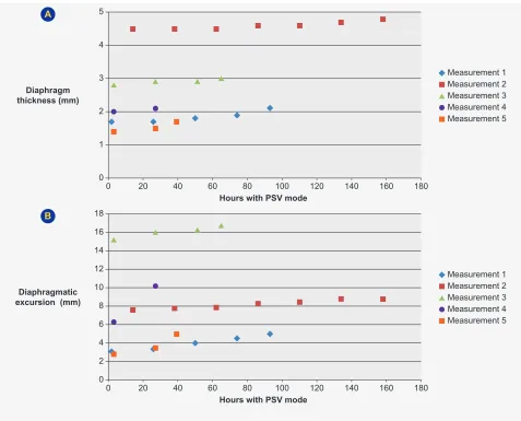
Five out of the 14 patients in pre-extubation with PSV remained with this ventilation mode for more than 48 hours. As the ultrasound assessments were carried out daily throughout that phase, two or more assessments of each parameter were obtained in those patients, allowing for the
analysis of clinical progression (Fig. 3). A mild trend towards an increasing thickness (Fig. 3A) and diaphragmatic excur-sion (Fig. 3B) were found as time with PSV mode increased. A 41,14 ± 11% average baseline value and 48.8 ± 3.7% average peak value of the diaphragmatic thickening fraction (DTf) have been found, with a 19 ± 6.6% average change in DTf.
Extubation failure has been found at values of DTf ≤ 35%, which has occurred within two out of the 17 pre-extu-bation periods that were evaluated (Fig. 4).
DISCUSSION
This is one the first studies in paediatric patients receiv -ing IMV aimed at the ultrasound assessment of diaphragm morphological and contractile activity.
Between 55 and 60% of the paediatric patients admitted to the CIPE of the Hospital Pediátrico de Coimbra were in need for invasive mechanical ventilation, which is one of the most frequently used techniques in the approach to critically ill paediatric patients.
The complications associated with IMV, namely VIDD, started immediately after the intubation, with a clear inter-ference on the outcomes, as suggested by different studies with adult patients.1,3,6,13 The systematic use of a sensitive
and specific diaphragm evaluation is therefore crucial, al -lowing for the early identification of VIDD.
As the constraints of the remaining modalities of dia-phragm assessment were overcome with diadia-phragm ultra-sound imaging, this was considered for the study.1,2,4
Ultrasound imaging allows for an accurate diaphragm morphological assessment, according to a systematic re-vision on its relevance in the approach to diaphragmatic dysfunction in critically ill patients,2 allowing for the
meas-urement of diaphragm thickness and the identification of at -rophy, one of the major characteristics of VIDD. In addition, functional evaluation is also allowed, with the measurement of the indices of contractility — diaphragmatic excursion
Figure 2 – Boxplot of diaphragm thickness (in mm) of patients during the first 10 days with pressure regulated volume control (PRVC)
mode
Day 0
Thickness (mm)
Days of invasive mechanical ventilation (PRVC) 0
1 2 3 4 5
0 1 2 3 4 5
Day 1 Day 2 Day 3 Day 4 Day 5 Day 6 Day 7 Day 8 Day 9
ARTIGO ORIGINAL
Table 2 – Comparison of the change in diaphragm thickness regarding the different variables
Characteristics
(n = 17) Change in Diaphragm Thickness (mm)Median (IQR) p(Mann-Whitney’s U-test)-value
Comorbidities
Yes (n = 7)
No 0.20 (0.20 – 0.70)0.40 (0.28 – 0.63)
0.475
Malnutrition
Yes (n = 3)
No 0.30 (0.20 – 0.63)0.20
0.432
Fentanyl
Yes (n = 8)
No 0.40 (0.23 - 0.68)0.30 (0.20 - 0.65)
0.481
Corticosteroids
Yes (n = 9)
No 0.30 (0.25 - 0.70)0.25 (0.20 - 0.58)
0.423
Aminoglycoside antibiotics
Yes (n = 2)
No 0.30 (0.20 - 0.60)0.45
0.824
Sepsis
Yes (n = 7)
No 0.70 (0.30 - 0.70)0.25 (0.20 - 0.35)
0.019
The change in diaphragm thickness (in mm) is presented as median and interquartile range (IQR). n: absolute number.
Figure 3 – Trend of ultrasound indices measurements with pressure support ventilation (PSV). (A) Change in diaphragm thickness (mm) over time (in hours), with PSV mode. (B) Change in diaphragmatic excursion (mm) over time (hours), with PSV mode.
0 0 1 2 3 4 5
40
20 60 80 100
Hours with PSV mode Diaphragm
thickness (mm)
120 140 160 180
Measurement 1 Measurement 2 Measurement 3 Measurement 4 Measurement 5
0 0 4 2 10 8 6 12 16 14 18
40
20 60 80 100
Hours with PSV mode Diaphragmatic
excursion (mm)
120 140 160 180
Measurement 1 Measurement 2 Measurement 3 Measurement 4 Measurement 5 A
ARTIGO ORIGINAL and thickness fraction. These measurements could be re-peated over time, allowing for the comparison of values
during follow-up.
Age-related reference values of diaphragm thickness in healthy children have been established by a cross-sectional prospective study.14 A 2.3 mm median baseline thickness has been found in our study, below the mean reference val-ue for any age group.14 Despite the differences in meas-ures of central tendency and dispersion that were used in both studies, this finding could be explained by the fact that some patients presented with a chronic disease/comorbid-ity (7/17) or malnutrition at admission (3/17), which could represent diaphragm fragility and therefore with reduced baseline thickness.
A 13% median decrease in diaphragm thickness has been found in this study throughout the time in which pa-tients were receiving IMV with PRVC mode, with nadir thick-ness found at a median four days of IMV, while the pres-ence of diaphragmatic atrophy was found in 14 patients. These findings are in line with those described by Glau et al.15 who had found a -13.8% median change in diaphragm thickness in one of only two studies carried out to date with paediatric patients, with a -3.4% daily atrophy rate. Rapid onset of diaphragmatic atrophy is shown by these findings, with an exponential decline in diaphragm thickness.
An 8.8% decrease in mean diaphragm thickness has also been described by Lee et al.16 within the first 24 hours upon intubation, with a 0.68% mean gradual daily decline up to day 7 of IMV. However, ventilation parameters that were used in the study do not correspond to lung protective ventilation strategies, except in patients admitted with acute respiratory distress syndrome, which is in contrast with our study, in which a 7 mL/kg median peak tidal volume has been found. Therefore, the discrepancies in these findings
could be explained by our small group of patients and by the differences regarding ventilation modes and parameters that were used.
In our study, patients mainly received IMV with PRVC mode up to pre-extubation, in which the patient’s inspira-tory effort is detected by the ventilator, assisting the patient in achieving a set tidal volume. As curarisation was a fre-quent procedure — 16/17 patients — the inspiratory effort developed by patients was reduced and subsequently the diaphragm remained ‘inactive’ most of the time. In addition, during pre-extubation, PSV mode was used in most pa-tients (14/15). PSV is frequently used in weaning from IMV, aimed at relieving the workload imposed to the respiratory muscles, preserving its spontaneous contraction and pre-venting atrophy.9
A trend towards a decrease in diaphragm thickness has been found during the use of PRVC. Instead, a small trend towards an increasing diaphragm thickness has been found with PSV in pre-extubation. These findings suggest that IMV-related diaphragm morphological changes could be reduced by titrating ventilation, using physiological levels of inspiratory effort.
An association between diaphragmatic activity and at-rophy in ventilated patients has been suggested by Gol -igher et al. in a large-scale observational study17 including the analysis of this parameter in patients receiving different ventilation modes for up to one week. Low diaphragmatic contractile activity was associated with quick reduction in diaphragm thickness, as shown in that study, while a high contractile activity was associated with an increased thick-ness as contractile activity is known to decrease with in-creasingly controlled ventilation modes.
Other factors apart from IMV may contribute to VIDD, including sepsis, multiple organ dysfunction and
Figure 4 – Pre-extubation diaphragmatic thickening fraction (DTf). Extubation failure occurred at values of DTf ≤ 35% (-). M: measurement.
0 10
M1 M2 M3 M4 M5 M6 M7 M8 M9 M10 M11 M12 M13 M14 M15 M16 M17 20
30 Thickening fraction (%)
Measurement of thickening fraction 40
ARTIGO ORIGINAL malnutrition.3,6 The administration of drugs including
corti-costeroids, aminoglycoside antibiotics and neuromuscular agents is also involved.3,5 A significantly higher change in diaphragm thickness has been found in the group of pa-tients with sepsis in our study. These findings are in line with those described in other studies3,13 in which sepsis was considered as one of the factors most strongly associated with diaphragmatic dysfunction.
As the assessment of diaphragm thickness regards only one of its morphological characteristics, which is not nec-essarily correlated to its functional capacity, the ultrasound indices of diaphragmatic contractility — diaphragmatic ex-cursion thickening fraction (DTf) were also evaluated.
DTf was shown by Umbrello et al.9 as the most reliable ultrasound indicator of change in the inspiratory muscle effort in response to modifications in the level of ventilation support, in a study aimed at assessing the performance of diaphrag-matic excursion and DTf when compared to the traditional indices of inspiratory muscle effort with IMV, as it is only influ -enced by active muscle contraction.
Additionally, different studies have been carried out to analyse the accuracy of ultrasound indices of diaphragmat-ic function — excursion and DTf — in predicting the suc-cess or failure of extubation, while DTf was considered as the most accurate index.2,4,18,19 In our study, extubation fail-ure was found at values of DTf ≤ 35%, in line with a recent systematic revision2 describing a range of DTf between
30% – 36% as predictive of extubation failure. Conflicting values regarding extubation failure were found in the study by Lee et al. in paediatric patients,16 at values of DTf <17%.
Up to date, there is no established approach to VIDD. The strategies for the use of PSV and earlier extubation need to be analysed and further studies aimed at deter-mining whether IMV could progress from lung-protective to muscle-protective.
Some limitations to our study are worth mentioning: the small group of patients does not allow for any inferences or robust conclusions; all the measurements of diaphragm thickness were carried out a few hours upon intubation and therefore no real baseline diaphragm thickness value was available; all ultrasound assessments were carried out by the same operator while intra and inter-observer reproduci-bility were not assessed.
Despite these limitations, this study could contribute to a better understanding on the presence of VIDD in paedi-atric patients and the usefulness of diaphragm ultrasound imaging in the approach to critically ill paediatric patients. It could also represent a model and forerunner for a larger scale study with wider inclusion criteria, namely duration of IMV >24 hours, allowing for stronger conclusions.
CONCLUSION
Diaphragm thickness and diaphragmatic thickening fraction could provide relevant data on the presence of di-aphragmatic atrophy and dysfunction while a 13% median decrease in thickness has been found in paediatric patients receiving PRVC ventilation, while nadir thickness was found at a median four days of IMV, with a higher decrease found in patients who developed sepsis. A mild trend towards an increasing diaphragm thickness has been found in pre-ex-tubation in patients receiving PSV ventilation and extuba-tion failure was found at values of DTf ≤ 35%.
These parameters could have been systematically as-sessed in critically ill paediatric patients in order to titrate ventilation, minimising the presence of diaphragmatic dys-function and extubation failure.
HUMAN AND ANIMAL PROTECTION
The authors declare that the followed procedures were according to regulations established by the Ethics and Clini-cal Research Committee and according to the Helsinki Dec-laration of the World Medical Association.
DATA CONFIDENTIALITY
The authors declare that they have followed the proto-cols of their work centre on the publication of patient data.
CONFLICTS OF INTER
The authors declare that there were no conflicts of inter -est in writing this manuscript.
FINANCIAL SUPPORT
The authors declare that there was no public or private financial support in writing this manuscript.
REFERENCES
1. Grosu HB, Lee YI, Lee J, Eden E, Eikermann M, Rose KM. Diaphragm muscle thinning in patients who are mechanically ventilated. Chest. 2012;142:1455–60.
2. Zambon M, Greco M, Bocchino S, Cabrini L, Beccaria PF, Zangrillo A. Assessment of diaphragmatic dysfunction in the critically ill patient with ultrasound: a systematic review. Intensive Care Med. 2017;43:29–38. 3. Dot I, Pérez-Teran P, Samper MA, Masclans JR. Disfunción
diafragmática: una realidad en el paciente ventilado mecánicamente. Arch Bronconeumol. 2017;53:150–6.
4. Dinino E, Gartman EJ, Sethi JM, McCool FD. Diaphragm ultrasound as a predictor of successful extubation from mechanical ventilation. Thorax. 2014;69:423–7.
5. Goligher EC, Dres M, Fan E, Rubenfeld GD, Scales DC, Herridge MS, et al. Mechanical ventilation-induced diaphragm atrophy strongly
impacts clinical outcomes. Communities. 2009;1–53.
6. Schepens T, Verbrugghe W, Dams K, Corthouts B, Parizel PM, Jorens PG. The course of diaphragm atrophy in ventilated patients assessed with ultrasound: a longitudinal cohort study. Crit Care. 2015;19:422.
7. Spadaro S, Grasso S, Mauri T, Dalla Corte F, Alvisi V, Ragazzi R, et al. Can diaphragmatic ultrasonography performed during the T-tube trial predict weaning failure? The role of diaphragmatic rapid shallow breathing index. Crit Care. 2016;20:1–11.
8. Sanchez De Toledo J, Munoz R, Landsittel D, Shiderly D, Yoshida M, Komarlu R, et al. Diagnosis of abnormal diaphragm motion after cardiothoracic surgery: Ultrasound performed by a cardiac intensivist vs. fluoroscopy. Congenit Heart Dis. 2010;5:565–72.
ARTIGO ORIGINAL
Diaphragm ultrasound as indicator of respiratory effort in critically ill patients undergoing assisted mechanical ventilation: A pilot clinical study. Crit Care. 2015;19:1–10.
10. Straney L, Clements A, Parslow RC, Pearson G, Shann F, Alexander J, et al. Paediatric index of mortality 3: an updated model for predicting mortality in pediatric intensive care. Pediatr Crit Care Med. 2013;14:673–81.
11. Martindale. Martindale: the complete drug reference, 34th ed. Choice
Curr Rev Acad Libr. 2005;42:1962.
12. Goligher EC, Laghi F, Detsky ME, Farias P, Murray A, Brace D, et al. Measuring diaphragm thickness with ultrasound in mechanically ventilated patients: feasibility, reproducibility and validity. Intensive Care Med. 2015;41:642–9.
13. Demoule A, Jung B, Prodanovic H, Molinari N, Chanques G, Coirault C, et al. Diaphragm dysfunction on admission to the intensive care unit. Prevalence, risk factors, and prognostic impact—a prospective study. Am J Respir Crit Care Med. 2013;188:213–9.
14. El-Halaby H, Abdel-Hady H, Alsawah G, Abdelrahman A, El-Tahan H.
Sonographic evaluation of diaphragmatic excursion and thickness in healthy infants and children. J Ultrasound Med. 2016;35:167–75. 15. Glau CL, Conlon TW, Himebauch AS, Yehya N, Weiss SL, Berg RA,
et al. Progressive diaphragm atrophy in pediatric acute respiratory failure. Pediatr Crit Care Med. 2018;19:406–11.
16. Lee EP, Hsia SH, Hsiao HF, Chen MC, Lin JJ, Chan OW, et al. Evaluation of diaphragmatic function in mechanically ventilated children: an ultrasound study. PLoS One. 2017;12:1–11.
17. Goligher EC, Fan E, Herridge M, Murray A, Vorona S, Brace D, et al. Evolution of diaphragm thickness during mechanical ventilation: impact of inspiratory effort. Articles A. 2010;1:1–56.
18. Matamis D, Soilemezi E, Tsagourias M, Akoumianaki E, Dimassi S, Boroli F, et al. Sonographic evaluation of the diaphragm in critically ill patients. Technique and clinical applications. Intensive Care Med. 2013;39:801–10.