Address for correspondence
Dr. Rajesh Kumar Mandal, Associate Professor Department of Dermatology, STD, and Leprosy, North Bengal Medical College,
Darjeeling, West Bengal, India
Email: [email protected]
Original Article
Clinical versus patho-microbial diagnosis of leprosy: a
comparative study in North Bengal population of
India
Introduction
Leprosy is a chronic infectious disease caused by Mycobacterium leprae. It principally affects the cooler parts of the body, mainly skin and peripheral nerves; it also involves muscles, eyes, bones, testis and internal organs.1 Leprosy continues to be an important public problem in
most parts of Asia, Africa and Latin America. In India, more than 125,000 of new leprosy cases were detected in the year 2014-15 which gives the annual new case detection rate (ANCDR) of 9.73 per 100,000 population. The prevalence rate (PR) was 0.69 per 10,000 populations. Though there has been slight decrease (2.5%) in ANCDR from previous year but there is 1.5% increase in PR from previous year (0.68%). West Bengal has already eliminated leprosy with a PR of 0.94.2 Out of 19 districts of the state, four districts still have PR more than two. Anushweta Singh, Rajesh Kumar Mandal*, Sabyasachi Banerjee**, Biswajit Halder, Manas Pratim Roy***
Department of Pathology, North Bengal Medical College, Darjeeling, West Bengal, India.
* Department of Dermatology, STD, and Leprosy, North Bengal Medical College, Darjeeling, West Bengal, India.
** Department of Dermatology, STD and Leprosy, Malda Medical College, West Bengal, India. *** Department of Pediatrics, Safdarjung Hospital, New Delhi, India
Abstract
Objective To correlate and compare clinical diagnosis with histopathological findings and acid-fast bacilli status in different types of leprosy to compare the efficacy of bacillary index in slit-skin smears and bacterial index of granuloma in biopsies for detecting the acid fast bacilli.Methods Detailed history was taken and thorough clinical examination was done. The findings were noted in a predesigned standard format. Slit-skin smear and skin biopsy were performed and evaluated. Subsequently, association was tested between the histopathological findings, bacteriological index and clinical classification.
Results Among the 72 study cases, diagnosis of leprosy was corroborated with histopathological and microbiological finding in 78% cases, among which LL (27.8%) constituted the major group, followed by TT (18.1%), BL (15.3%), BT (12.5%) with the least being BB (4.2%). Another 22.2% cases were non-leprosy histopathologically and diagnosed as nonspecific dermatitis (NSD).
Conclusion Histopathological examination along with bacteriological index of skin biopsy should be carried out in all cases of leprosy to arrive at a definite diagnosis of leprosy and to classify the type of the disease.
Key words
Diagnosis of leprosy is based on different clinical parameters which involve detailed examination of skin lesions and peripheral nerves.3 Demonstration of acid-fast bacilli in slit-skin smears by Ziehl-Neelsen (ZN) staining also aids in diagnosis of leprosy.4 Leprosy clinically mimics numerous other skin diseases. Therefore, a reliable diagnosis hinges around a good histopathological examination and demonstration of bacilli in histopathological sections.5,6 Histopathology not only aids in diagnosis of difficult cases, it also helps to determine the type of leprosy and thus, provides information about prognosis and treatment. Current study is performed in a tertiary care hospital of North Bengal to know the correlation between clinical and histolpathological diagnosis.
Methods
The study was carried out in tertiary care hospital over a period of one year (2014-2015). All the new patients clinically diagnosed as leprosy without any prior treatment attending dermatology OPD were included in the study. Formal permission from the Institutional Ethics Committee was obtained.
Detailed history was taken and thorough clinical examination was done. The findings were noted in a predesigned standard format. Slit-skin smear (SSS) were made and stained with ZN stain. Skin biopsies with 4-6 mm size punch needle were taken in the same sitting from the most representative lesion and stained with hematoxylin-eosin, ZN and modified Fite’s stain and evaluated under light microscope.
With histopathological features and bacteriological status (bacteriological index), the diagnosis of leprosy was confirmed and grouped histopathologically as per the criteria formulated by Ridley and Jopling and WHO.3 Subsequently,
association was tested between the clinical and histopathological diagnosis. Continuous variables were expressed using mean and percentages. Categorical variables were analyzed by chi-square test and Fisher’s exact test. P value <0.05 was considered significant. The data were analyzed using Microsoft Excel 2010 data analysis tool and SPSS (Statistical Programme for Social Science) version 16.0 for Windows.
Results
The present study included 72 patients who were clinically diagnosed to have leprosy. Most of the patients (34.7%) were in the 2nd to 3th decades of age. Male to female ratio was1.6:1. Male predominance was seen in all types of leprosy except in BB type where male: female ratio was one. Overall, 93.1% patients presented with hypopigmented skin lesions and rest with erythematous skin lesions. Majority (84.7%) patients suffered from loss of sensation and 36.1% cases had thickened nerve.
According to Ridley and Jopling’s classification, 34.7% cases clinically belonged to lepromatous leprosy (LL) followed by tuberculoid (TT) (25%), borderline tuberculoid (BT) (19.5%), borderline lepromatous (BL) (15.3%), and borderline (BB) (5.6%) whereas according to WHO classification, 70.8% cases were multibacillary (MB) and 29.2% were paucibacillary (PB).
Histopathologically, LL (27.8%) constituted the major group, followed by TT (18.1%), BL (15.3%), BT (12.5%) with the least being BB (4.2%). In 22.2% cases, histopathological diagnosis was not possible due to nonspecific changes (Table 2).
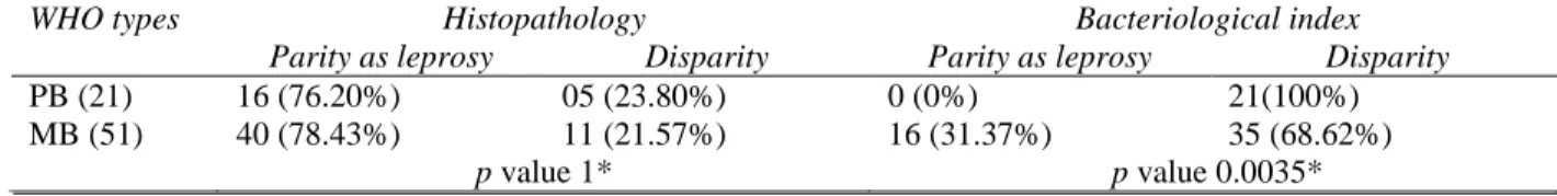
(p=1). When we compared BI with WHO types, patients with MB leprosy were more prone to be diagnosed bacteriologically and the association was statistically significant (p= 0.0035), (Table 3).
On histopathological study, 72.2% cases showed thinning of epidermis where as 13.9% cases each showed thickening and unremarkable changes in epidermis. All cases of BT, BB, BL and LL group showed thinning of epidermis. On the contrary, thickening of epidermis was not
present in any of histopathologically diagnosed leprosy cases.
Out of histopathologically confirmed leprosy cases, SSS was found positive for M. leprae
only in LL and BL types (22.2% cases). Among LL, SSS were positive in 70% cases and among BL, only 18.2% cases were positive. Only in BB, BL and LL skin smears and biopsies were found to be positive for acid fast bacilli (Table 4). None of the paucibacillary cases were positive for acid fast bacilli in slit skin smear or skin biopsy.
Table 1 Comparison between Clinical and histological diagnosis Clinical
groups
Histological groups
Total
TT BT BB BL LL PN Nonspecific
TT 11 2 0 0 0 0 5 18
BT 2 7 0 0 0 0 5 14
BB 0 0 2 0 0 0 2 4
BL 0 0 1 6 2 0 2 11
LL 0 0 0 5 18 0 2 25
Total 13 9 3 11 20 0 16 72
BB= borderline, BL= borderline lepromatous, BT= borderline tuberculoid, LL= lepromatous leprosy, PN= pure neural, TT= tuberculoid.
Table 2 WHO Classification & Histopathological correlation
WHO Histopathological types and number
TT BT BB BL LL NSD
PB (21) 13 (61.90%) 03 (14.28%) 0 0 0 05 (23.80%)
MB (51) 0 06 (11.76%) 03 (05.88%) 11 (21.56%) 20 (39.21%) 11 (21.56%)
BB= borderline, BL= borderline lepromatous, BT= borderline tuberculoid, LL= lepromatous leprosy, NSD= nonspecific dermatitis, PN= pure neural, TT= tuberculoid.
Table 3 Overall Parity between clinical and histopathological types according to WHO classification
WHO types Histopathology Bacteriological index
Parity as leprosy Disparity Parity as leprosy Disparity
PB (21) 16 (76.20%) 05 (23.80%) 0 (0%) 21(100%)
MB (51) 40 (78.43%) 11 (21.57%) 16 (31.37%) 35 (68.62%)
p value 1* p value 0.0035*
*Fisher’s exact test
Table 4 Comparison between histopathological type and mode of diagnosis
Histopathological types Slit-skin smear Skin biopsy AFB (in HP slide)
LL (20) 14 (70%) 20 (100%)
BB (03) 0 3 (100%)
BL (11) 2 (18%) 11 (100%)
Total 16 34
BB= borderline, BL= borderline lepromatous, BT= borderline tuberculoid, LL= lepromatous leprosy, TT= tuberculoid.
Discussion
Disease occurrence in leprosy is often related to age at detection rather than age at onset of disease. It is known to occur at all ages ranging from early infancy to very old age.7 In the present study, the highest incidence was seen in the age group of 21-30 years (34.7%) followed by age group 31-40 years (22.2%). Similar observations were made by majority of the studies.8-11 However, Kaur et al.12 observed 4th decade as the major age group involved. Although exact reason cannot be given for this age distribution, variable and long incubation period may be considered as an explanation.12
Generally, leprosy is believed to be more common in males than in females.13 In this study male to female ratio was 1.6:1 and this is in accordance with the previous researches.10,14-16 It may be because of many factors like industrialization, urbanization and more opportunities for contact in males. Social customs and taboos may also account for the smaller number of females reporting for treatment to the hospital.10
Like previous studies, we found sensory loss and hypopigmented patches as the most common features of clinical presentation.16-17 Since skin and nerves are the common habitats of lepra bacillus, the signs and symptoms related to them are common.
Similar to Jindal et al.18 we also found LL as the most common type. In another study, Arora et al.19 found BB and BL in 45% of patients. We found most of the cases as multibacillary, according to WHO classification. This result was similar to the findings of Tiwari et al.20 Giridhar et al.21 on the contrary, found paucibacillary in majority of the cases. These differences can be attributed to regional
variation and different socioeconomic and immune status in population studied.
In our study, histopathological diagnosis of leprosy was corroborated in 77.8% cases. The proportion is less than previous studies.21-23The discrepancy seems to be due to clinical overdiagnosis of leprosy and misinterpretation of many skin conditions presenting with hypopigmented patch as leprosy. Selection of the site for biopsy plays an important role in the histopathological diagnosis since clinically dissimilar lesions biopsied from the same patient can show different types of histopathology. When considered type-wise, maximum clinico-histological correlation was observed in LL cases (72%). Moorthy et al.17 and Mitra et al.24 also noted such correlation in LL patients.
The variation in different studies may be due to different criteria used to select the cases and difference in number of cases of each type. Various factors also influence the histopathological diagnosis such as differences in sample size, choice of the biopsy site, age of the lesion, immunological and treatment status of the patient at the time of biopsy.
The finding that SSS is positive for M. leprae
was done by Ridley himself in 1955 in which he observed higher values of BI in skin biopsies compared to SSS.28 Higher BI in the skin biopsies indicates that the BI of SSS only reflects density of the bacilli in a foci, while BI takes in to account both size of foci and bacterial density. In other words, BI is more accurate in assessing the bacterial status of the tissue specimen.28
Conclusion
Clinical diagnosis of early leprosy lesions offer difficulties even to experienced dermatologists and leprologists. Moreover, a sizable proportion of leprosy cases are in a continuously changing immunological spectrum and histological classification gives a better indication for any recent shift of a case position in the spectrum. As there can be some degree of overlap between different types of leprosy, both clinically and histopathologically, correlation of clinical and histopathological features along with bacteriological index appears to be more useful for accurate typing of leprosy than considering any of the single parameters alone. Taking serial biopsies from the same lesion, or from paired lesions, should be studied for a better clinico-histopathological relationship.
We conclude from our study that histopathological examination along with bacteriological index of skin biopsy should be carried out in all cases of leprosy to arrive at a definite diagnosis of leprosy and to classify the type of the disease. Since number of new cases of leprosy has been greatly reduced of late in our country and it is thought to be on the verge of elimination, this task should not regarded as an unnecessary burden considering the necessity of early, accurate diagnosis and treatment of the disease in order to prevent its long term complications.
References
1. Park K. Epidemiology of communicable diseases. In: Park K, Editor. Preventive and Social Medicine. 21st edn. Jabalpur: Banarsidas Bhanot; 2011. pp.288-303. 2. Ridley DS, Jopling WH. Classification of
leprosy according to immunity: a five group system. Int J Leprol. 1996;34:255-77. 3. National Leprosy eradication programme.
Progress report 2014-15. Available on http://nlep.nic.in/data.html. Accessed on 7th November, 2015.
4. Jopling WH, McDougall AC, editors. Handbook of Leprosy. 5th edn. New Delhi: CBS Publisher. 1996.
5. Lucus SB, Ridley DS. Use of histopathology in leprosy diagnosis and research. Lepr Rev. 1989;60:257-62.
6. Nayak SV, Shivarudrappa AS, Nagarajapa AH, Ahmed SM. Role of modified rapid AFB method in histopathological sections of Hansen’s disease. Indian J Dermatol Venereol Leprol. 2003;69:173-4.
7. Shetty VP, Doshi RP. Detection and classification of leprosy: Future needs and strategies. Indian J Lepr. 2008;80:139-47. 8. Guha PK, Pandey SS, Singh G, Kaur P. Age
of Onset of Leprosy. Lepr India. 1981;53:83-7.
9. Kaur S, Kumar B, Roy SN. Endemicity of leprosy in union territory of Chandigarh and surrounding states. Lepr India. 1982;54 :428-40.
10. Sehgal VN, Ghorpade A, Saha K. Urban leprosy an appraisal from Northern India. Lepr Rev. 1984;55:159-66.
11. Mathur MC, Ghimire RBK, Shrestha P, Kedia SK. Clinicohistopathological correlation in leprosy. Kathmandu Univ Med J. 2011;36:248-51.
12. Kaur I, Indira D, Dogra S, Sharma VK, Das A, Kumar B. Relatively spared zones in leprosy: A clinicopathological study of 500 patients. Int J Lepr. 2003;71:227-9.
13. Gupte MD. Leprosy: Epidemiology. In: Valia RG, Valia AR, editors. Textbook and Atlas of Dermatology. 2nd edn Mumbai: Bhalani Publishing House; 2001. P. 1543-52.
15. Nandarni NS, Rege VL. Significance of histopathological classification in leprosy. Indian J Lepr. 1999;71:325-9.
16. Verma OP. Some epidemiological features of leprosy in a rural area in Hoogly district. Lepr India. 1976;48:371-81.
17. Moorthy BN, Kumar P, Chatura KR, Chandrasekhar HR, Basavaraja PK. Histopathological correlation of skin biopsies in leprosy. Indian J Dermatol Venereol Leprol. 2001;67:299-301.
18. Jindal N, Shanker V, Tegta GR, Gupta M, Verma GK. Clinico-epidemiological trends of leprosy in Himachal Pradesh. Indian J Lepr. 2009;81:173-9.
19. Arora M, Katoch K, Natrajan M, Kamal R, Yadav VS. Changing profile of disease in leprosy patients diagnosed in tertiary care centre during years 1995-2000. Indian J Lepr. 2008;80:257-65.
20. Tiwary PK, Kar HK, Sharma PK, Gautam RK, Arora TC, Naik H et al. Epidemiological trends of leprosy in an urban leprosy centre. Indian J Lepr. 2011;83:201-8.
21. Giridhar M, Arora G, Lajpal K, Chahal KS. Clinicohistopathological concordance in Leprosy – A Clinical, Histopathological and Bacteriological study of 100 cases. Indian J Lepr. 2012;84:217-25.
22. Singh PA, Agarwal R, Misra V, Gupta SC, Bajaj AK. Clinico-histopathological
concordance in leprosy. Trop Doct. 2000;30:228-31.
23. Bhatia AS, Katoch K, Narayanan RB, Ramu G, Mukherjee A, Lavania RK. Clinical and histopathological correlation in the classification of leprosy. Int J Lepr Other Mycobact Dis. 1993;61:433-8.
24. Mitra K, Biswas S, Saha B, Dasgupta A. Correlation between clinical and histopathological criteria for the classification of leprosy. Indian J Dermatol. 2001;46(3):135-7.
25. Manandhar U, Adhikari RC, Sayami G. Clinico-histopathological correlation of skin biopsies in leprosy. J Pathol Nepal. 2013;3:452-8.
26. Bal A, Mohan H, Dhami GP. Infectious granulomatous dermatitis: A clinicopathological study. Indian J Dermatol. 2006;51:217-20.
27. Bhushan P, Sardana K, Koranne RV, Choudhary M, Manjul P. Diagnosing multibacillary leprosy: A comparative evaluation of diagnostic accuracy of slit skin smear, bacterial index of granuloma and WHO operational classification. Indian J Dermatol Venereol Leprol. 2008;74:322-6. 28. Ridley DS. The bacteriological