Increased neural and pupillary reactivity to emotional
faces in adolescents with current and remitted major
depressive disorder
Katie L. Burkhouse,
1,2Max Owens,
3Cope Feurer,
1Effua Sosoo,
4Anastacia Kudinova,
1and Brandon E. Gibb
11
Center for Affective Science, Department of Psychology, Binghamton University, Binghamton, NY 13902,
USA,
2Department of Psychiatry, University of Illinois at Chicago, Chicago, IL 60608, USA,
3Department of
Psychology, University of South Florida, St Petersburg, FL 33701, USA and
4Department of Psychology,
University of North Carolina, Chapel Hill, NC 27514, USA
Correspondence should be addressed to Katie L. Burkhouse, Department of Psychiatry, University of Illinois at Chicago, 1747 W Roosevelt Road, Chicago, IL 60608, USA. E-mail: [email protected]
Abstract
This study combined multiple levels of analysis to examine whether disrupted neural and pupillary reactivity to emotional faces serves as a state- or trait-like marker of adolescent major depressive disorder (MDD). The study examined differences in pupil dilation and the event-related potential (ERP) late positive potential (LPP) component to emotional faces before and after a negative mood induction between 71 adolescents (age 11–18 years) with (i) a current diagnosis of MDD, (ii) a past epi-sode of MDD currently in full remission and (iii) no lifetime history of any Axis I disorder. Relative to healthy control (HC) youth, adolescents with current or remitted MDD exhibited an enhanced LPP and pupillary response to all emotional facial expressions (fearful, happy and sad). This difference in reactivity between remitted depressed and HC adolescents persisted following the negative mood induction. Results also revealed that LPP and pupillary responses to emotional faces were sig-nificantly related, but only among the currently depressed adolescents. This study suggests that increased physiological and neural activation in response to social-emotional stimuli may not only characterize currently depressed adolescents, but also remains following MDD remission, potentially serving as a mechanism of risk for future depression relapse.
Key words:adolescence; major depressive disorder; emotional reactivity; pupil dilation; late positive potential
Adolescents with a previous history of major depressive dis-order (MDD) are at extremely high risk of recurrence, with a cu-mulative probability of recurrence of 40% by 2 years and 70% by 5 years (Rao, 2006; Avenevoliet al., 2015). This high rate of recur-rence almost certainly reflects the presence of stable vulnerabil-ity factors. Discriminating between state- and trait-like markers of adolescent MDD may be the first step to help identify specific vulnerabilities for the illness.
One potential trait-like vulnerability marker for adolescent depression may involve disrupted processing of emotionally sa-lient stimuli. Specifically, behavioral studies suggest that
adolescent depression is characterized by an attention and in-terpretation bias for depression-relevant or negative stimuli (for a review, see Plattet al., 2016). There is also some evidence that youth with MDD exhibit biased processing for positive stimuli at the behavioral level (Kyte et al., 2005; Harrison and Gibb, 2015), though others have failed to replicate this finding (for a review, see Plattet al., 2016). Notably, there is some indication that these emotion processing biases represent vulnerability factors implicated in adolescent MDD, as behavioral studies have shown that they are present in youth of depressed par-ents, a population at high-risk for developing MDD (Joormann
Received:1 July 2016;Revised:1 November 2016;Accepted:12 December 2016
VCThe Author (2017). Published by Oxford University Press. For Permissions, please email: [email protected]
783
doi: 10.1093/scan/nsw184
et al., 2007; Gibbet al., 2009; Kujawaet al., 2011; Connellet al., 2013), and among adolescents in full remission from MDD (Hankinet al., 2010). When compared with behavioral findings, neural and psychophysiological measures can provide relatively objective measures of emotion processing and offer insight into the brain circuitry underlying vulnerability. There is also evi-dence that neural measures account for unique variance in pre-dicting future behavior, including clinical status and response to treatment, above and beyond that accounted for by clinical and behavioral measures (Gabrieliet al., 2015), raising the possi-bility that these measures may aid in identifying vulnerapossi-bility factors implicated in adolescent MDD.
Researchers have utilized pupillometry, a psychophysio-logical measure capturing the assessment of changes in pupil dilation, to study emotion processing patterns and depression risk. The pupil dilates in response to stimuli requiring greater cognitive load and attentive responses and to stimuli of greater emotional intensity and remains dilated as long as processing persists, providing a peripheral index of brain activation in re-sponse to a specific stimulus (for a review, see Laenget al., 2012). Studies show that adults with current MDD exhibit increased pupil dilation to negative stimuli, compared with healthy controls (HCs) (Siegleet al., 2001, 2003a). Other studies suggest that adults in remission from depression exhibit an enhanced pupillary response to negative words prior to a nega-tive mood induction; however, following an instruction to think of a sad event in their lifetime, remitted depressed adults expe-rienced a blunted pupillary response to negative words relative to HC adults (Steidtmannet al., 2016). A heightened pattern of emotional reactivity has also been observed among adolescent populations deemed at high risk for depression by virtue of hav-ing a depressed mother. Specifically, greater pupil dilation to sad faces among these high risk youth prospectively predicted depression onset over a 2-year longitudinal follow-up period (Burkhouseet al., 2015). No studies to date, however, have exam-ined whether a similar pattern exists among adolescents in remission from MDD, representing a potential trait-like psycho-physiological marker of risk for future depressive relapse.
Researchers have also utilized event-related potentials (ERPs) to capture emotion processing styles among clinical populations given their excellent temporal resolution and abil-ity to capture multiple stages of emotional processing. The late positive potential (LPP) ERP component, in particular, is a prom-ising measure of emotional processing, as it has been shown to be enhanced in response to emotional stimuli, including faces, pictures, and words (Hajcaket al., 2010). Both an early and a late LPP component have been identified in previous studies, and it is suggested that the late portion may be better at distinguish-ing clinical groups (Hajcak and Olvet, 2008). Notably, in a sample of adolescents, females with current MDD exhibited a larger early and late LPP response to negative words, relative to non-depressed female youth (Auerbachet al., 2015). Studies examin-ing the LPP among youth of depressed mothers, however, tend to yield inconsistent patterns. Specifically, whereas one study showed that children of depressed mothers exhibit an enhanced LPP response to negative words (Speedet al., 2016), others have shown that these high risk youth exhibit a blunted LPP response to emotional faces and images, regardless of va-lence (Kujawaet al., 2012; Nelsonet al., 2015).
Taken together, these previous studies suggest that differ-ences in pupillary and neural (i.e. LPP) reactivity to emotional stimuli serve as a correlate of current depression in adolescents and adults, and can predict future depression onset among chil-dren deemed at high risk for depression. However, one critical
unanswered question is whether these biases are also present in adolescents with remitted MDD (rMDD), a population at high risk for depressive relapse (Avenevoliet al., 2015). In this study, we sought to examine whether disrupted neural and psycho-physiological reactivity (measured via the LPP and pupil dila-tion, respectively) to emotional faces serves as a trait-like marker of adolescent MDD by examining these responses among youth with current MDD, rMDD, and no history of any psychiatric disorder.
Consistent with cognitive models of depression (Clark and Beck, 1999) suggesting that biases remain latent until activated by a negative stressor, this study examined pupillary and LPP responses to emotional faces before and after a standardized negative mood induction. Based on previous studies examining the relation between emotion processing and depression risk (Siegleet al.2003a; Auerbachet al., 2015; Burkhouseet al., 2015; Speedet al.2016), we predicted that, prior to the negative mood induction, the currently depressed adolescents would exhibit greater pupil dilation and an enhanced LPP response relative to rMDD and HC adolescents, and that this would be specific for negative (sad and fearful),vspositive (happy) faces. Consistent with disrupted physiological and electrocortical reactivity to emotional stimuli serving as a trait-like marker of adolescent MDD, we predicted that after, but not before, the negative mood induction, the rMDD adolescents would exhibit greater pupil dilation and an enhanced LPP response relative to HC adoles-cents, and that this would be specific for negative,vspositive, faces.
Finally, exploratory analyses were conducted to examine re-lations between pupil dilation and LPP responses prior to the mood induction. Although previous studies suggest that these two measures capture similar processes, only one study has dir-ectly tested this and it failed to find a significant relation be-tween the two measures when healthy participants viewed natural scenes (Ferrariet al., 2016). In this study, we examined relations between these two measures in response to emotional faces, and examined whether adolescents’ depression status moderated the relation between pupil dilation and LPP re-sponses to emotional faces.
Methods
Participantspsychotropic medication. Table 1 displays participant charac-teristics separated by diagnostic status.
Measures
Diagnoses.The K-SADS-PL (Kaufmanet al., 1997) was used to as-sess for Axis I psychopathology. Among the 18 currently de-pressed adolescents, 9 had a current anxiety disorder, 5 had a current behavioral disorder and 5 had a history of recurrent MDD. Among the 27 rMDD adolescents, 9 had a current anxiety dis-order, 7 had a current behavioral disdis-order, and 4 had a history of recurrent MDD. The average number of days since last MDD epi-sode among the remitted depressed group was 593 (s.d.¼531, range¼67–2217). Interrater reliability for the K-SADS-PL, based on 20% of the sample interviews, was excellent (j¼1.00).
Depressive and anxious symptoms. Adolescent’s symptoms of depression and anxiety were assessed using the Children’s Depression Inventory (CDI; Kovacs, 1981) and the Multidimensional Anxiety Scale for Children (MASC; Marchet al.
1997), respectively. In this study, both the CDI and MASC ex-hibited excellent internal consistency (a ¼ 0.94 and 0.93, respectively).
Emotional faces task. Participants completed an emotional faces task where they viewed fearful, happy, sad and neutral youth faces presented one at a time on a computer screen. The stimuli in the paradigm were taken from the NIMH Child Emotional Faces Picture Set, comprised of emotional and neu-tral faces of youth aged 10–17-years old (Egger et al., 2012). Participants were instructed to identify the emotional valence of the face by pressing a corresponding button on a keypad. For each trial of the task, participants first viewed a fixation cross for 1000 ms, followed by the presentation of the face for 5000 ms, followed by a fixation cross for 3000 ms. The inter-trial interval varied randomly between 500 and 750 ms. The task included 22 fearful, 22 happy, 22 sad and 22 neutral faces. Pupil dilation and EEG were recorded while participants completed the task.
Pupillometry recording and processing. A Tobii T60XL eye tracker was used to monitor pupil dilation. The eye-tracker con-sisted of a video camera and infrared light source that was used to track the location and size of participants’ pupils during each task. Pupil was recorded at 60 Hz (every 16.7 ms). Blinks were identified as large changes in pupil dilation occurring too rap-idly to signify actual dilation or contraction. Trials comprised of over 50% blinks were removed from consideration. Participants with>50% of rejected trials were removed from analyses. This resulted in the following number of subjects being removed pre-mood induction: HC¼1, rMDD¼1, current MDD¼0. Of the 51 rMDD and HC adolescents who had clean pre-mood induc-tion pupil data, 9 subjects were removed for the post-mood in-duction data analysis due to poor data quality (HC¼4, rMDD¼
5). The number of subjects removed from analyses due to
excessive blinks was not significantly related to adolescents’ diagnostic status (lowestP¼0.42). The average pupil diameter over the 333 ms preceding the onset of the stimulus was sub-tracted from pupil diameter after stimulus onset to produce stimulus-related pupil dilation. Consistent with previous stud-ies (Silket al., 2008, 2009), average stimulus-related pupil dila-tion was calculated by taking the average early (2–5 s) and late (5–8 s) pupil response for each stimulus across all trials for each valence (fearful, happy, sad, neutral).
Electroencephalogramrecording and processing. Continuous electroencephalogram (EEG) was recorded using a custom cap and the BioSemi ActiveTwo system. The signal was pre-amplified at the electrode with a gain of 1600 and the EEG was digitized at 24-bit resolution with a sampling rate of 512 Hz. Recordings were taken from 34 scalp electrodes based on the 10/20 system. The electrooculogram was recorded from four fa-cial electrodes. Data were processed offline using Brain Vision Analyzer software (Brain Products, Gilching, Germany). All data were re-referenced to the average of the left and right mastoid electrodes, and filtered with high- and low- pass filters of .1 and 30 Hz, respectively. Continuous EEG data were segmented be-ginning 100 ms before stimulus onset and continuing for 2000 ms after onset. Eyeblinks were corrected using the method by Gratton and colleagues (Gratton et al., 1983) and semi-automated artifact rejection procedures removed artifacts with the following criteria: voltage step of more than 50 uV between sample points, a voltage difference of 300 uV within a trial, and a maximum voltage difference of<0.5 uV within 100 ms inter-vals. Participants were required to have a minimum of 12 artifact-free trials in each condition to be included in analyses (Moranet al., 2013). This resulted in the following number of subjects being removed pre-mood induction: HC¼6, rMDD¼7, current MDD¼1. Of the 40 rMDD and HC adolescents who had clean pre-mood induction ERP data, 4 were removed for the post-mood induction data analysis due to poor data quality (HC
¼2, rMDD¼2). The number of subjects removed from analyses due to excessive artifacts was not significantly related to ado-lescents’ diagnostic status (lowestP¼0.26). Trials were baseline corrected using the 200 ms prior to stimulus onset and averaged across each face condition. Consistent with previous studies measuring LPP responses in youth (Kujawaet al., 2012, 2015; Auerbachet al., 2015) and based on visual inspection of the cur-rent data to determine where the LPP was maximal, the LPP was scored as the mean activity from 400 to 1000 ms (early segment) and 1000–2000 ms (late segment) at a cluster of parietal-occipital sites: P3, P4, PO3, PO4, Pz, O1, O2 and Oz.
Mood induction.The rMDD and HC adolescents completed a standardized negative mood induction in which the youth watched a 165 second clip from the movieThe Champ(a scene in which a badly injured boxer summons his young son to the ring).
State mood ratings.A visual analog scale (VAS; Killgore, 1999) was used to measure state sadness before and after the Table 1.Demographic and clinical characteristics of participants
Controls (n¼26) Remitted (n¼27) Current (n¼18)
Age (M, s.d.) 14.23a(1.75) 14.04a(2.23) 14.39a(1.88)
CDI (M, s.d.) 2.57a(2.48) 8.15b, 6.06 22.33c, 10.30
Family income (median) $70 001–$75 000a $35 000–$40 000b $45 000-$50 000b
Gender (n, % Female) 11, 42% 15, 56% 14, 77%
Race (n, % Caucasian) 23, 88% 22, 82% 15, 83%
negative mood induction. Participants marked how they were feeling on a scale with anchors of very happy and very sad. The scale measured 100 mm and the distance from the left side of each scale was measured, with higher scores indicating greater state sadness.
Procedure
Informed consent and assent were collected upon arrival to the laboratory. Adolescents were administered the K-SADS-PL by a research assistant and then completed the CDI and MASC. Following this, adolescents completed the faces task. The rMDD and HC adolescents completed the negative mood induction and then repeated the faces task. The current MDD adolescents did not receive the negative mood induction or post-mood in-duction computer tasks. Participants completed the VAS before and after the negative mood induction. During this time, the adolescent’s primary guardian was administered the K-SADS-PL. The University’s Institutional Review Board approved all study procedures.
Analysis plan
To examine physiological and neural reactivity (pupil dilation and LPP) elicited during the faces task prior to the negative mood induction across all participants, two separate Emotion (fearful, happy, sad, neutral)Segment [early (400–1000 ms for LPP, 2000–5000 ms for pupil dilation), late (1000–2000 ms for LPP, 5000—8000 ms for pupil dilation)] repeated measures ANOVAs were conducted, with LPP or pupil dilation serving as the de-pendent variables. Greenhouse-Geisser corrections were used in cases in which the sphericity assumption was violated.
To examine group differences in behavioral performance [accuracy and response time (RT)] during the faces task, GroupEmotion repeated measures ANOVAs were conducted with accuracy or RT serving as the dependent variables and sep-arate analyses were conducted for responses before and follow-ing the mood induction.
To examine group differences in LPP and pupillary reactivity, consistent with previous research (Kujawa et al., 2012; Kappenmanet al., 2013), difference scores were first calculated to reflect each participant’s reactivity to each emotional stimu-lus compared with neutral (i.e. emotion—neutral). Following this, GroupEmotionSegment repeated measures ANCOVAs were conducted, with LPP or pupil dilation serving as the de-pendent variables. Given the significant group difference in family income, this variable was included as a covariate in all analyses. To examine group differences in LPP and pupillary re-activity following the negative mood induction, similar ANCOVAs as described earlier were conducted with the addition of Time (pre- and post-MI) as a within-subjects factor, and the Group factor now including only remitted depressed and control adolescents. Bonferroni procedures were used to correct for multiple comparisons. Specifically, an adjusted P-value of 0.0125 was used to interpret significant group effects, and post-hoccomparisons were conducted using Bonferroni correction in SPSS.
Finally, to examine relations between pupil dilation and the LPP to emotional faces prior to the mood induction, linear mixed modeling was used with an autoregressive (AR1) covari-ance structure. Data were included for LPP responses for each emotion at each segment, with subject treated as a random ef-fect and emotion and segment treated as repeated measures. LPP served as the dependent variable, and predictors in the
analysis were depression group, pupil dilation, emotion, seg-ment and all interactions.
Results
Preliminary analyses
Demographic and clinical characteristics separated by diagnosis are presented in Table 1. Preliminary analyses were conducted to examine LPP and pupil responses to the faces task prior to the mood induction across all participants. Results from the re-peated measures ANOVAs are presented in Table 2. We should also note that the groups did not differ in their LPP or pupillary response to neutral faces at pre- or post-mood induction (lowest
P¼0.29) justifying the use of difference scores to examine dif-ferences in LPP and pupil dilation to emotional stimuli.
Pre-mood induction
Behavior—faces.Participants displayed high detection accuracy on the task overall (M¼92.5, s.d.¼4.47). No significant group main effects or interactions were observed for accuracy (lowest
P¼0.35) or RT (lowestP¼0.20).
Late positive potential.Results revealed a significant main ef-fect of depression group,F(2, 53)¼5.71,P<0.01,np2¼0.18, with no other significant main effects or interactions (lowest P ¼
0.15).Post-hocpairwise comparisons revealed that, across emo-tions, the current (M¼1.30, SE¼1.34,p¼0.04, Cohen’sd¼0.64) and remitted (M¼2.33, SE¼1.20,P<0.01, Cohen’sd¼0.86) de-pressed adolescents exhibited a greater LPP response across emotional expressions, relative to HC adolescents (M¼ 3.24, SE
¼1.14). The current and remitted depressed adolescents did not differ significantly (P¼1.00; Cohen’sd¼0.20). The scalp topog-raphies and waveforms depicting this finding are presented in Figure 1. The group difference remained significant after statis-tically controlling for the influence of children’s anxiety symp-toms (MASC) and diagnoses (P¼0.04), suggesting that the findings were at least partially independent of adolescent’s cur-rent and past anxiety. This finding was also maintained when statistically controlling for the influence of adolescents’ psycho-tropic medication status (highestP¼0.04).
Pupil dilation.When examining the influence of depression status on adolescents’ pupillary response to emotional faces, results revealed a main effect of group,F(2, 64)¼3.91,P¼0.01, n2
p ¼0.12.Post-hocpairwise comparisons revealed that, across emotions, the HC adolescents (M¼ 0.002, SE¼0.002) exhibited significantly less pupil dilation than remitted depressed adoles-cents (M¼0.004, SE¼0.002,P¼0.01; Cohen’sd¼0.61). The dif-ference between the HC and currently depressed adolescents was a non-significant trend (M¼0.003, SE¼0.002, P¼0.08; Cohen’sd¼0.41) and there was no difference between current and remitted depressed adolescents (P¼1.00; Cohen’sd¼0.10). This finding is illustrated in Figure 2. The main effect of group was maintained when statistically controlling for the influence of children’s current anxiety symptoms and diagnoses and psy-chotropic medication status (highestP¼0.03). None of the two-or three-way interactions with group were significant (lowestP
¼0.10).
Post-mood induction
State mood ratings.To examine the influence of depression sta-tus on adolescents’ ratings of state sadness following the mood induction, we conducted a Group (rMDD, HC)Time (Pre-vs
Table 2.Repeated measures ANOVA results and means and s.d. of LPP and pupil dilation responses to emotional faces prior to the mood induction across all participants
Fear
M(s.d.)
Happy
M(s.d.)
Sad
M(s.d.)
Neutral
M(s.d.)
Emotion
F(P-value)
Segment
F(P-value)
EmotionSegment
F(P-value)
LPP 6.80a(0.99) 7.40a(0.99) 7.40a(0.98) 6.23a(0.97) 2.41 (0.16) 200.74 (<0.001) 1.43 (0.24)
Early 10.82a(1.07) 10.69a(1.05) 10.84a(1.06) 8.92a(1.05)
Late 2.78a(1.11) 4.11a(1.11) 3.79a(1.11) 3.55a(1.05)
Pupil 0.010a(0.002) 0.007b(0.001) 0.009a,b(0.002) 0.007b(0.001) 4.45 (0.04) 57.15 (<0.001) 1.05 (0.37)
Early 0.017a(0.003) 0.013a(0.002) 0.016a(0.002) 0.015a(0.002)
Late 0.003a(0.001)
0.001a(0.001) 0.001a(0.001)
0.001a(0.001)
Note:Means with different superscripts differ significantly. LPP, late positive potential. LPP early¼400–1000 ms, LPP late¼1000–2000 ms, Pupil early¼2000–5000 ms, Pupil late¼5000–8000 ms.
sadness ratings as the dependent variable. Results revealed a main effect of time,F(1, 46)¼7.71,P<0.001,np2¼0.52, with par-ticipants exhibiting a significant increase in sadness from base-line (M¼3.04, SE¼0.24), to after the mood induction, (M¼5.17, SE¼.30). The GroupTime interaction was nonsignificant,
F(1, 46)¼0.09,P¼0.76,np2¼0.01.
Behavior—post-mood induction. Neither the main effects of group or time, nor the GroupTime interaction was significant in predicting accuracy (lowestP¼0.43) or RT (lowestP¼0.26) during the faces task.
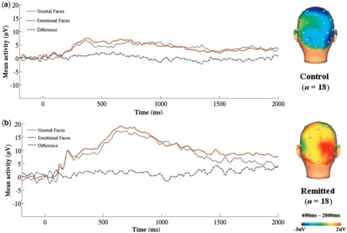
LPP—post-mood induction.When examining the influence of group and the mood induction on adolescents’ LPP response to emotional faces, results revealed a significant main effect of group,F(1, 34) ¼7.71,P ¼0.01, np2¼0.19. Across both time points, the remitted depressed adolescents exhibited a greater LPP response to emotional faces (M¼1.35, SE¼0.97) relative to HC adolescents (M¼-1.70, SE¼0.86; Cohen’s d¼0.50). The scalp topographies and waveforms depicting this finding are presented in Figure 3. The main effect of group was maintained when statistically controlling for the influence of children’s cur-rent anxiety symptoms, history of anxiety diagnoses, and psy-chotropic medication status (highestP¼0.04). In contrast, there were no significant main effects of time or emotion or any sig-nificant interactions (lowestP¼0.09).
Pupil dilation—post-mood induction.When investigating the influence of diagnostic status on adolescents’ pupillary re-sponse to emotional faces, results revealed a significant main effect of group,F(1, 40)¼10.90,P<0.01,np2¼0.22. Across both time points, remitted depressed adolescents (M¼0.001, SE¼
0.001) exhibited greater pupil dilation to emotional faces rela-tive to HC adolescents (M ¼ 0.005, SE ¼0.001; Cohen’sd ¼
0.70). This effect remained significant after statistically control-ling for the influence of children’s current anxiety symptoms, history of anxiety diagnoses and psychotropic medication sta-tus (highest P ¼ 0.02). In contrast, there were no significant main effects of time or emotion or any or significant inter-actions (lowestP¼0.10).
Exploratory analyses
Follow-up analyses were conducted to determine whether any of the group findings described earlier were moderated by chil-dren’s age, gender or family income. None of these analyses were significant (lowestP¼0.09). We also conducted additional follow-up analyses to determine if the pmood induction re-sults replicated when excluding subjects who did not have
post-mood induction pupil or LPP data. All of the significant effects were maintained and the pattern of results was identical.
Relation between LPP and pupil dilation
Finally, a linear mixed model was conducted to examine the ex-tent to which LPP and pupil dilation were correlated prior to the mood induction, and whether this would be moderated by ado-lescents’ depression status. Results from the LMM with LPP as the dependent variable revealed significant main effects of group,F(2, 106) ¼4.43,P ¼0.01, and segment,F(1, 261) ¼
39.76,P<0.001, and a significant grouppupil interaction,F(2, 404)¼2.79,P¼0.03. Further examination of the grouppupil interaction revealed that, among currently depressed adoles-cents, pupil dilation and LPP were significantly and positively related,t(119)¼2.96,P<0.01. However, this relation was not significant among the rMDD,t(147)¼1.30,P¼0.55 or HC,t(135)
¼0.88,P ¼0.83, adolescents. These findings are depicted in Figure 4. None of the other main effects or interactions were sig-nificant (lowestP¼20).
Discussion
The goal of this study was to examine whether disrupted physiological (pupillary) and neural (LPP) reactivity to emotional stimuli serves as a state- or trait-like marker of adolescent de-pression. First, we examined differences in pupillary reactivity and LPP responses to emotional faces between adolescents with current MDD, rMDD or no history of any psychiatric disorder. The current and remitted depressed adolescents exhibited an enhanced LPP and pupillary response to all emotional facial ex-pressions (fearful, happy, and sad) relative to HCs. Notably, this finding was at least partially independent of adolescent’s cur-rent levels of anxiety, history of anxiety disorders, differences in family income, and use of psychotropic medication.
Second, we examined whether pupillary and LPP responses to emotional faces in remitted and never depressed adolescents changed following a negative mood induction. Although adoles-cents in both groups exhibited a significant increase in sadness from baseline to after the mood induction, this increase in sad-ness was equivalent across groups and there were no signifi-cant changes in physiological or electrocortical reactivity for either group. Although the precise reason for this finding re-mains unclear, one possibility is that the remitted depressed adolescents exhibited a ceiling effect in their reactivity to emo-tional stimuli prior to the negative mood induction (i.e. these biases were already apparent prior to the negative mood induc-tion). On the other hand, a second, alternative explanation could be that the movie clip used to induce a negative mood may have not been salient enough to elicit changes in physio-logical or neural activity despite the significant increase in state sadness, as it focused predominately on the parent–child rela-tionship. That is, during adolescence, there is a shift in social and affective dynamics of parent–child interactions (Nelson
et al., 2005; Crone and Dahl, 2012), as youth orient more towards peers and individualization. Thus, a negative mood induction that elicits social exclusion among peers may be more success-ful in changing physiological and neural responses in adoles-cent populations.
In combination, the current results suggest that adolescents with current or past MDD exhibit similar levels of pupillary and neural reactivity to facial displays of emotion. To the extent that emotion processing biases are evident in formerly de-pressed adolescents even in the absence of a negative stressor, Fig. 2.Differences in average pupil dilation to emotional facial expressions
increased physiological and neural activation in response to emotional faces may be a trait-like marker of risk for depressive relapse among this population. Although, because of the cross-sectional design of this study, we could not determine whether this reactivity actually increases risk for first onsets or recur-rence of MDD, there is evidence from previous prospective stud-ies showing that increased pupil dilation to negative faces predicts future depressive episodes among children of de-pressed mothers (Burkhouse et al., 2015). Thus, heightened physiological and neural activation to emotional facial expres-sions may be a predictor of future depressive relapse among adolescents.
When compared with previous adolescent depression stud-ies (e.g. Auerbachet al., 2015), this study found limited evidence for enhanced physiological and neural reactivity being specific
to negative stimuli. Instead, adolescents with current and rMDD exhibited increased reactivity to both positive and negative fa-cial expressions. Notably, the Auerbachet al.(2015) study uti-lized self-referential words, rather than faces, with a sample of females only. Thus, there may be important factors (e.g. gender, stimulus type) that moderate the relation between emotional reactivity and youth depression risk. Moreover, although behav-ioral studies have suggested that adolescent depression is char-acterized by an attention bias specifically for negative stimuli, this finding has not been consistently replicated across depres-sion risk studies, and the direction of this negative bias (i.e. preferential attentionvs attentional avoidance) has also been inconsistent across studies (for a review, see Plattet al., 2016). This may be a result of the low reliability for behavioral meas-ures of attentional biases across studies (Schmukle, 2005; Fig. 3.Waveforms and scalp topographies depicting the LPP in response to emotional faces (average of fearful-neutral, happy-neutral, sad-neutral) from 400 to 2000 ms following stimulus onset for the(a)control (n¼18) and(b)remitted depressed (n¼18) groups after the negative mood induction.
Kappenmanet al., 2015). Future studies that utilize multiple lev-els of measurement (e.g. eye-tracking, pupil dilation, ERPs) with larger samples are needed to help shed light on the direction and specificity of these biases for youth depression risk.
It is also noteworthy that although enhanced pupillary and LPP responses to facial expressions appear to be evident among adolescents with current and rMDD, these reactivity measures were only significantly correlated among the currently de-pressed adolescents. This could be the result of schematic inter-connectedness (Dozois, 2014), which suggests that a negative cognitive schema is activated among individuals with current depression and, as a result, their emotional and attentional biases can be observed and connected at multiple levels of ana-lysis. According to this theory, relations among multiple meas-ures of reactivity to emotional stimuli are less likely to be observed among individuals with lower depressive symptoms, as their negative cognitive schema is not currently activated. It is also possible that we did not observe relations among these measures for the remitted depressed and HC adolescents due to their underlying neural circuitry. Studies show that the LPP is correlated with activity in the occipital and parietal neural re-gions (Keilet al., 2002; Sabatinelliet al., 2007), and it has been suggested that the increased occipital activation, in particular, may be a result from projections from the amygdala (Bradley
et al., 2003). Therefore, enhanced LPP responses may reflect sus-tained attention to emotion and index downstream processes resulting from hyperactivation of the amygdala (Hajcaket al., 2010). On the other hand, pupil dilation has been less reliably linked with amygdala, and instead linked to activity in brain areas associated with the processing and regulation of emotion, including the dorsolateral prefrontal cortex and ACC (Siegle
et al., 2003b; Urryet al., 2009). Moreover, there is evidence that pupil dilation is a reliable index of activity in the locus coeruleus-noradrenergic neuromodulatory system, which is es-sential for a broad range of cognitive and emotional processes (Murphyet al., 2014). Thus, although both pupil dilation and LPP represent areas of emotion processing, the LPP may be more tightly linked with sustained attentional responses, whereas pupil dilation may be more broadly associated with the regula-tion of emoregula-tions. Future studies using neuroimaging are needed to investigate the underlying neural circuits associated with these enhanced physiological and neural responses to emo-tional facial expressions among adolescents with depression.
This study benefited from several strengths, including the use of diagnostic interviews to assess adolescent depression history and the multi-method assessment of reactivity to emo-tional stimuli. This study also extends prior research by exam-ining trait-like markers of adolescent depression, and benefited from the inclusion of adolescents with current and rMDD. Despite these strengths, there were also limitations. First, pro-spective designs are needed to determine if increased physio-logical and neural activation in response to emotional facial expressions in adolescents in remission from MDD is present prior to the first onset of MDD, thereby representing a putative vulnerability factor, or is the result of a previous episode of de-pression, representing a potentially scar effect. Second, the remitted depressed adolescents had a high rate of psychiatric comorbidity though this may have resulted in a more generaliz-able sample given the high rates of comorbidity among adoles-cents with MDD (Costelloet al., 2003). Third, the sample size of the current study may have precluded the detection of impor-tant moderators (e.g., gender, age). Given the gender difference that emerges during the adolescent period with females being twice as likely to experience MDD compared with males
(Hankinet al., 1998), future studies with larger sample sizes are needed to examine whether neural and physiological activation to emotional faces assessed prior to puberty may help to iden-tify which adolescents are at greatest risk for developing de-pression and help to explain the emergence of gender differences in depression during this time.
Despite these limitations, this study suggests that increased physiological and neural activation in response to social-emotional stimuli may serve as a trait-like marker that remains following remission of MDD, which could be one mechanism by which these adolescents are at such increased risk for depres-sion in the future. To the extent that this is true, it may have im-portant clinical implications. For example, pupillometry is an inexpensive tool that could be administered in clinical settings, such as pediatricians’ offices. In addition, compared to other neural measures, such as fMRI, EEG measures are more eco-nomical, easily administered, and easily transportable to clin-ical settings to inform prevention and treatment planning. Thus, the current findings could lead to targeted prevention and intervention efforts among adolescents.
Funding
This project was supported by an Elizabeth Munsterberg Koppitz Child Psychology Graduate Student Fellowship from the American Psychological Foundation awarded to K.L. Burkhouse, dissertation awards from the American Psychological Association and the Society for a Science of Clinical Psychology awarded to K. L. Burkhouse, and National Institute of Mental Health grant MH098060 awarded to B. E. Gibb. KLB is supported by National Institute of Mental Health Grant T32- MH067631 (Training in the Neuroscience of Mental Health; PI: Mark Rasenick).
Acknowledgements
We would like to thank Katelynn Champagne, Jennifer Bertello, and Jillian Kaufman for their help in conducting as-sessments for this project.
Conflict of interest. None declared.
References
Auerbach, R.P., Stanton, C.H., Proudfit, G.H., Pizzagalli, D.A. (2015). Self-referential processing in depressed adolescents: A high-density event-related potential study.Journal of Abnormal Psychology,124(2), 233.
Avenevoli, S., Swendsen, J., He, J.P., Burstein, M., Merikangas, K.R. (2015). Major depression in the national comorbidity survey–adolescent supplement: Prevalence, correlates, and treatment.Journal of the American Academy of Child & Adolescent Psychiatry,54(1), 37–44.
Bradley, M.M., Sabatinelli, D., Lang, P.J., Fitzsimmons, J.R., King, W., Desai, P. (2003). Activation of the visual cortex in motivated attention.Behavioral Neuroscience,117(2), 369–80.
Burkhouse, K.L., Siegle, G.J., Woody, M.L., Kudinova, A.Y., Gibb, B.E. (2015). Pupillary reactivity to sad stimuli as a biomarker of depression risk: evidence from a prospective study of children.
Journal of Abnormal Psychology,124(3), 498–506.
Clark, D.A., Beck, A.T. (1999).Scientific Foundations of Cognitive Theory and Therapy of Depression. New York: John Wiley & Sons. Connell, A.M., Patton, E., Klostermann, S.,et al. (2013). Attention
depressive symptoms moderated by emotion regulation and affective dynamics during family interactions. Cognition and Emotion27(8), 1522–34.
Costello, E.J., Mustillo, S., Erkanli, A., Keeler, G., Angold, A. (2003). Prevalence and development of psychiatric disorders in child-hood and adolescence. Archives of General Psychiatry, 60(8), 837–44.
Crone, E.A., Dahl, R.E. (2012). Understanding adolescence as a period of social – affective engagement and goal flexibility.
Nature,13(9), 636–50.
Dozois, D.J.A. (2014). Schema change in cognitive therapy for de-pression. In: Cooper, S., Ratele, L., editors.Psychology Serving Humanity: Proceedings of the 30th International Congress of Psychology, Vol.2, p. 166, Western Psychology: London. Egger, H.L., Pine, D.S., Nelson, E.,et al. (2012). The NIMH child
emotional faces picture set (NIMH-ChEFS): A new set of chil-dren’s facial emotion stimuli.International Journal of Methods in Psychiatric Research,20(3), 145–56.
Ferrari, V., De Cesarei, A., Mastria, S.,et al. (2016). Novelty and emotion: pupillary and cortical responses during viewing of natural scenes.Biological Psychology,113, 75–82.
Gabrieli, J.D.E., Ghosh, S.S., Whitfield-gabrieli, S. (2015). Prediction as a humanitarian and pragmatic contribution from human cognitive neuroscience.Neuron,85(1), 11–26.
Gibb, B., Benas, J., Grassia, M., Mcgeary, J. (2009). Children’s atten-tional biases and 5-HTTLPR genotype: potential mechanisms linking mother and child depression.Journal of Clinical Child and Adolescent Psychology,38(3) ,415–26.
Gratton, G., Coles, M.G.H., Donchin, E. (1983). A new method for off-line removal of ocular artifact.Electroencephalography and Clinical Neurophysiology,55(4), 468–84.
Hajcak, G., MacNamara, A., Olvet, D.M. (2010). Event-related po-tentials, emotion, and emotion regulation: an integrative re-view.Developmental Neuropsychology,35(2), 129–55.
Hajcak, G., Olvet, D.M. (2008). The persistence of attention to emotion: brain potentials during and after picture presenta-tion.Emotion,8(2), 250–5.
Hankin, B.L., Abramson, L.Y., Moffitt, T.E., Silva, P.A., McGee, R., Angell, K.E. (1998). Development of depression from preadoles-cence to young adulthood: Emerging gender differences in a 10-year longitudinal study. Journal of Abnormal Psychology, 107(1), 128.
Hankin, B.L., Gibb, B.E., Abela, J.R.Z., Flory, K. (2010). Selective at-tention to affective stimuli and clinical depression among youths: Role of anxiety and specificity of emotion.Journal of Abnormal Psychology,119(3), 491–501.
Harrison, A.J., Gibb, B.E. (2015). Attentional biases in currently depressed children: an eye-tracking study of biases in sus-tained attention to emotional stimuli.Journal of Clinical Child and Adolescent Psychology,44(6), 1008–14.
Joormann, J., Talbot, L., Gotlib, I.H. (2007). Biased processing of emotional information in girls at risk for depression.Journal of Abnormal Psychology,116(1), 135–43.
Kappenman, E.S., Farrens, J.L., Luck, S.J., Proudfit, G.H. (2015). Behavioral and ERP measures of attentional bias to threat in the dot-probe task: poor reliability and lack of correlation with anxiety.Frontiers in Psychology,5, 88–95.
Kappenman, E.S., MacNamara, A., Proudfit, G.H. (2013). Electrocortical evidence for rapid allocation of attention to threat in the dot-probe task. Social Cognitive and Affective Neuroscience,10(4), 577–83.
Kaufman, J., Birmaher, B., Brent, D.,et al. (1997). Schedule for Affective Disorders and Schizophrenia for School-Age Children-Present and Lifetime Version (K-SADS-PL): initial
reliability and validity data.Journal of the American Academy of Child and Adolescent Psychiatry,36(7), 980–8.
Keil, A., Bradley, M.M., Hauk, O., Rockstroh, B., Elbert, T., Lang, P.J. (2002). Large-scale neural correlates of affective picture processing.Psychophysiology,39(5), 641–9.
Killgore, W.D. (1999). The visual analogue mood scale: can a single-item scale accurately classify depressive mood state?.
Psychological Reports,85, 1238–43.
Kovacs, M. (1981). Rating scales to assess depression in school-aged children. Acta Paedopsychiatrica: International Journal of Child & Adolescent Psychiatry,46(5–6), 305–15.
Kujawa, A., Hajcak, G., Torpey, D., Kim, J., Klein, D.N. (2012). Electrocortical reactivity to emotional faces in young children and associations with maternal and paternal depression.
Journal of Child Psychology and Psychiatry and Allied Disciplines, 53(2), 207–15.
Kujawa, A., Macnamara, A., Fitzgerald, K.D., Monk, C.S., Phan, K.L. (2015). Enhanced neural reactivity to threatening faces in anxious youth: evidence from event-related potentials.Journal of Abnormal Child Psychology,43(8), 1493–501.
Kujawa, A., Torpey, D., Kim, J.,et al. (2011). Attentional biases for emotional faces in young children of mothers with chronic or recurrent depression. Journal of Abnormal Child Psychology, 39(1), 125–35.
Kyte, A., Goodyer, I.M., Sahakian, B.J. (2005). Selected executive skills in adolescents with recent first episode major depres-sion.Journal of Child Psychology and Psychiatry,9, 995–1005. Laeng, B., Sirois, S., Gredeb€ack, G. (2012). Pupillometry:
A Window to the Preconscious?.Perspectives on Psychological Science,7, 18–27.
March, J.S., Parker, J., Sullivan, K., Stallings, P., Conners, C. (1997). The Multidimensional Anxiety Scale for Children (MASC): fac-tor structure, reliability, and validity. Journal of the American Academy of Child and Adolescent Psychiatry,36(4), 554–65. Moran, T.P., Jendrusina, A.A., Moser, J.S. (2013). The
psychomet-ric properties of the late positive potential during emotion pro-cessing and regulation.Brain Research,1516, 66–75.
Murphy, P.R., O’Connell, R.G., O’Sullivan, M., Robertson, I.H., Balsters, J.H. (2014). Pupil diameter covaries with BOLD activity in human locus coeruleus.Human Brain Mapping,35(8),4140–54. Nelson, B., Perlman, G., Hajcak, G., Klein, D., Kotov, R. (2015). Familial risk for distress and fear disorders and emotional re-activity in adolescence: an event-related potential investiga-tion.Psychological Medicine,45(12), 2545–56.
Nelson, E.E., Leibenluft, E., McClure, E.B., Pine, D.S. (2005). The so-cial re-orientation of adolescence: a neuroscience perspective on the process and its relation to psychopathology.
Psychological Medicine,35, 163–74.
Platt, B., Waters, A.M., Schulte-Koerne, G., Engelmann, L., Salemink, E. (2016). A review of cognitive biases in youth de-pression: attention, interpretation and memory.Cognition and Emotion1–22.
Rao, U. (2006). Development and natural history of pediatric de-pression: Treatment implications.Clinical Neuropsychiatry,3(3), 194–204.
Sabatinelli, D., Lang, P.J., Keil, A., Bradley, M.M. (2007). Emotional perception: correlation of functional MRI and event-related potentials.Cerebral Cortex,17(5), 1085–91.
Schmukle, S.C. (2005). Unreliability of the dot probe task.
European Journal of Personality,19(7), 595–605.
Siegle, G.J., Steinhauer, S.R., Carter, C.S., Ramel, W., Thase, M.E. (2003a). Do the seconds turn into hours? Relationships be-tween sustained pupil dilation in response to emotional infor-mation and self-reported rumination. Cognitive Therapy and Research,27(3),365–82.
Siegle, G.J., Steinhauer, S.R., Stenger, V.A., Konecky, R., Carter, C.S. (2003b). Use of concurrent pupil dilation assessment to inform in-terpretation and analysis of fMRI data.NeuroImage,20(1), 114–24. Silk, J.S., Dahl, R.E., Ryan, N.D.,et al. (2008). Pupillary reactivity to
emotional information in child and adolescent depression: links to clinical and ecological measures.American Journal of Psychiatry,164(12), 1873–80.
Silk, J.S., Siegle, G.J., Whalen, D.J., Ostapenko, L.J., Ladouceur, C.D., Dahl, R.E. (2009). Pubertal changes in emotional informa-tion processing: Pupillary, behavioral, and subjective evidence
during emotional word identification. Development and Psychopathology,21(1), 7–26.
Speed, B.C., Nelson, B.D., Auerbach, R.P., Klein, D.N., Hajcak, G. (2016). Depression risk and electrocortical reactivity during self-referential emotional processing in 8 to 14-year-old girls.
Journal of Abnormal Psychology,125(5), 607–19.
Steidtmann, D., Ingram, R.E., Siegle, G.J.,et al. (2016). Pupil re-sponse to negative emotional information in individuals at risk for depression Pupil response to negative emotional infor-mation in individuals at risk for depression. Cognition and Emotion,24(3), 480–96.