Prolactin alters blood pressure by modulating the
activity of endothelial nitric oxide synthase
Albert S. Changa, Ruriko Granta, Hirofumi Tomitaa, Hyung-Suk Kima, Oliver Smithiesa,1, and Masao Kakokia,1
aDepartment of Pathology and Laboratory Medicine, University of North Carolina, Chapel Hill, NC 27599
Contributed by Oliver Smithies, September 12, 2016 (sent for review March 31, 2016; reviewed by Jason D. Heaney and Akihiro Tojo)
Increased levels of a cleaved form of prolactin (molecular weight 16 kDa) have been associated with preeclampsia. To study the effects of prolactin on blood pressure (BP), we generated male mice with a single-copy transgene (Tg; inserted into the hypoxanthine-guanine phosphoribosyltransferase locus) that enables inducible hepatic production of prolactin and its cleavage product. The Tg is driven by the indole-3-carbinol (I3C)-inducible rat cytochrome P450 1A1 promoter. When the Tg mice were fed normal chow (NC), plasma prolactin concentrations were comparable to those in female WT mice in the last third of pregnancy, and BP was lower than in WT mice (∼95 mm Hg vs.∼105 mm Hg). When the Tg mice were fed chow containing IC3, plasma prolactin concentrations increased three-fold, BP increased to ∼130 mm Hg, and cardiac function became markedly impaired. IC3 chow did not affect the WT mice. Urinary ex-cretion of nitrite/nitrate and the amount of Ser1177-phosphorylated endothelial nitric oxide (NO) synthase (eNOS) were significantly greater in the Tg mice fed NC than in WT mice, as they are during pregnancy. However, when I3C was fed, these indicators of NO pro-duction became significantly less in the Tg mice than in WT mice. The effects of increased plasma prolactin were abolished by a genetic absence of eNOS. Thus, a threefold increase in plasma prolactin is sufficient to increase BP significantly and to markedly impair cardiac function, with effects mediated by NO produced by eNOS. We sug-gest that pregnant women with abnormally high prolactin levels may need special attention.
hypertension
|
lactation|
protein kinase B/AktP
rolactin, a 23-kDa polypeptide hormone, is a potentmulti-functional cytokine with a broad range of biological effects, including water and salt balance, lactogenesis, cell proliferation and differentiation, testicular Leydig cell function, T-cell
im-munity, pancreatic β-cell function, hematopoiesis, and
adipo-genesis (1). Prolactin is physiologically secreted mainly from the anterior lobe of pituitary gland. The secretion is negatively regu-lated by dopamine and positively reguregu-lated by prolactin-releasing peptide (2) synthesized in the hypothalamus. The levels of pro-lactin in the serum, urine, and amniotic fluids are significantly higher in patients with preeclampsia than in subjects with normal
pregnancy (3–5), suggesting that prolactin is involved in the
pathogenesis of pregnancy-associated hypertension. However, animal experiments have shown inconsistent effects of prolactin on blood pressure (BP). Thus, acute i.v. infusion of prolactin in-creased BP in rabbits (6), but, when ovine prolactin was chronically given i.p. via an osmotic minipump in rats, BP was decreased (7). It has also been reported that chronic prolactin infusion caused an increase in urinary sodium, potassium, and water excretion, but no significant changes in arterial pressure, in rats (8).
To study the chronic effects of different plasma concentrations of prolactin on BP and general well-being, we have generated male mice with a single-copy transgene [Tg; inserted into the
hypoxanthine-guanine phosphoribosyltransferase (Hprt) locus]
that enables hepatic production of prolactin and its cleavage product when the mice are fed indole-3-carbinol (I3C), a xeno-biotic agent naturally present at high levels in broccoli and similar vegetables. The Tg is leaky, and, when the mice are fed normal chow (NC), it leads to basal prolactin levels comparable
to those in the plasma of WT female mice in the last third of a normal pregnancy, accompanied by a decrease in BP and an in-crease in nitric oxide (NO) production, likewise similar to those occurring in females during pregnancy. When the Tg mice are fed a diet containing IC3, plasma levels become threefold basal, BP in-creases, and cardiac insufficiency develops. In the absence of en-dothelial NO synthase (eNOS), these changes did not occur.
Results
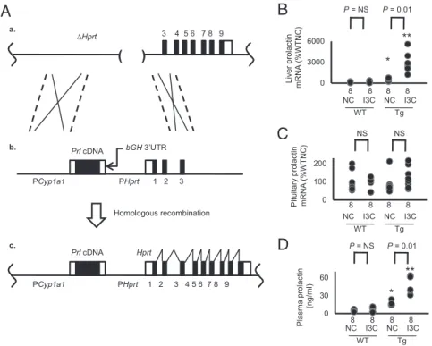
Generation of Tg Mice Producing Prolactin in the Liver. Gene tar-geting, as described previously (9), was used to insert a single-copy
Tg into theHprtlocus of the X chromosome. The Tg is comprised
of a copy of mouse cDNA for prolactin (Prl cDNA; Fig. 1A) driven
by the rat cytochrome P450 1A1 promoter (PCyp1a1) (10), with the
3′-UTR of the bovine growth hormone gene (bGH) in place of its
natural 3′-UTR. Male Tg mice fed NC have greater than WT ex-pression of prolactin mRNA in the liver and a higher concentration
of prolactin in the plasma (16.1±1.1 ng/mL vs. 5.1±0.6 ng/mL),
indicating that the inducible Tg is leaky (Fig. 1BandD). This basal
plasma prolactin concentration is close to that we observe in female
WT mice in the last third of pregnancy (15.3±0.6 ng/mL). When
the Tg mice were fed I3C, hepatic prolactin mRNA levels were markedly increased, and the plasma prolactin concentration
be-came approximately three times basal (Fig. 1BandD). The
ex-pression of prolactin in the pituitary gland was not significantly
altered by the Tg or by the I3C diet (Fig. 1C).
Opposite Changes in BP in the Tg Mice with Modestly and Substantially Increased Plasma Prolactin Levels.We found that the Tg mice fed NC
had a significantly lower BP than WT mice fed NC [WT (n=9),
108±1 mm Hg vs. Tg (n=11), 97±3 mm Hg;P<0.05; Fig. 2A
and Fig. S1A], but the heart rate was not significantly different
Significance
Prolactin is a hormone secreted by the pituitary gland that controls changes in the breast to enable milk production after the baby is born. In some mothers with pregnancy-related high blood pressure (BP), the concentration of prolactin in the blood is higher than normal, but whether this causes the high BP or is a consequence of it is uncertain. To answer this question, we have generated experimental mice that produce prolactin in the liver when we feed them a substance, indole-3-carbinol (IC3), that is found in broccoli. When fed normal chow, the mice are well, but, when fed IC3, they develop high BP and heart problems. This suggests that pregnant women with abnor-mally high prolactin levels may need special attention.
Author contributions: M.K. designed research; A.S.C., R.G., H.T., H.-S.K., and M.K. per-formed research; H.-S.K. and M.K. analyzed data; and O.S. and M.K. wrote the paper. Reviewers: J.D.H., Baylor College of Medicine; and A.T., University of Tokyo.
The authors declare no conflict of interest.
Freely available online through the PNAS open access option.
1To whom correspondence may be addressed. Email: [email protected] or [email protected].
This article contains supporting information online atwww.pnas.org/lookup/suppl/doi:10.
between Tg and WT [WT (n=9), 635±39 beats per minute (bpm)
vs. Tg (n=11), 561±17 bpm; Fig. 2BandFig. S1A].
When the Tg mice were fed chow containing 3.0% I3C (wt/wt), their BP was markedly higher than that in WT mice fed the I3C diet
[WT (n=9), 106±2 mm Hg vs. Tg (n=11), 132±3 mm Hg;P<
0.01; Fig. 2A], but their heart rates were not significantly different
[WT (n=9), 555±39 bpm vs. Tg (n=11), 630±25 bpm; Fig. 2B].
Urinary Excretion of Nitrite/Nitrate and Renal Expression of eNOS in the Tg Mice.Renal excretion of nitrite/nitrate in the Tg mice fed
NC was significantly higher than in WT mice fed NC [WT (n=8),
1.60±0.27μmol/d vs. Tg (n=8), 3.44±0.49μmol/d;P<0.05; Fig.
2C]. However, when I3C was fed, the Tg mice had significantly less
renal excretion of nitrite/nitrate than the WT mice [WT (n=8),
1.55±0.17μmol/d vs. Tg (n=8), 0.56±0.15μmol/d;P<0.01;
Fig. 2C].
Urinary nitrite/nitrate has both dietary and endogenous origins (10, 11). Because food consumption is not different in the WT and
Tg mice with or without I3C (Fig. 2D), we conclude that the
dif-ference in urinary excretion of nitrite/nitrate is a result of difdif-ferences in the endogenous production of NO. Water intake and creatinine
clearance were not different among the 4 groups (Fig. S1BandC).
Renal mRNA and protein levels of eNOS were not changed by
the presence of the Tg or by the I3C diet (Fig. 2EandF).
How-ever, the protein levels of Serine 1177-phosphorylated eNOS, which is the active form of eNOS, were increased in the Tg mice
fed NC and decreased in the Tg mice fed the I3C diet (Fig. 2F).
Increased Expression of 16-kDa Prolactin in the Prolactin Tg Mice Fed
I3C.To determine total plasma prolactin (full-length plus 16 kDa),
we performed an immunoblot with an antibody raised against the N-terminal peptide of prolactin. The result showed that total prolactin is greater in the Tg mice fed NC than in WT mice fed
NC. The I3C diet increased total prolactin in the Tg mice but not
in the WT mice (Fig. 2G). A faint band corresponding to the
16-kDa form of prolactin is visible in the plasma sample from the Tg mouse fed IC3, but whether this is because the cleaved form is now a greater proportion of the total or is just a reflection of the higher total amount is not clear.
This uncertainty was resolved by determining the renal ex-pressions of genes for inducible NO synthase (iNOS) and plas-minogen activator inhibitor-1 (PAI-1), both of which are induced
by the 16-kDa prolactin but not by full-length prolactin (12–14).
We found that the expressions of PAI-1 and iNOS were
signif-icantly increased only in the Tg mice fed IC3 (Fig. 2HandI). We
conclude that only the Tg mice fed IC3 express the 16-kDa fragment. Because it has been shown that the 16-kDa prolactin, but not the full-length protein, decreases the activity of eNOS (15, 16), this result suggests that 16-kDa prolactin plays a direct role in the increase in BP that occurs in the Tg mice fed IC3 by decreasing eNOS production of NO.
Deficiency of eNOS Abolishes the Effects of Prolactin on BP.To study the role of eNOS in the regulation of BP by prolactin, we crossbred the prolactin Tg mice with mice deficient in eNOS (17). We found that the eNOS deficiency by itself did not
sig-nificantly change plasma levels of prolactin (Fig. 3A), but the
lack of eNOS abolished the differences in BP in the WT and Tg
mice with and without I3C (Fig. 3BandC).
These findings show that eNOS plays a critical role in the decrease in BP observed in the Tg mice fed NC and the increase in BP seen in the Tg mice fed IC3.
Cardiac Dysfunction in the Mice with high Plasma Levels of Prolactin. We used echocardiography.to determine the effects of the different levels of plasma prolactin on cardiac function and structure. We
P = 0.01
Liver prolac
ti
n
m
R
NA
(%W
T
NC)
Plasm
a
pr
olactin
(ng/
m
l)
a.
B
A
D
b.c.
( )
3 4 5 6 7 8 9
ΔHprt
1 2 3 PHprt
PrlcDNA bGH 3’UTR
PCyp1a1
1 2 3 4 5 6 7 8 9 PHprt
PCyp1a1 PrlcDNA
Homologous recombination
Hprt
*
P = NS
P = NS P = 0.01
** **
6 8 8
P
it
u
it
ary prolac
ti
n
mR
N
A
(
%
W
T
N
C
)
C
NS NS*
WT Tg NC I3C NC I3C 0
3000 6000
0 100 200
WT Tg NC I3C NC I3C
0 30 60
WT Tg NC I3C NC I3C
8 8 8 8
8 8 8 8
8 8 8 8
Fig. 1. Generation of drug-inducible hypermorphic mice for prolactin. (A) The gene-targeting strategy (*P<0.05 and **P<0.01 vs. WT; NS, not significantly different). (a) The target locus, into which the exogenous Tg is introduced by homologous recombination, is the mutatedHprtgene (ΔHprt) in the E14TG2a cells. (b) The targeting vector contains the ratCyp1a1promoter, mouse prolactin (Prl) cDNA, and the 3′-UTR ofbGH. (c) The resulting locus after homologous recombination. The prolactin expression now is controlled by theCyp1a1promoter, which can be induced by I3C. Coding sequences of the prolactin gene are shown as black columns. (B) The mRNA levels of prolactin in the liver of WT and the Tg mice with and without I3C at the age of 12 wk. Coding sequences of the prolactin gene are shown as black columns. (C) The mRNA levels of prolactin in the pituitary gland are not changes by the Tg or by the administration of I3C. (D) Plasma levels of prolactin of WT and the Tg mice with and without I3C.
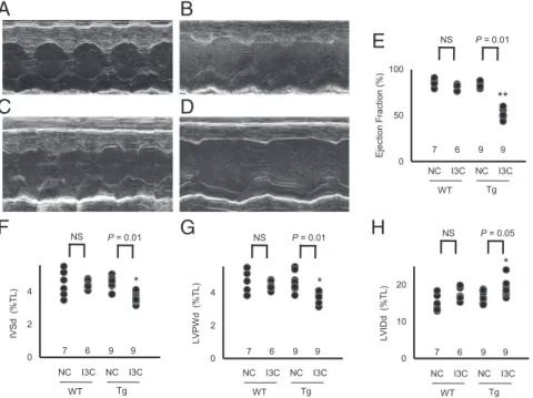
MEDICAL
found that the ejection fraction of the left ventricle, an indicator of
cardiac function, was decreased from∼85% to ∼50% in the Tg
mice fed IC3, but was not changed in the Tg mice fed NC (Fig. 4
A–E). The thicknesses of the intraventricular septum and the left
ventricular posterior wall were also significantly reduced in the
Tg mice fed I3C (Fig. 4FandG). Thus, a threefold increase in
plasma prolactin concentration is sufficient to severely impair cardiac function and structure.
A
B
C
D
E
F
I
H
G
Fig. 2. Bidirectional change in arterial pressure by overexpression of prolactin is associated with changes in urinary excretion of nitrite/nitrate and in the expression of 16K prolactin and its downstream genes (*P<0.05 and **P<0.01 vs. WT; NS, not significantly different). (A) sBP in WT and prolactin Tg male mice on NC or on the I3C diet determined with a tail-cuff method. (B) Heart rate in WT and prolactin Tg mice fed NC or the I3C diet. (C) Urinary nitrite/nitrate excretion. (D) Food consumption. (E) Renal expression of eNOS in WT and the prolactin Tg mice without and with I3C administration. (F) Immunoblot for Ser1177-phosphorylated and total eNOS in the liver of WT and Tg mice without and with I3C administration. (G) Immunoblot for the full-length (23K) prolactin and 16K prolactin in the plasma of WT and the Tg mice without and with I3C administration. (H) Renal expression of PAI-1 in WT and prolactin Tg mice without and with I3C administration. (I) Renal expression of iNOS in WT and prolactin Tg mice without and with I3C administration.
0 50 100 150
A
P = NSSy
sto
lic BP
(m
m
H
g
)
P = NS
P
u
ls
e rat
e
(bpm
)
B
C
P = NS P = NSeNOS - /- eNOS -
/-eNOS -
/-P = 0.01
* P = NS
**
)l
m/
g
n(
nit
c
al
or
p
a
m
s
al
P 5 5 6 5
NC I3C NC I3C
WT Tg
0 300 600
5 5 6 5
NC I3C NC I3C
WT Tg
0 20 40
6 6 6 6
NC I3C NC I3C
WT Tg
Discussion
In the present study, we have generated male Tg mice that have a
leaky prolactin Tg at theHprtlocus. The Tg is driven by a
pro-moter that is inducible by feeding chow containing IC3. When the Tg mice are fed NC without IC3, their basal plasma prolactin concentration is approximately twice that of WT mice fed NC, their BP is approximately 10 mm Hg less than in WT mice, and their urinary excretion of nitrate/nitrite is approximately three times that of WT mice. When the Tg mice are fed chow containing IC3, their plasma prolactin concentration increases to approximately three times basal, their BP becomes approximately 25 mm Hg higher than in WT mice, and their cardiac function and structure are markedly compromised. In the absence of eNOS, feeding IC3 has no effects. We show that the effects of increased plasma concentrations of prolactin are mediated by its short form (16 kDa).
In humans, several studies have shown that increased levels of prolactin are associated with elevated arterial pressure (18, 19).
In the opposite direction, a loss-of-function SNP in theGPR10
gene, which codes for the prolactin-releasing peptide receptor, is associated with reduced BP (20). Recently, a cohort study has demonstrated that a higher daytime plasma prolactin level is associated with an increased risk of incident hypertension among postmenopausal women (21). These results imply that circulating prolactin levels are positively correlated with BP in humans.
In animal experiments, whether changes in the expression of prolactin cause different BP levels has been debatable. Acute i.v. infusion of prolactin increases BP in rabbits (6). When ovine prolactin was chronically given i.p. via an osmotic minipump in rats, BP was decreased, which was associated with increased extracellular fluid volume (7). It has also been reported that chronic prolactin infusion caused an increase in urinary sodium, potassium, and water excretion, but no significant changes in arterial pres-sure in rats (8). A dopamine agonist lergotrile mesylate, which suppresses prolactin secretion (7), and a rabbit antiserum to rat prolactin significantly lowered BP in rats (22). In contrast,
prolactin has been shown to reduce BP in rabbits with pro-gesterone-induced hypertension (23) and in spontaneously hy-pertensive rats (24).
Prolactin increases NO production (25) by increasing in-tracellular calcium in mouse mammary epithelial cells (26) and by increasing the expression of carboxypeptidase-D, which
re-leases the NOS substrate L-arginine from the C terminus of
polypeptides (27, 28). Prolactin causes endothelium-dependent vasodilation via prolactin receptors in the rat aorta (29).
It has recently been demonstrated that the 16-kDa form of prolactin, which is cleaved out of prolactin by cathepsin-D and matrix metalloproteinases, has antiangiogenic effects, whereas the full-length prolactin has angiogenic effects (13, 30). Over-expression of the 16 kDa impairs cardiac function (31). The 16-kDa prolactin has also been demonstrated to inhibit eNOS ac-tivity by modulating intracellular calcium mobilization (15) and activating protein phosphatase 2A, leading to dephosphorylation and inactivation of endothelial NOS (16). The 16 kDa levels are elevated in plasma, urine, and amniotic fluid obtained from women with preeclampsia (4).
WT mice and healthy women have a decline in BP in
preg-nancy, but mice deficient in eNOS do not (32–34), suggesting
that eNOS is involved in the pregnancy-induced decrease in BP. In the last third of pregnancy and during lactation, prolactin levels in female mice normally become three to four times higher than those in virgin females (32). Our Tg males fed NC have plasma prolactin concentrations comparable to this, and, like the pregnant mice, their BPs are less than in WT mice fed NC, and their cardiac parameters are normal. The situation is completely different when they are fed IC3; their plasma prolactin concen-trations are threefold those of the Tg mice fed NC, their BPs are in the hypertensive range, and their hearts are damaged.
The changes in BP we observed in our Tg mice were accom-panied by the changes in urinary excretion of NO metabolites nitrite/nitrate despite no changes in dietary intake of nitrite/nitrate, suggesting that endogenous production of NO is changed by the
0 2 4
P = 0.05
Ejection
Fraction
(
%
)
LV
ID
d (%
TL)
B
A
C
NS NS P = 0.01
* **
D
E
)
L
T
%(
d
S
VI
P = 0.01 NS
F
G
H
* *
LV
P
W
d
(%
TL)
P = 0.01 NS
7 6 9 9
NC I3C NC I3C
WT Tg
0 2 4
7 6 9 9
NC I3C NC I3C
WT Tg
7 6 9 9
NC I3C NC I3C
WT Tg
0 10 20
7 6 9 9
NC I3C NC I3C
WT Tg
0 50 100
Fig. 4. Cardiac dysfunction by marked overexpression of prolactin (*P<0.05 and **P<0.01 vs. WT; NS, not significantly different; TL, tibia length). (A–D) M-mode echocardiography in WT and prolactin Tg mice with and without the I3C diet. (A) WT mice fed NC. (B) WT mice fed I3C diet. (C) Tg mice fed NC. (D) Tg mice fed I3C diet. (E) Ejection fraction of the left ventricle. (F) Thickness of the intraventricular septum in diastole (IVSd). (G) Thickness of the left ventricular posterior wall in diastole (LVPWd). (H) Left ventricular internal diameter in diastole (LVIDd). MEDICAL
overproduction of prolactin. The total amount of eNOS was not changed by the higher plasma concentrations of prolactin. However, in agreement with published work showing that eNOS is activated when its Ser1177 is phosphorylated by Akt-kinase and that prolactin
phosphorylates Akt-kinase (35–37), Ser1177-phosphorylated eNOS
was increased in the Tg mice fed NC. In contrast, Ser1177 phos-phorylation was decreased in the Tg mice fed I3C, in agreement with work showing that 16-kDa prolactin decreases phosphorylation of Ser1177 (16). These findings, together with our finding that a lack of eNOS almost totally abolished the alterations in BP by changes in plasma prolactin levels, lead us to conclude that the full-length and cleaved 16-kDa prolactin both change BP mainly via modulating activity of eNOS. We also observed an increase in iNOS expression, although lack of iNOS has no significant effects on plasma nitrite/ nitrate levels (38), and the changes we observed in iNOS expression are minor compared with those that others have found to cause decreases in BP (39, 40).
The levels of 16-kDa prolactin in the plasma of the Tg mice fed I3C are more than those in WT, but are much less than those of the full-length prolactin. This situation is similar to the pre-vious finding that the levels of 16-kDa prolactin are increased in the plasma, urine, and amniotic fluid of the patients with pre-eclampsia compared with those of women with normal preg-nancy, although, as in our mice, the amount of 16-kDa prolactin is much less than that of the full-length prolactin (4). Why the hypertensive effect of the 16-kDa form can overcome the de-pressor effect of the full-length prolactin is poorly understood. The two forms clearly differ in many ways. Thus, PAI-1 and iNOS are downstream of 16-kDa prolactin but not of full-length
prolactin (12–14). The 16-kDa form, but not the full-length form,
requires the formation of a complex with PAI-1 to be active (41), and their intracellular signaling pathways differ (42).
Postpartum cardiomyopathy occurs within the ninth month of pregnancy to the fifth month postpartum, and is manifested by hypertrophy or dilatation with a low ejection fraction (43). Our findings that increased levels of full-length and 16-kDa prolactin cause hypertension and cardiac dysfunction are in agreement with the previously suggested link between the 16-kDa prolactin and preeclampsia/postpartum cardiomyopathy.
In summary, we find that different degrees of elevated plasma levels of prolactin have opposite effects: a decrease in BP caused by increased production of NO when plasma prolactin is mod-estly above normal, and an increase in BP together with cardiac malfunction caused by decreased production of NO when plasma prolactin is substantially increased. These findings, together with the known association of greater-than-normal prolactin levels with preeclampsia, suggest that pregnant women with high levels of prolactin may need special attention.
Materials and Methods
Animals.We generated prolactin hypermorphic mice by inserting the single-copy Tg comprising the ratCyp1a1promoter, mouse prolactin cDNA (do-nated by Daniel I. H. Lintzer, Northwestern University, Evanston, IL), and the
3′-UTR ofbGHinto theHprtlocus in ES cells of the E14TG2a strain using homologous recombination (Fig. 1A) (9). After positive selection with hy-poxanthine (16μg/mL), aminopterin (0.175 mg/mL), and thymidine (4.8 mg/mL), the gene targeting was confirmed by Southern blot by using genomic DNA digested by BamHI (9). After generation of the animals, genotyping of prolactin hypermorphic mice was performed based on the presence or absence of the junction between prolactin cDNA and the 3′-UTR ofbGHwith conventional PCR using the toe DNA as a template. The primers used for the genotyping are as follows: forward, 5′-CAAGGTCATCAATGACTGCC-3′; reverse, 5′ -ACAGTGG-GAGTGGCATCTT-3′. The targeted mice were backcrossed into the C57BL/6 strain at least eight times before use.
IC3. Normal mouse chow supplemented with 0.3% IC3 was obtained from Envigo.
Measurement of Biological Parameters.We measured systolic BP (sBP) and pulse rate with the tail-cuff method (44). Plasma levels of prolactin were studied with ELISA (Mouse Prolactin DuoSet; R&D Systems). Urine nitrite/ nitrate levels were studied with the Griess method (Nitrite/Nitrate Colori-metric Assay; Cayman). Metabolic balance studies were performed by using metabolic cages (Solo Mouse Metabolic Cage; Tecniplast). All animal pro-tocols have been approved by the University of North Carolina Institutional Animal Care and Use Committee.
Immunoprecipitation/Western Blot.The kidney was homogenated in immu-noprecipitation buffer (50 mM Tris·HCl, pH 7.4, 150 mM NaCl, 50 mM β-glycerophosphate, 30 mM NaF, 2 mM EDTA, 2 mM EGTA, 30 mM Na4P2O7, 2 mM Na3VO4, 1% Triton X-100) and protease inhibitors (1 mM PMSF, 5μg/mL leupeptin, 5μg/mL antipain, 5μg/mL pepstatin A, and 10μg/mL aprotinin). The homogenate was centrifuged at 1,000×gto pellet the nucleus and debris. Equal amounts of protein were incubated with an appropriate antiserum and protein G agarose for 16 h at 4 °C. The proteins were fractionated by SDS/PAGE (Criterion; Bio-Rad) and detected with Western blot by chem-iluminescence (Supersignal West Pico; Thermo). The polyclonal antibody against the N terminus of prolactin (Cell Signaling) and the monoclonal antibody against GAPDH (Cell Signaling) were used for Western blot.
Quantitative RT-PCR.Total RNA was extracted from different tissues, and the mRNAs were assayed by quantitative RT-PCR as previously described (45). The primers and probes used to measure mRNAs are shown inTable S1.
Echocardiography.For analysis of heart function in mice, we used the Vevo 2100 v1.1.2 ultrasonograph system (VisualSonics) and a 30-MHz transducer. Diastolic and systolic wall thickness, end-diastolic dimensions, and end-systolic chamber dimensions of the left ventricle were measured by using a parasternal short-axis view. All measurements were done in the University of North Carolina Rodent Advanced Surgical Models Core according to the American Society of Echocardiography guidelines.
Statistical Analysis.Data are expressed as means±SEs. To compare groups, we used one-factor ANOVA. Post hoc pairwise comparisons were performed by Tukey–Kramer honestly significance differences test with JMP software (version 5.1.2; SAS). When the number of animals was fewer than five, a nonparametric Kruskal–Wallis test was used to compare the groups.
ACKNOWLEDGMENTS. This work was supported by NIH Grants HL49277, HL70523, and HL71266 and by Career Development Award 2006-102 from the Juvenile Diabetes Research Foundation.
1. Bole-Feysot C, Goffin V, Edery M, Binart N, Kelly PA (1998) Prolactin (PRL) and its receptor: Actions, signal transduction pathways and phenotypes observed in PRL re-ceptor knockout mice.Endocr Rev19(3):225–268.
2. Hinuma S, et al. (1998) A prolactin-releasing peptide in the brain.Nature393(6682): 272–276.
3. Leaños-Miranda A, et al. (2008) Urinary prolactin as a reliable marker for pre-eclampsia, its severity, and the occurrence of adverse pregnancy outcomes.J Clin Endocrinol Metab93(7):2492–2499.
4. González C, et al. (2007) Elevated vasoinhibins may contribute to endothelial cell dysfunction and low birth weight in preeclampsia.Lab Invest87(10):1009–1017. 5. Nakajima R, et al. (2015) Elevated vasoinhibin derived from prolactin and cathepsin D
activities in sera of patients with preeclampsia.Hypertens Res38(12):899–901. 6. Horrobin DF, Manku MS, Burstyn PG (1973) Effect of intravenous prolactin infusion on
arterial blood pressure in rabbits.Cardiovasc Res7(5):585–587.
7. Mills DE, Buckman MT, Peake GT (1981) Effect of prolactin administration and sup-pression on blood pressure and body fluid compartments in the rat.Endocrinology
109(5):1590–1596.
8. Ibarra F, et al. (2005) Prolactin, a natriuretic hormone, interacting with the renal dopamine system.Kidney Int68(4):1700–1707.
9. Bronson SK, et al. (1996) Single-copy transgenic mice with chosen-site integration.
Proc Natl Acad Sci USA93(17):9067–9072.
10. Kantachuvesiri S, et al. (2001) Controlled hypertension, a transgenic toggle switch reveals differential mechanisms underlying vascular disease.J Biol Chem276(39): 36727–36733.
11. Wang J, Brown MA, Tam SH, Chan MC, Whitworth JA (1997) Effects of diet on measurement of nitric oxide metabolites.Clin Exp Pharmacol Physiol24(6):418–420. 12. Lee H, Struman I, Clapp C, Martial J, Weiner RI (1998) Inhibition of urokinase activity by the antiangiogenic factor 16K prolactin: Activation of plasminogen activator in-hibitor 1 expression.Endocrinology139(9):3696–3703.
13. Struman I, et al. (1999) Opposing actions of intact and N-terminal fragments of the human prolactin/growth hormone family members on angiogenesis: An efficient mechanism for the regulation of angiogenesis.Proc Natl Acad Sci USA96(4):1246–1251. 14. Corbacho AM, et al. (2000) Proteolytic cleavage confers nitric oxide synthase inducing
15. Gonzalez C, et al. (2004) 16K-prolactin inhibits activation of endothelial nitric oxide synthase, intracellular calcium mobilization, and endothelium-dependent vaso-relaxation.Endocrinology145(12):5714–5722.
16. García C, et al. (2008) Vasoinhibins prevent retinal vasopermeability associated with diabetic retinopathy in rats via protein phosphatase 2A-dependent eNOS inactivation.
J Clin Invest118(6):2291–2300.
17. Shesely EG, et al. (1996) Elevated blood pressures in mice lacking endothelial nitric oxide synthase.Proc Natl Acad Sci USA93(23):13176–13181.
18. Tanaka S, et al. (1985) Plasma prolactin levels in patients with essential hypertension, malignant hypertension and secondary hypertension.Jpn J Med24(1):19–23. 19. Oney T, Bellmann O, Kaulhausen H (1988) Relationship between serum prolactin
concentration, vascular angiotensin sensitivity and arterial blood pressure during third trimester pregnancy.Arch Gynecol Obstet243(2):83–90.
20. Bhattacharyya S, et al. (2003) Association of polymorphisms in GPR10, the gene en-coding the prolactin-releasing peptide receptor with blood pressure, but not obesity, in a U.K. Caucasian population.Diabetes52(5):1296–1299.
21. Zhang L, Curhan GC, Forman JP (2010) Plasma prolactin level and risk of incident hypertension in postmenopausal women.J Hypertens28(7):1400–1405.
22. Mills DE, Buckman MT, Peake GT (1982) Neonatal treatment with antiserum to pro-lactin lowers blood pressure in rats.Science217(4555):162–164.
23. Mati JK, Mugambi M, Odipo WS, Nguli K (1977) Prolactin and hypertension.Am J Obstet Gynecol127(6):616–619.
24. Ryszka F, et al. (1984) Hypotensive action of prolactin in rats with spontaneous hy-pertension.Acta Physiol Pol35(3):215–217.
25. Meli R, et al. (1996) Recombinant human prolactin induces protection against Salmo-nella typhimurium infection in the mouse: Role of nitric oxide.Immunopharmacology
34(1):1–7.
26. Bolander FF, Jr (2002) Prolactin activation of mammary nitric oxide synthase: Mo-lecular mechanisms.J Mol Endocrinol28(1):45–51.
27. Abdelmagid SA, Too CK (2008) Prolactin and estrogen up-regulate carboxypeptidase-d to promote nitric oxide production and survival of mcf-7 breast cancer cells.Endocrinology
149(10):4821–4828.
28. Thomas LN, Morehouse TJ, Too CK (2012) Testosterone and prolactin increase car-boxypeptidase-D and nitric oxide levels to promote survival of prostate cancer cells.
Prostate72(4):450–460.
29. Gonzalez C, et al. (2015) The prolactin family hormones regulate vascular tone through NO and prostacyclin production in isolated rat aortic rings.Acta Pharmacol Sin36(5):572–586.
30. Macotela Y, et al. (2006) Matrix metalloproteases from chondrocytes generate an antiangiogenic 16 kDa prolactin.J Cell Sci119(pt 9):1790–1800.
31. Hilfiker-Kleiner D, et al. (2007) Acathepsin D-cleaved 16 kDa form of prolactin me-diates postpartum cardiomyopathy.Cell128(3):589–600.
32. Guillou A, et al. (2015) Assessment of lactotroph axis functionality in mice: Longitu-dinal monitoring of PRL secretion by ultrasensitive-ELISA.Endocrinology156(5): 1924–1930.
33. Reckelhoff JF, Hennington BS, Moore AG, Blanchard EJ, Cameron J (1998) Gender differences in the renal nitric oxide (NO) system: Dissociation between expression of endothelial NO synthase and renal hemodynamic response to NO synthase inhibition.
Am J Hypertens11(1 pt 1):97–104.
34. Hefler LA, Tempfer CB, Moreno RM, O’Brien WE, Gregg AR (2001) Endothelial-derived nitric oxide and angiotensinogen: Blood pressure and metabolism during mouse pregnancy.Am J Physiol Regul Integr Comp Physiol280(1):R174–R182.
35. Al-Sakkaf KA, Mooney LM, Dobson PR, Brown BL (2000) Possible role for protein ki-nase B in the anti-apoptotic effect of prolactin in rat Nb2 lymphoma cells.
J Endocrinol167(1):85–92.
36. Bailey JP, et al. (2004) Prolactin and transforming growth factor-beta signaling exert opposing effects on mammary gland morphogenesis, involution, and the Akt-fork-head pathway.Mol Endocrinol18(5):1171–1184.
37. Fulton D, et al. (1999) Regulation of endothelium-derived nitric oxide production by the protein kinase Akt.Nature399(6736):597–601.
38. Cook S, et al. (2003) Increased eNO and pulmonary iNOS expression in eNOS null mice.
Eur Respir J21(5):770–773.
39. MacMicking JD, et al. (1995) Altered responses to bacterial infection and endotoxic shock in mice lacking inducible nitric oxide synthase.Cell81(4):641–650.
40. Yamaguchi N, et al. (2006) Time-dependent expression of renal vaso-regulatory molecules in LPS-induced endotoxemia in rat.Peptides27(9):2258–2270.
41. Bajou K, et al. (2014) PAI-1 mediates the antiangiogenic and profibrinolytic effects of 16K prolactin.Nat Med20(7):741–747.
42. Halkein J, et al. (2013) MicroRNA-146a is a therapeutic target and biomarker for peripartum cardiomyopathy.J Clin Invest123(5):2143–2154.
43. Futterman LG, Lemberg L (2000) Peripartum cardiomyopathy: An ominous compli-cation of pregnancy.Am J Crit Care9(5):362–366.
44. Krege JH, Hodgin JB, Hagaman JR, Smithies O (1995) A noninvasive computerized tail-cuff system for measuring blood pressure in mice.Hypertension25(5):1111–1115. 45. Kim HS, Lee G, John SW, Maeda N, Smithies O (2002) Molecular phenotyping for
analyzing subtle genetic effects in mice: Application to an angiotensinogen gene titration.Proc Natl Acad Sci USA99(7):4602–4607.
MEDICAL