ADVANCES IN TREATMENTS AND ANIMAL MODELS OF PEANUT ALLERGY
Kelly A. Orgel
A dissertation submitted to the faculty at the University of North Carolina at Chapel Hill in partial fulfillment of the requirements for the degree of Doctor of Philosophy in the Department of Cell
Biology and Physiology in the School of Medicine.
Chapel Hill 2018
Approved by: A. Wesley Burks Mohanish Deshmukh Kurt Gilliland
ABSTRACT
Kelly A. Orgel: Advances in Treatments and Animal Models of Peanut Allergy (Under the direction of A. Wesley Burks)
Food allergies are a growing health concern affecting approximately 6-8% of the US population. In particular, peanut allergy has an estimated prevalence of greater than 1% of the population and is uncommonly outgrown, making it a life-long disease. Ingestion of allergens can lead to a variety of allergic symptoms ranging from hives or gastrointestinal symptoms to constriction of the airways and anaphylactic shock. Because there is currently no FDA-approved treatment for food allergy, these patients are managed with education and strict allergen avoidance. However, even with the most careful avoidance, accidental ingestion does occur and can lead to life-threatening anaphylaxis. As a result, treatment options are needed. Treatments currently under investigation in clinical trials include peanut oral immunotherapy (OIT), sublingual immunotherapy (SLIT), and epicutaneous immunotherapy (EPIT), though mechanisms of these therapies remain unclear. While results from these trials are promising, limitations include daily dosing, adverse effects, and limited long-term efficacy after therapy is discontinued. Thus, there remains an urgent need for improved therapy options. The work in this dissertation provides the foundation for future drug discovery. First, IgG-mediated basophil inhibition was elucidated as a mechanism of OIT and SLIT and was shown to be associated with long-lived protection. Understanding this mechanism further may result in a targeted therapy option. Separately, a therapy targeting inhibitory receptors on antigen-specific B cells was developed for the prevention of sensitization in a mouse model of peanut allergy.
Here, we describe the use of the genetically diverse Collaborative Cross to identify
CC027/GeniUnc as a more relevant mouse strain that exhibits a severe reaction following oral sensitization and challenge. Together, this work provides a platform for better understanding the mechanisms of food allergy and its treatments, as well as the development of new
To the peanut-allergic children and their families that I have had the privilege of meeting throughout this process, thank you for always being an inspiration.
ACKNOWLEDGEMENTS
I would not be where I am without the help and support of so many truly amazing people. First, I would like to acknowledge my family, especially my husband, Ryan for his constant help and encouragement. He has always helped me to laugh through the busy times in graduate school. Words can not express how grateful I am to have him in my life. Next, I would like to thank our beautiful baby boy, Aiden, for loving me unconditionally. His smiles are always timed for when I need them most. I am so lucky to have been chosen to be his mom. He won’t remember this time in his life, but he was very much a part of the work in this dissertation. I would also like to acknowledge my parents, Keith and Linda for always pushing me to be the best version of myself. Between these two, I have learned the meaning of hard work, to always ask questions about the world around me, and to make time for the most important thing in life: family. These are values I will carry with me forever. Thank you to my brothers, Chris and Andrew, for being the best big brothers a girl could ask for.
I would next like to acknowledge the many faculty mentors who have helped me throughout this journey, especially my research mentors Drs. Wesley Burks and Mike Kulis. I could not have asked for better mentors to be trained under. Their combined compassion, critical thinking, and encouragement are unlike any I have seen. Dr. Burks has always made training me a priority and helped me to grow as a researcher and as a person throughout the last four years. Mike, better known in the lab as “Kilos,” I can’t thank you enough for everything you have done. Thank you for the continuous laughs throughout the many hours in the mouse room. Thank you for always challenging me to think critically about ideas and data, for
could not have asked for a better lab family. I was fortunate to have wonderful mentors outside of the lab as well. I would like to thank my clinical mentors and friends, Drs. Kenya McNeal-Trice and Michelle Hernandez. I do not know how I got so lucky to have such amazing women to look up to. Thank you for all of the “check-ins” during graduate school and for your constant encouragement. I wish that every student had people like you two cheering them on along the way. I would like to thank my committee members for taking time out of their busy lives to talk with me individually not only about science, but also about balancing family and my future training on several occasions. I am so grateful for all of their advice.
TABLE OF CONTENTS
LIST OF TABLES ... x
LIST OF FIGURES ... xi
LIST OF ABBREVIATIONS ... xii
CHAPTER 1: INTRODUCTION ... 1
1.1 Public Health Concern ... 1
1.2 Pathogenesis ... 2
1.3 Etiology and Risk Factors ... 4
1.4 Peanut Allergens ...10
1.5 Diagnosis ...12
1.6 Prevention ...14
1.7 Treatment ...15
1.8 Animal Models ...27
1.9 Topics Addressed ...34
1.10 Tables ...35
1.11 Figures ...36
CHAPTER 2: BLOCKING ANTIBODIES INDUCED BY PEANUT ORAL AND SUBLINGUAL IMMUNOTHERAPY SUPPRESS BASOPHIL ACTIVATION AND ARE ASSOCIATED WITH SUSTAINED UNRESPONSIVENESS ...38
3.1 Introduction ...38
3.3 Results ...43
3.4 Discussion ...47
3.5 Figures ...51
3.6 Supplementary Figures ...47
CHAPTER 3: SIGLEC-ENGAGING TOLERANCE-INDUCING ANTIGENIC LIPOSOMES (STALS) IN THE PREVENTION OF PEANUT ALLERGY ...58
3.1 Introduction ...58
3.2 Materials and Methods ...59
3.2 Results ...62
3.3 Discussion ...64
3.4 Figures ...67
CHAPTER 4: GENETIC DIVERSITY BETWEEN MOUSE STRAINS ALLOWS IDENTIFICATION OF CC027/GENIUNC AS AN ORALLY REACTIVE MODEL OF PEANUT ALLERGY ...70
4.1 Introduction ...70
4.2 Materials and Methods ...72
4.3 Results ...76
4.4 Discussion ...80
4.5 Figures ...84
4.6 Supplementary Figures ...90
CHAPTER 5: CONCLUSIONS AND FUTURE DIRECTIONS ...92
5.1 Mechanisms of Immunotherapy ...93
5.2 STALs ...95
5.3 Oral Challenge Model ...97
5.4 Concluding Remarks ... 100
LIST OF TABLES
LIST OF FIGURES
Figure 1-1 Schematic of peanut allergy pathogenesis ...37
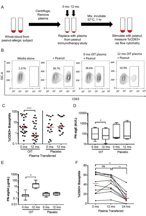
Figure 2-1 Basophil activation following pre- and post-immunotherapy plasma transfer ...51
Figure 2-2 Inhibition capacity of IgG-depleted plasma ...52
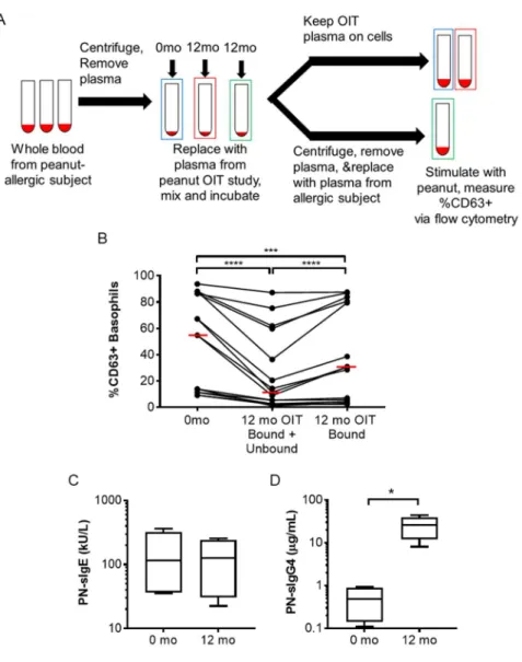
Figure 2-3 Assessment of cellular bound- and unbound-mediated plasma inhibition ...53
Figure 2-4 OIT and SLIT plasma blocking capabilities ...55
Figure 2-5 OIT and SLIT clinical outcomes in relation to basophil inhibition ...56
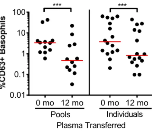
Figure 2-S1 Blocking capability of pooled plasma compared to individual plasma ...57
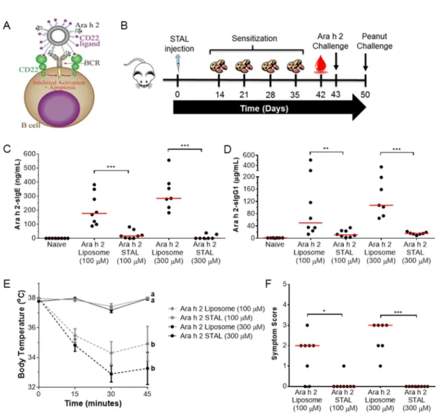
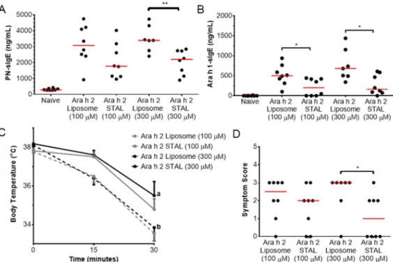
Figure 3-1 Ara h 2-specific immune responses are prevented in mice administered Ara h 2 STALs ...67
Figure 3-2 Ara h 2 STALs specificity ...68
Figure 3-3 Immune responses to peanut and Ara h 1 in mice treated with Ara h 2 STALs. ...69
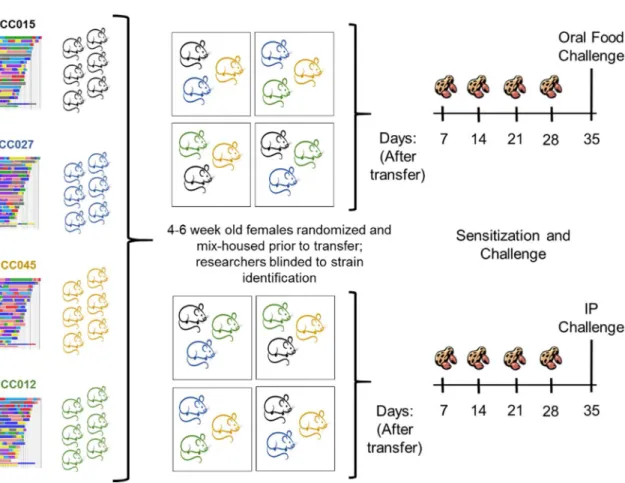
Figure 4-1 Collaborative Cross Screening Approach ...84
Figure 4-2 Anaphylaxis in peanut-sensitized Collaborative Cross strains following peanut challenge. ...85
Figure 4-3. Immune response of CC027/GeniUnc to peanut extract relative to that of C3H/HeJ and C57BL/6J mice ...86
Figure 4-4 Cellular responses in CC027/GeniUnc, C3H/HeJ, and C57BL/6J. ...87
Figure 4-5 Enumeration of effector cells in CC027/GeniUnc, C3H/HeJ, and C57BL/6J ...88
Figure 4-6 Post-OFC serum levels of mast cell degranulation marker and the major peanut allergen Ara h 2. ...89
Figure 4-S1 Post-sensitization peanut-specific immunoglobulins and reaction severity correlation ...90
LIST OF ABBREVIATIONS
Alum aluminum hydroxide
ARA-LAMP-vax peanut Lysosomal Associated Membrane Protein DNA Plasmid vaccine
BCR B cell receptor
CC Collaborative Cross
CHILD Canadian Health Infant Longitudinal Development CoFAR Consortium of Food Allergy Research
CRD component resolved diagnostics
CT cholera toxin
DBPCFC double-blind, placebo-controlled food challenge
DO Diversity Outbred
EPIT epicutaneous immunotherapy FAB facilitated antigen binding FAHF-2 Food Allergy Herbal Formula-2 FVIII factor VIII
Gata3 Gata-3 gene
GRP genetic reference panel
GWAS genome wide association assay
HLA human leukocyte antigen
HRP horseradish peroxidase
IgA immunoglobulin A
IgE immunoglobulin E
IgG immunoglobulin G
IgG1 immunoglobulin G1
IgG4 immunoglobulin G4
IL10 interleukin-10 gene IL-10 Interleukin-10
IL12 interleukin-12 gene
IL-12p40 interleukin-12 subunit p40 IL-13 interleukin-13
IL-4 interleukin-4
IL-5 interleukin-5
IP intraperitoneal
ITIMs immunoreceptor tyrosine-based inhibitory motifs
IV intravenous
LEAP Learning Early About Peanut Allergy MMCP-1 mucosal mast cell protease-1
NOD non-obese diabetic
OFC oral food challenge
OIT oral immunotherapy
OVA ovalbumin
Ox40L Ox40 ligand gene
Ox40L Ox40 ligand
PN-sIgE peanut-specific IgE PN-sIgG4 peanut-specific IgG4
PRROTECT Peanut Reactivity Reduced by Oral Tolerance in an Anti-IgE Clinical Trial SCID severe combined immunodeficient
SEB Staphylococcal Enterotoxin B
sIgE specific IgE
Siglecs sialic acid-binding immunoglobulin-type lectins
SLIT sublingual immunotherapy SNP single nucleotide polymorphism
SPDP succinimidyl 3-(2-pyridyldithio) propionate
SPT skin prick test
STAL siglec-engaging tolerance-inducing antigenic liposome
SU sustained unresponsiveness
Tfh T follicular helper
Th T helper
TIM-4 T cell immunoglobulin-domain and mucin domain-4 TLR toll-like receptor
TNFα tumor necrosis factor α Treg T regulatory cell
CHAPTER 1: INTRODUCTION
1.1 Public Health Concern
IgE-mediated food allergies occur in 6-8% of children under four years old and are estimated to affect 15 million Americans.1-3 The foods most commonly associated with food allergy in the US include milk, egg, peanut, tree nuts, wheat, soy, fish, and shellfish.4 Not only are food allergies common, but they are also increasing in prevalence.5-7 In a 2013 report, the US Centers for Disease Control and Prevention estimated an increase in food allergy
prevalence from 3.4% to 5.1% over a 14 year period.8 A survey study estimated that peanut allergies in the US increased from 0.4% in 1997 to 0.8% in 2002 and then to 1.4% in 2008.9 This increase in prevalence places a larger number of US children at risk for life-threatening anaphylaxis.
Food-induced anaphylaxis accounts for about one third of cases of anaphylaxis seen in hospital emergency departments.10 It is estimated that approximately 30,000 food-induced anaphylactic events are seen in US emergency departments each year with about 200 of these reactions resulting in death.11,12 The majority of fatal reactions are associated with ingestion of peanuts or tree nuts.11,12 During an allergic reaction, symptoms can range from mild urticaria to life-threatening anaphylaxis, and most frequently include involvement of the skin,
food.13 In fact, the stress and anxiety provoked by food allergies, reach beyond the affected patient and truly impact the entire family.14 Despite the severe nature of the disease, large public health concern, and drastic lifestyle implications, the lack of FDA-approved treatments and understanding of the etiology of food allergy leave significant knowledge gaps.
1.2 Pathogenesis
The pathogenesis of food allergy can be divided into two phases which are summarized in Figure 1-1. In the first phase, the sensitization phase, an allergen, defined by the American Academy of Asthma, Allergy and Immunology as a typically harmless substance that is capable of triggering an allergic reaction, enters the body and leads to the production of antigen-specific IgE. In the second phase, the reaction phase, a later exposure to the allergen causes allergic symptoms including symptoms related to the skin, gastrointestinal and respiratory tracts. In the sensitization phase, antigen is taken up and presented by antigen presenting cells to naïve CD4+ T cells. Allergic individuals develop a CD4+ T helper (Th) 2 skewed immune response. During such a response, CD4+ T cells secrete Th2-type cytokines including 4, 5, and IL-13.15 These cytokines trigger B cells to class switch and produce antigen-specific IgE.16 This antigen-specific IgE binds to the high affinity IgE receptor, FcεRI, on effector cells including mast cells and basophils. At this point, the effector cells are primed for a future reaction. It is important to note that unlike B cells and T cells, mast cells and basophils are not specific for a single antigen. It is estimated that up to 500,000 IgE molecules can blanket the mast cell surface at one time, and these molecules have different specificities.17
degranulation event, and this work has been reviewed elsewhere.19 During degranulation, mast cells release pre-formed granules containing mediators such as histamine, proteoglycans, and proteases including tryptase.20 The most readily available mediator, histamine has several physiological effects including vasodilation, increased vascular permeability,
bronchoconstriction, and mucus production that are responsible for the symptoms observed during an allergic reaction.19 Because of the quick release of pre-formed histamine, the resulting symptoms of itching, swelling, throat tightness, and difficulty breathing have an acute onset, usually within minutes of ingestion of the antigen. Histamine has a short half-life, resulting in its quick clearance and resolution of symptoms. In addition to the release of pre-formed mediators, activated mast cells also synthesize and release cytokines, leukotrienes, prostaglandins and platelet activating factor, all of which further act on smooth muscle and vasculature.17
During mast cell development, progenitor mast cells migrate from the bone marrow to tissues, where they take up residence and complete their maturation.20 As a result, human mast cells have proven difficult to study. Basophils are effector cells that, similar to mast cells, express FcεRI and release granules containing histamine.21 Despite their many similarities, basophils also have many characteristics that distinguish them from mast cells. For example, basophils are short-lived, with a life span of days, while mast cells remain in tissues for
months.21 Basophils also have larger and fewer granules than mast cells and have low tryptase content.21 Unlike mast cells, mature basophils circulate through the vasculature system, making them easier to obtain from subjects for research purposes. Because of their similarities in IgE-activation, basophil activation following ex vivo stimulation with antigen has been widely studied, as will be discussed in Chapter 2.
the initial reaction have resolved.22 These late-phase reactions peak 4-8 hours following
allergen ingestion.23 One study of emergency room records from two Canadian hospitals found that 14.7% of patients who were seen for an allergic reaction to food experienced a biphasic reaction.24 In separate studies, examination of affected tissues including skin, nasal mucosa, and lung revealed infiltration by eosinophils, neutrophils, CD4+ T cells, and basophils during late-phase reactions.23 It is not known which patients are likely to develop late-phase reactions and monitoring guidelines remain inconsistent.25
1.3 Etiology and Risk Factors
Much remains unknown about the initial phase of sensitization in food allergic patients, which is ultimately a failure of oral tolerance mechanisms. Oral tolerance, described as a state of active inhibition of immune responses to ingested food proteins, occurs in the majority of people.26 While the mechanisms of oral tolerance are not completely understood, it is well accepted that T cell anergy, T cell deletion, or induction of T regulatory cells (Tregs) all mediate the induction of oral tolerance.26 A breach in any of these mechanisms can result in the
development of food allergy, yet the risk factors that make an individual susceptible remain unclear. Research on this topic has focused primarily on the potential routes of initial exposure, genetics, and the gut microbiome. Research in each of these areas is discussed briefly below.
1.3.a Routes of Sensitization
For unknown reasons, patients with food allergies have a defect in either their ability to develop or maintain oral tolerance to a food antigen. Interestingly, in most cases of food allergy, the first reaction is observed during the first consumption of the eliciting food. Thus, the timing and route of the first exposure remain an intriguing question. Some researchers have
hypothesized that sensitization may happen in utero. One study of 503 infants with likely egg or milk allergy found that peanut consumption during pregnancy was a predictor for the
Avon Longitudinal Study of Parents and Children, a large birth cohort study in which 14,000 pregnant women were enrolled, to investigate this question.28 This study reported that of 23 preschool-aged children with confirmed peanut allergy, there was no evidence of sensitization from maternal diet and cord blood had no detectable peanut-specific IgE (PN-sIgE). A separate study showed that while IgE was detectable in cord blood samples, it appeared to be of
maternal origin, as it matched the specificity of IgE in maternal blood, and was not found in blood samples from these infants at 6 months of age.29 These reports suggest that sensitization does not occur in utero, though there is clearly conflicting data. Other studies have investigated whether peanut exposure occurs through breast milk, and the results are similarly inconclusive. Peanut proteins Ara h 1, Ara h 2, and Ara h 6 have been detected in breast milk as soon as one hour after consumption, making breast milk a potential source of allergens for sensitization.30-32 Three large studies did not find any association between consumption of peanut in lactating moms and the development of peanut allergy in their infants.27,28,33 Despite the
recommendation that pregnant and lactating mothers of children at risk for developing atopic diseases avoid antigen consumption, a meta-analysis found that this avoidance did not protect against allergic diseases, including food allergy.34
There is a well-established link between atopic dermatitis, or eczema, and food allergy.35 Reports have documented that about one third of patients with moderate to severe atopic
dermatitis have a food allergy.36 Because eczema results in a defective skin barrier, it has been proposed that children with eczema are sensitized to food allergens epicutaneously.
Environmental peanut protein is detectable in house dust37 as well as in schools,38 and was found to increase the risk of peanut allergy in a dose-dependent manner,37 further increasing the plausibility of this hypothesis. However, the results of several studies suggest that a
of food allergies.40,41 However, because food allergies and eczema are so closely linked, it is hard to distinguish the contribution of fillagrin mutations to the development of food allergy, independent of its role in eczema pathogenesis. Several small animal models, which will be discussed further in Section 1.8.b, have demonstrated that mice can be sensitized to food allergens epicutaneously.42,43 Together, this work suggests that environmental exposure to food allergens may ultimately lead to sensitization in a select, atopic population.
Recently, work has investigated whether peanut protein in the environment is capable of sensitizing children through the airway. Detecting peanut protein in air samples has proven difficult, but it was detectable by a sampler head placed directly above peanuts actively being shelled.37,44 In a recent study, peanut-responsive Th cells (indicated by increased CD154 expression following ex vivo stimulation with peanut) from peanut-allergic subjects had
increased expression of CCR4, a skin and airway chemokine receptor, compared to non-allergic subjects.45 However, they did not have different expression of the skin chemokine receptor, CCR10, or the skin homing antigen, CLA. These results suggest that peanut sensitization may, at least partly, occur through the airways. Another study found that both BALB/c and C57BL/6 mice developed peanut allergy after four weeks of airway exposure to peanut.46 Altogether, these findings suggest epicutaneous and airway exposures may result in sensitization in addition to presumed sensitization via oral exposure.
1.3.b Genetics
There is little dispute that a person’s genetics are a contributing factor to the
pairs: mother-child, father-child, child-sibling. Later, a twin study, which found that there was a higher concordance of peanut allergy among monozygotic twins, reported the heritability of peanut allergy to be 81.6%.48 Several studies have specifically evaluated the association of human leukocyte antigen (HLA) alleles with the development of peanut allergy, and these are thoroughly reviewed elsewhere.49 Briefly, there is conflicting data on the association between HLA loci and peanut allergy. In one study by Howell, et al., the frequency of HLA-DRB1*08 and DQB1*04 were increased in peanut-allergic subjects compared to controls; however, no
significant HLA class II associations were found when comparing peanut-allergic subjects to their non-allergic siblings.50 As a result, these alleles are likely associated with atopy rather than specific to peanut allergy. Shreffler and colleagues were unable to replicate these findings and concluded that there is not an association between HLA alleles and the development of peanut allergy,51 while a recent 2017 study was able to replicate the original findings.52 Based on a similar study, Dreskin, et al., concluded that HLA-DRB1*08 is a marker of families that have an increased propensity for developing peanut allergy.53 In 2015, the first genome wide association assay (GWAS) of a food allergy cohort was performed to identify associations between genetic variables and food allergy.54 This study found two single nucleotide polymorphisms (SNPs), one intergenic between HLA-DQB1 and HLA-DQA2 and one in the HLA-DRA gene product, to be associated with peanut allergy. Further, the association between these SNPs and peanut allergy was at least partially due to DNA methylation. Overall, these studies demonstrate that the relationship between HLA and peanut allergy is not
straightforward.
peanut allergy: clade B serpin gene cluster, the cytokine gene cluster at 5q31.1, the fillagrin gene, C11orf30, and HLA.56 These loci are all involved in immune regulation and epithelial barrier function. Interestingly, this study found that the effect of the fillagrin mutation (discussed previously in Section 1.3.a) was independent from the development of eczema. In summary, the genetic component of food allergy is complex, but studies such as the ones mentioned above give rise to potential risk identifiers as well as insight into disease mechanisms.
1.3.c Gut Microbiome
The rise in food allergy prevalence observed over the last 50 years has led to immense speculation about potential causes, with special attention to changes in lifestyle and
environment. The hygiene hypothesis, which says that lack of appropriate microbial exposure early in life increases the risk of allergic disease,57 was the first to suggest an association between the microbiome and food allergy. Consistent with this hypothesis, children with older siblings as well as children raised in rural areas have a lower incidence of allergic diseases.58-60 These factors are each known to influence the microbiome of an individual.57 To further
Infant Longitudinal Development (CHILD) study, and fecal samples were collected at three months and 12 months for 16s rRNA sequencing. They found that low microbial richness at three months was associated with food sensitization (confirmed by skin prick test) at one year. Further, Enterbacteriaceae were overrepresented and Bacteroidaceae were underrepresented in samples from food-sensitized infants. The Enterbacteriaceae/Bacteroidaceae ratio was elevated both at three months and one year, suggesting that this colonization occurs early in infancy and persists. In addition to studies investigating microbiota associated with
sensitization, one study has found that gut microbiome composition can predict whether
subjects will outgrow a milk allergy.63 Together, these findings from human studies demonstrate a clear association between gut microbiome and both the development and resolution of food allergies.
Mechanistic microbiome studies have been conducted in mouse models of food allergy. Initial studies demonstrated that animals lacking gut colonization through either antibiotic
treatment64 or germ-free housing,65 were more susceptible to peanut allergy. These results suggest that microbes play a protective role in the development of food allergy. Germ-free mice were then specifically colonized with different microbes to identify the one(s) exhibiting a
protective effect. Several mouse studies have now demonstrated that colonization with Clostridia species is sufficient to suppress peanut sensitization. One study demonstrated that colonization with Clostridia results in increased IL-22 expression and decreased Ara h 2 and Ara h 6 absorption into the blood stream following oral gavage with peanut.65 Use of an IL-22
identify other microbes involved may allow for future manipulation of the microbiome for the prevention and treatment of food allergy.
1.4 Peanut Allergens
Extensive work has been done to identify and characterize the peanut allergens responsible for eliciting the allergic response. The International Union for Immunological Societies recognizes 16 peanut allergens. These allergens are named Ara h 1-17, with the exception of Ara h 4, using the Genus and Species classification for the peanut (Arachis
hypogaea) plant. It currently remains unknown what is responsible for the allergenicity of these
proteins and why proteins with similar structures from other foods do not elicit an allergic response. A 2010 study showed that peanut-allergic individuals in different parts of the world mount an IgE response to peanut allergens to differing extents.67 In this study, they
demonstrated that IgE specificity differed in patients from Spain, the United States, and Sweden and hypothesized that the results were due to differences in dietary habits and pollen
exposures. Each allergen contains several IgE-binding epitopes as well as T cell-binding epitopes. Epitopes can be linear (dependent on the neighboring amino acids) or conformational (dependent on the tertiary structure of the protein). Understanding these allergens and their epitopes has been the target of investigational diagnostics and therapeutics, discussed in Sections 1.5 and 1.7, respectively. Ara h 1, 2, 3, 6, and 9 are considered major allergens, accounting for 75% of peanut protein content,68 and will be discussed further, along with Ara h 8, which has been implicated in Oral Allergy Syndrome.
Ara h 1 is a member of the vicilin seed storage family.69 This family of proteins are typically disc-shaped and comprised of trimers.70 The 63.5 kDa monomers combine to form a 180 kDa trimer.70 As a glycoprotein, Ara h 1 has the potential to be post-translationally
reactivity to Ara h 1.71 Twenty-four linear IgE-binding epitopes, ranging from 6-25 amino acids in length, have been identified.71,72 Interestingly, the IgE-binding epitopes have been shown to cluster in the contact points of the trimer components. The stability of Ara h 1 when subjected to enzymatic digestion remains unclear, as results have varied widely, perhaps due to differing digestion conditions.73
Ara h 2 and Ara h 6 are both 2S albumins that have gained a great deal of attention as major peanut allergens. Ara h 2 has two isoforms with molecular masses of 16.7 kDa and 18 kDa and contains 10 binding epitopes, whereas Ara h 6 is 15.0 kDa and contains 7 IgE-binding epitopes.70 These glycoproteins contain four helices held together by four disulfide bonds.70 Ara h 6 and Ara h 2 share 55% homology in their amino acid sequences and have known cross-reactivity.74 Both Ara h 2 and Ara h 6 have been shown to be resistant to heat and enzymatic digestion.75 Disruption of the disulfide bonds in either allergen has been shown to decrease their allergenicity, suggesting that the conformation of Ara h 2 and 6 are essential for eliciting an allergic response.76 Most peanut-allergic individuals mount an immune response to Ara h 2 and 6. In one study, 100% of children with a peanut allergy had IgE to Ara h 2 and 80% had IgE to Ara h 6.77 This response was exploited in a mouse study where immunotherapy of Ara h 2 and Ara h 6 was sufficient to desensitize peanut-allergic mice.78
Ara h 3 belongs to the legumin family and is a hexameric protein in which each subunit is 60 kDa and contains a disulfide bond.70,79 Four IgE-binding epitopes have been shown to be important in peanut allergy.80 In one study, 42% of peanut-allergic subjects were reported to have Ara h 3-specific IgE.81 Cross-reactivity between Ara h 1, Ara h 2, and Ara h 3 has been noted.82 Further, researchers believe that the similarities of Ara h 3 to other legumins in tree nuts may account for some cross-reactivity observed between allergens.
Ara h 8, a homologue of the birch pollen allergen, Bet v 1, is thought to be responsible for cross-reactivity between birch pollen and peanut.83 Patients with isolated Ara h 8
This local reaction is due to a phenomenon referred to as Oral Allergy Syndrome in which pollen-allergic individuals experience a local reaction confined to the mouth, lips, and throat upon ingestion of a cross-reacting food allergen.84 Thus, identification of isolated Ara h 8 sensitization may be suggestive of a less severe allergy. Ara h 9, while still of significant importance, is less well-studied than the previously mentioned allergens. Ara h 9 has been of particular interest in peanut-allergic patients in the Mediterranean.85 It is a nonspecific lipid transfer protein, thought to require sensitization to the peach allergen, Pru p 3, prior to the development of allergic symptoms to peanut.86
1.5 Diagnosis
The diagnosis of food allergies is based on a thorough clinical history that will guide any further studies. Questions regarding the possible triggers, symptoms of the reaction, timing of symptoms following ingestion of the eliciting allergen, and a family history of atopy can be useful in guiding testing. Two types of routine testing are typically done.87 The first, skin prick testing (SPT), measures mast cell degranulation to a specific antigen. In this testing, an epicutaneous prick is used to place the antigen under the skin and within minutes, local mast cells may release their mediators resulting in wheal and flare formation. SPT has a negative predictive value of greater than 90%, making it a useful test for excluding food allergy.88 However, this testing only has a 50% positive predictive value that has been shown to vary based on age.89 Therefore, use of SPT needs to be chosen carefully to avoid unnecessary avoidance of foods. The second testing type involves measurement of serum antigen-specific IgE levels.
Due to the discrepancy between sensitization and allergy, an oral food challenge (OFC) remains the gold standard for diagnosis of food allergies.91 This test involves having the patient or research subject ingest gradually increasing doses of food containing the allergen while a supervising medical professional observes for signs of an allergic reaction. An OFC is typically done unblinded and without a placebo control. The OFC has many obstacles including the length of time required, cost, often anxiety for the patient and family, and potential risk of an allergic reaction, including anaphylaxis. Because of safety concerns, OFCs should only be performed in clinical settings that have experienced personnel with proper medications to treat potential reactions.
Research has led to several advancements in the diagnostics described above. Lately, a great deal of attention has been given to component resolved diagnostics (CRD). CRD measures serum IgE against individual peanut allergens. The hope of using CRD over
peanut allergen, Ara h 2.96 They used patient sera to determine immunogenicity of these epitopes and hope that this will become a future diagnostic tool.
1.6 Prevention
Landmark studies completed in the last five years have drastically altered the American Academy of Pediatrics recommendations regarding the timeframe for introduction of peanut. Previously, parents were told to avoid feeding their children peanut until 3 years of age.97 However, in 2008, researchers in the United Kingdom (UK) noted that the prevalence of peanut allergy amongst Jewish children in the UK was 10 times higher than in Jewish children from Isreal.98 Further, their surveys indicated that peanut was introduced earlier and eaten in larger quantities in Israel than in the UK. These results suggested that the increased early
consumption of peanut may lead to prevention of peanut allergy.
Du Toit, et al. tested this hypothesis in the Learning Early About Peanut Allergy (LEAP) trial. In this study, 640 infants at risk for developing peanut allergy due to a history of severe eczema and/or egg allergy, but skin prick test <4 mm were randomized to either early peanut consumption or peanut avoidance starting between four and 11 months of age.99 The 60 month outcomes were broken down into subjects with a negative skin prick test and positive skin prick test at the start of the study. For the intention-to-treat analysis, of the infants that had negative skin prick tests, 13.7% of the avoidance group and 1.9% of the consumption group (p<0.001) developed a peanut allergy. Similarly, in the intention-to-treat analysis of infants with a positive skin prick test, 35.3% of the avoidance group and 10.6% of the consumption group developed a peanut allergy. These results confirm that early and regularly ongoing weekly exposure to peanut in a high-risk population prevents the development of a peanut allergy.
discontinuing peanut consumption for 12 months.100 After avoiding peanut for 12 months, the intention-to-treat analysis revealed that the prevalence of peanut allergy was 18.6% in the peanut-avoidance group and 4.8% in the peanut-consumption group when both the skin prick positive and negative cohorts were combined (p<0.001). These results demonstrate that the protection conferred during the 60 months of peanut-consumption was long-lasting, even after consumption was discontinued. Immunologic assessment showed that PN-sIgE and Ara h 2-specific IgE were higher in the avoidance group and that peanut-2-specific IgG4 (PN-sIgG4) levels continued to be higher in the consumption group, despite a decline that started around 30 months, while subjects were still consuming peanut. As a result, an addendum was created to the 2010 Guidelines for the Diagnosis and Management of Food Allergy in the United States. This addendum now recommends introducing peanut into infants’ diets as early as 6 months of age.
1.7 Treatment
Recent work in the field has focused on the use of peanut immunotherapies to prevent life-threatening reactions. These immunotherapies include oral immunotherapy (OIT),
sublingual immunotherapy (SLIT), and more recently, epicutaneous immunotherapy (EPIT). Characteristics of OIT, SLIT, and EPIT are summarized in Table 1-1. While there are many differences between these therapies, the idea behind each of them is the same. The subject is exposed to escalating doses of allergen, with the goal being that after treatment, a peanut-allergic individual may tolerate an accidental ingestion of the allergen. Two clinical endpoints are often discussed: desensitization and sustained unresponsiveness (SU). A subject
experiences desensitization when they are able to tolerate ingestion of a predetermined amount of allergen while still on therapy. During SU, however, the subject can still tolerate ingestion of the allergen after therapy has been discontinued for a period of time, suggesting that the immune changes induced by therapy are more long-lived. While these terms are useful in discussing clinical trial outcomes, it is important to note that the OFC doses used to determine desensitization and SU as well as the time off therapy before designating someone as
experiencing SU are not consistent between studies. Numerous paramount, well-designed studies have been conducted on the use of OIT, SLIT, and EPIT to treat peanut allergy and these will be reviewed below.
1.7.a Oral Immunotherapy
of the 29 subjects successfully consumed 3,900 mg peanut with no more than mild symptoms. Only one subject required the use of epinephrine during the OFC. Interestingly, desensitization appeared to occur much earlier than expected. By 6 months of therapy, skin prick test reactivity was smaller and basophils were less reactive to ex vivo peanut stimulation. By 18 months of therapy PN-sIgE was decreased from baseline and PN-sIgG4 was increased. Additionally, the frequency of CD4+CD25+FoxP3+ Tregs was transiently increased in subjects on peanut immunotherapy, leaving changes in their function unknown.
In 2011, the first double-blind placebo-controlled OIT trial was reported by Varshney, et al.103 In this study, subjects aged 1 to 16 years old were randomized 2:1 to receive peanut flour or placebo for 12 months. After an initial day escalation and build-up phase, subjects received a 4,000 mg maintenance dose for one month before undergoing a double-blind,
placebo-controlled food challenge (DBPCFC) with a maximum cumulative dose of 5,000 mg of peanut protein. Three peanut OIT subjects withdrew from the study due to adverse symptoms while dosing, but all of the remaining 16 subjects in the treatment arm ingested the 5,000 mg of protein. Subjects in the placebo arm, however, ingested a median cumulative dose of 280 mg protein. In comparison to the placebo group, the immune changes in the treatment group were similar to those described previously by Jones and colleagues. This trial further showed that therapy resulted in decreased IL-5 and IL-13, and an increased ratio of FoxP3hi:FoxP3intermediate CD4+CD25+ T cells. This study demonstrated that unlike placebo, peanut OIT safely results in the desensitization of most subjects.
Later, a study sought to compare the effect of peanut OIT to peanut avoidance, which is the current standard of care.104 Peanut-allergic subjects between the ages of 7 and 16 years old were randomized 1:1 to active OIT or avoidance for the first phase of the study. Following 6 months, the subjects initially randomized to avoidance were crossed-over to 6 months of active treatment in the second phase of the study. The primary end-point of the study was
DBPCFC. Of the 39 subjects in the active treatment group who completed the study, 69% were successfully desensitized while none in the control intervention group were desensitized. After the second phase of the study in which the avoidance group was crossed-over to active
treatment, 54% of the subjects tolerated the 1,400 mg DBPCFC. Most side effects reported during OIT were mild, and affected the gastrointestinal tract, with only one subject requiring the use of intramuscular epinephrine. Importantly, this study also showed that OIT improved quality of life scores in subjects, suggesting that the positive effect of OIT reaches beyond immune changes.
Together, the previously mentioned studies show that peanut OIT can be used to desensitize subjects, but the longevity of this affect remained in question. In a follow-up study to the 2009 Jones paper described above, Vickery, et al. investigated the ability of OIT to induce SU, defined as the persistence of tolerance to an antigen after stopping therapy for a period of time.105 To test this, eligible subjects from the previous study underwent two additional food challenges of 5,000 mg. The first, termed “desensitization food challenge”, was completed at the end of therapy, while the second, termed “SU food challenge” was completed one month after therapy was discontinued. The authors found that of the 24 subjects who completed the study, 50% achieved SU and peanut was subsequently re-introduced into their diet. These subjects had decreased skin prick test reactivity and lower levels of peanut-specific, Ara h 1-specific, and Ara h 2-specific IgE both at baseline and at completion of the study. PN-sIgG4 levels and function, as determined by facilitated antigen binding (FAB) assay, were not different between groups.
of 5,000 mg, during which 81% of subjects on OIT were found to be desensitized. SU was assessed 4 weeks after discontinuing therapy by a second DBPCFC of 5,000 mg peanut protein. Results were compared to that of 154 standard of care controls retrospectively collected from a pediatric allergy clinic database. Authors found that in the intention-to-treat analysis, 81% of the study population passed the desensitization challenge, and 78% of the 300 mg arm and 85% of the 3,000 mg arm achieved SU. Adverse events were common in both arms of the study, affecting 95% of the subjects, though slightly more frequent within the group receiving the higher maintenance dose. However, side effects were mostly mild, with only one subject requiring treatment with epinephrine. Compared to baseline, PN-sIgE levels decreased while PN-sIgG4 levels increased following treatment with OIT. These results suggest that in younger peanut-allergic children, a shorter regimen of OIT with a lower maintenance dose is sufficient to induce desensitization and SU.
Other clinical studies have investigated the use of peanut OIT in combination with other treatments. In one such clinical trial, Tang, et al. determined the efficacy of probiotics in combination with OIT to desensitize subjects with peanut allergy.107 In this 2014 double-blind, placebo-controlled trial, 62 children between the ages of 1 and 10 were enrolled in an 18 month study in which they were randomized to either OIT combined with probiotics or placebo. On the last day of treatment, a DBPCFC of 3,000 mg was conducted to assess desensitization. The subjects who were desensitized discontinued therapy for 2 to 5 weeks, and then underwent a second DBPCFC of 3,000 mg to determine SU status. Results showed that 82.1% of the treatment group and only 3.6% of the placebo group achieved SU. Similar to OIT studies reviewed above, treatment with probiotics combined with OIT led to decreased skin prick test reactivity and PN-sIgE levels but increased PN-sIgG4 levels compared to placebo-treated controls. Importantly, this study lacked an OIT-treated group that received peanut in the
Separately, a study published in 2016 investigated the use of omalizumab, an anti-IgE monoclonal antibody, in conjunction with OIT to treat peanut allergy.108 In the Peanut Reactivity Reduced by Oral Tolerance in an Anti-IgE Clinical Trial (PRROTECT), 37 subjects between the ages of 7 and 25 with known peanut allergy were randomized to either omalizumab or placebo in combination with peanut OIT. Subjects received study drug (omalizumab or placebo) for 12 weeks before undergoing a one-day rapid desensitization with peanut OIT. They then
continued weekly up-dosing until reaching 2,000 mg peanut protein. The omalizumab-treated group tolerated a median peanut dose of 250 mg while the placebo-treated group tolerated a median of 22.5 mg on the initial desensitization day, suggesting that treatment with omalizumab allows for rapid desensitization. After discontinuing study drug for 6 weeks, 79% of the subjects in the omalizumab arm and only 12% of the placebo arm were able to tolerate ingestion of 2,000 mg peanut protein, demonstrating that this desensitization is long-lasting in many subjects.
1.7.b Sublingual Immunotherapy
Unlike OIT, which involves the ingestion of the allergen, SLIT requires the allergic subject to place a liquid extract of the allergen under their tongue and hold it in place for a few minutes, allowing for antigen uptake by tolerogenic Langerhans cells in the oral mucosa. SLIT is FDA-approved to treat ragweed and grass pollen allergy but is still investigational for food allergy. In the past 10 years, several reports have investigated the ability of SLIT to desensitize peanut-allergic subjects. In 2011, Kim, et al. conducted the first double-blind,
vivo basophil activation, PN-sIgE levels, and IL-5 levels but increased PN-sIgG4 levels. However, they did not find any changes in levels of IL-13, IFNγ, Tregs, or IL-10. Furthermore, this study demonstrated that the use of peanut SLIT to desensitize peanut-allergic subjects is safe. The study had no dropouts and symptoms reported for 11.5% of peanut doses and 8.6% of placebo doses. The symptoms observed on peanut SLIT were mild, mostly consisting of oropharyngeal itching, and none required treatment with epinephrine.
At the same time as the study discussed above, the Consortium of Food Allergy Research (CoFAR) conducted a randomized, double-blind, placebo-controlled trial in a larger, older study population to investigate the safety and efficacy of SLIT for the treatment of peanut allergy.110 In this study, 40 peanut-allergic subjects between the ages of 12 and 37 were treated with 44 weeks of peanut SLIT or placebo before undergoing a DBPCFC of up to 5 g peanut powder (2.5 g peanut protein). Responders were defined by successful consumption of 5 g peanut powder or a 10-fold increase in the consumed dose compared to their baseline OFC. Seventy percent of subjects on active treatment were considered responders with the median consumption dose increasing from 3.5 mg to 496 mg. Eight of the 14 responders still consumed less than 500 mg at the 44 week OFC. Fifteen percent of the placebo group were classified as responders. After the 44 week challenge, the placebo group crossed over to receive 44 weeks of high dose peanut SLIT and then underwent a second OFC. Of the 16 subjects who were evaluated at this OFC, seven (44%) were responders, however, four responders still consumed less than 500 mg at the OFC. Similar trends in PN-sIgE, PN-sIgG4, basophil activation, and SPT reactivity to the Kim 2011 paper were observed. Also similar to the 2011 study, most dosing symptoms involved oral/pharyngeal itching; however, one subject required treatment with epinephrine and dosing was discontinued. Overall, these initial two studies demonstrated that SLIT is safe and moderately effective in desensitizing subjects with peanut allergy.
unanswered. To address this question, Burks and colleagues in the CoFAR network continued following the subjects from the 2013 cross-over study described above.111 Subjects remained on peanut SLIT for a total of up to three years. By the two-year OFC with 10 g peanut powder, 4 out of 37 (10.8%) of the subjects were desensitized, with no difference in median consumed dose between the high dose SLIT cross-over group and the low dose group. All four of these subjects experienced SU as was determined by a 10 g challenge 8 weeks after discontinuing therapy. Again, this study demonstrated that peanut SLIT was safe with about 18% of the doses eliciting mild dose-related symptoms. The study was limited by a large number of subjects who chose to drop out, mainly due to difficulties with daily dosing, but showed that SLIT has a modest effect at inducing SU in peanut-allergic subjects.
Recent work has sought to directly compare OIT to SLIT in terms of both safety and efficacy in the treatment of peanut allergy. In a double-blinded study, Narisety, et al. randomized a total of 21 subjects to either active SLIT/placebo OIT or placebo SLIT/active OIT.112 SLIT doses reached 3.7 mg while OIT doses reached 2,000 mg, and then subjects underwent two DBPCFCs after 6 months and 18 months of therapy. Both arms of the study experienced a greater than 10-fold increase in tolerated OFC dose, though the median tolerated dose in the active OIT group and active SLIT groups were 141-fold higher and 22-fold higher, respectively. More adverse events were reported in active OIT compared to active SLIT. A similar study was conducted to compare the safety and efficacy of OIT and SLIT in the
treatment of cow’s milk allergy.113 All subjects underwent an initial SLIT escalation before being randomized to continuation of SLIT, low dose OIT or high dose OIT. Consistent with the
1.7.c Epicutaneous Immunotherapy
EPIT is an emerging investigational treatment modality for peanut allergy. EPIT involves application of the antigen via a patch that is applied to the skin. Preclinical studies have shown that the antigen is taken up by dendritic cells in the dermis and does not enter the circulation, suggesting that EPIT may be a safer option for the delivery of antigen.114 In clinical studies, the Viaskin patch, created by DBV Technologies, is 26 mm in diameter containing dried peanut extract.115 In a Phase 1 clinical study, Jones, et al. demonstrated that the Viaskin patch is a safe delivery system for peanut in allergic individuals.116 In this double-blind placebo-controlled study, 100 subjects were randomized to patches containing a range of peanut doses or placebo for two weeks. Eighty-four percent of the peanut-treated subjects experienced at least one local adverse event, though 60% of the placebo group did as well, suggesting that the symptoms may be due to the adhesive itself. Importantly, symptoms were mostly mild to moderate, including erythema and pruritus.
age group (6-11 years), in which the response rate was 34.2%. Adverse events in this study were common but mild, as was seen in the other EPIT studies. While peanut EPIT doesn’t seem to be as effective as OIT or SLIT it is possible that a subset of allergic patients may benefit from this technology or that future adjustments in dose and/or duration of treatment may improve outcomes.
1.7.d Other Investigational Therapies
Allergen-specific immunotherapies have stolen the spotlight when it comes to investigational therapies for food allergies, especially peanut allergy. However, many other modes of treatment are under investigation both in clinical and pre-clinical studies. Food Allergy Herbal Formula (FAHF-2) is one such investigational treatment. FAHF-2 is a botanical
investigational new drug consisting of 9 herbs based on a classical Chinese herbal formula.118 Pre-clinical studies showed promising results in murine models of food allergy.119 Peanut-sensitized mice were treated with FAHF-2 for 7 weeks and then challenged at several time intervals. FAHF-2 completely blocked anaphylaxis in the treated mice. Further, Th2 cytokine production and PN-sIgE levels were decreased in treated mice. Recently, a Phase 2 clinical study on the safety and efficacy of FAHF-2 in the treatment of food allergy was completed.118 This study randomized 68 subjects with known allergies to peanut, tree nut, sesame, fish, and/or shellfish to treatment with FAHF-2 or placebo for 6 months of therapy before completing a DBPCFC. The treatment was safe with no serious adverse events, but the therapy was less successful than placebo at desensitizing subjects. In fact, 45.5% of the placebo-treated
resulting in longer protection than OIT alone in the treatment of food allergies.120 These findings, though yet to be shown in human studies, provide another possible use of FAHF-2 in food allergy treatment.
Because food allergy is an IgE-mediated disease, researchers have tried to treat peanut allergic subjects with the anti-IgE monoclonal antibody, omalizumab. Sampson, et al.
investigated the use of omalizumab independent of OIT to reduce the risk of peanut allergic reactions compared to before treatment.121 Subjects were treated with either omalizumab or placebo for 20 to 22 weeks and then underwent an OFC. The study was stopped early because of several severe anaphylactic reactions during the entry OFC. Of the 14 subjects who
completed the study prior to its discontinuation, the subjects on active treatment tended to have greater improvements in tolerated peanut dose. Omalizumab has since been studied in
combination with peanut OIT as discussed previously in Section 1.7.a.
The use of recombinant peanut proteins that maintain their ability to bind T cells but lack the ability to bind IgE would provide a safer form of immunotherapy. To accomplish this,
or mutated Ara h 2 and then challenged.123 The mice treated with the mutated Ara h 2 had less severe symptom scores and lower plasma histamine levels following challenge than either of the other two groups. Surprisingly, human IgE-binding assays identified several peanut-allergic subjects in whom binding to IgE was not altered with mutated Ara h 2. The mutated
recombinant allergens were tested in a Phase 1 trial of E. coli-encapsulated recombinant modified peanut proteins, Ara h 1, Ara h 2, and Ara h 3.124 Five of the 10 peanut-allergic
subjects enrolled in the trial experienced adverse reactions that prevented them from continuing dosing. These results suggest that either not all IgE-binding epitopes were identified or the binding in these subjects depends on more than just the amino acids mutated.
Similar in rationale to the use of mutated recombinant proteins, researchers have tried to develop peptide immunotherapy for the treatment of peanut allergy. This strategy uses short synthetic peptides that contain the sequences of T cell epitopes, but are not long enough to cross-link IgE, thus should not elicit allergic symptoms. Intranasal or subcutaneous
administration of a vaccine containing 30 overlapping Ara h 2 peptides, 20 amino acids in length, reduced symptoms of anaphylaxis as well as serum levels of Ara h 2-specific IgE in a C3H/HeJ murine model of peanut allergy.125 One study sought to develop short T cell epitope-based peptides that target Ara h 2-specifc CD4+ T cells but can’t cross-link Ara h 2-specific IgE.126 By using T cells from 16 HLA-diverse patients, five dominant CD4+ T cell epitopes were identified in Ara h 2. Three short peptide variants, each less than 18 amino acids long and containing these epitopes, were created. Peptides were modified to have serines replace cysteines in order to increase stability. Experiments using sera from these subjects confirmed that none of the peptides bound IgE. ELISPOT cytokine assays demonstrated that the peptides maintained their ability to stimulate T cells to produce IL-4, IL-5, and IFNγ. This methodology has yet to be developed further for treatment in human studies.
injection of mouse skeletal muscle with DNA or RNA expression vectors resulted in protein expression that was detectable in the muscle for up to 2 months after injection.127 Additionally, it has been long known that such DNA vaccines produce a humoral response and that this
response can be boosted with subsequent doses of the vaccine.128 Of particular interest for the treatment of food allergy, a Th2-skewed process, DNA vaccines result in a Th1-skewed immune response.129 The use of genetic vaccination and the rationale for its use to treat peanut allergy in pre-clinical models have been extensively reviewed elsewhere.130 Briefly, the potential of an Ara h 2 DNA vaccine has been investigated in several mouse strains to treat peanut
allergy.131,132 A study where C3H/HeSn, AKR/J, and BALB/c mice received intramuscular injections with the vaccine demonstrated that the immune response induced varied by strain. These results suggest that there will be similar variability in humans. A mouse study found that treatment with a single multivalent peanut (Ara h 1, 2, and 3) Lysosomal Associated Membrane Protein DNA Plasmid Vaccine (ARA-LAMP-vax; Astellas Pharma Inc.) protected
peanut-sensitized mice from allergic reactions following challenge.133 Currently, a Phase 1 clinical trial is enrolling subjects to test the safety and tolerability of ARA-LAMP-vax for the treatment of peanut allergy in humans.
While several of the treatment modalities listed here have shown promise in animal and/or human studies, they have not been investigated in human studies at all or to the same extent as peanut OIT, SLIT, and EPIT discussed previously.
1.8 Animal Models
As with all diseases, animal models of peanut allergy have been paramount in
default immune response to antigens in the gastrointestinal tract is tolerance. Thus, to induce an allergy, researchers first need to break oral tolerance. This has been done in numerous ways including the use of adjuvants and manipulation of the epithelial barrier. Because oral tolerance is the natural response, some researchers believe that sensitization occurs through the skin, and mouse models that are sensitized through this route have been developed. Further manipulation including the creation of humanized mice have been used to model food allergy. These models as well as large animal models will be discussed here.
1.8.a Th2-skewing Adjuvants
Th2-skewing adjuvants have been used to break oral tolerance in animal models of food allergy. Cholera toxin (CT), one such Th2-skewing adjuvant, has been used extensively in food allergy models. In models of both cashew allergy and peanut allergy, oral co-administration of CT and food antigen to either BALB/cJ or C3H/HeJ mice on days 1, 8, 15 and 22 induces allergen-specific IgE and IgG1 production.134,135 C3H/HeJ mice contain a Toll-like receptor (TLR) 4 mutation, leaving the receptor defective, believed to be at least partly responsible for the Th2-skewing of these animals.136 Upon subsequent challenge via intraperitoneal (IP) injection with the antigen, mice experience anaphylaxis. Reactions can be measured
including our own. As a result, most groups have to challenge these mice by IP injection, leaving the need for an orally reacting mouse model of peanut allergy.
The mechanisms by which CT breaks oral tolerance have yet to be confirmed, though such findings may help to shed light on the break-down of oral tolerance in humans. Studies have shown that in vitro treatment with CT leads to maturation of macrophages and dendritic cells and upregulation of their costimulatory molecules as well as chemokine receptors.138 Specifically, CT was shown to increase OX40L expression in CD11c+ dendritic cells located in the mesenteric lymph nodes, and neutralizing antibodies against OX40L abrogated the CT-induced Th2 response.139 Several experiments have shown that treatment with CT results in increased migration of dendritic cells to lymph nodes and to the T cell area of the Peyer’s patch.139-141 Further, these antigen presenting cells were then able to prime naïve
CD4+CD45RA+ T cells and drive their polarization towards a Th2 phenotype. Separately, studies have found that treatment of mice with CT results in increased levels of IL-1 in the gastrointestinal tract and that activating dendritic cells with IL-1 results in Th2 skewing.142,143 Collectively, these findings suggest that alterations in dendritic cells may play an important role in the break-down of oral tolerance and resulting sensitization to foods.
TIM-4 with a polyclonal antibody dampened the Th2 response in these mice, suggesting a potential mechanism for SEB-mediated sensitization. Similar to the unreproducible findings above in C3H/HeJ mice with CT, the oral reactions following this SEB sensitization scheme have proven unreproducible in unpublished work by our group and others. Overall, these data demonstrate that SEB exposure in mice can lead to allergic sensitization in some published reports.
The last adjuvant commonly used to break oral tolerance in mice is aluminum hydroxide (alum). Using this model, mice were successfully sensitized to tree nuts.135 Tree nut extracts and alum were administered by IP injections over four weeks. Mice sensitized according to this protocol mount an IgE response to tree nuts. Following IP challenge, mice experienced
hypothermia with body temperatures decreasing greater than five degrees compared to baseline. The use of alum to break oral tolerance has been expanded to sensitize mice to peanut in a similar manner.146
1.8.b Epicutaneous Sensitization
Food allergy is closely associated with eczema in humans. Often the development of eczema precedes the development of a food allergy, and with the impaired skin barrier present in eczema, it seems possible for patients to be sensitized through the skin. Strid, et al.
mice and culturing them with CD4+ T cells, researchers showed that these cells were stimulated to produce Th2 cytokines, IL-4 and IL-5. These results suggest that the dendritic cells are sufficient to induce Th2 priming. Interestingly, this study also found that peanut acted as an adjuvant in the epicutaneous sensitization to OVA. These studies along with several
others148,149 that demonstrate epicutaneous sensitization give rise to a possible initial exposure site in humans.
1.8.c Airway Sensitization
Recently, the first animal model of peanut sensitization following airway exposure was developed.46 In this study, BALB/c mice and C57BL/6 mice were exposed to peanut flour in the absence of an adjuvant for four weeks by inhalation. After the four week sensitization period, both strains produced PN-sIgE. Mice IP challenged with peanut extract elicited symptoms of an allergic reaction including hypothermia. In this model, airway exposure initiates peanut allergy by involving the IL-1 pathway and IL-4- and IL21-secreting T follicular helper (Tfh) cells. Further research is needed to determine if the contribution of Tfh cells is unique to airway sensitization.
1.8.d Humanized Mouse Models
While work is ongoing to improve mouse models of peanut allergy, the concern remains that differences between rodent and human physiology limit understanding of disease. Recently two humanized mouse models have been created to ameliorate this problem. In the first,
containing human constant regions. Additionally, Burton, et al., engrafted NOD-SCID mice carrying a human stem cell factor transgene with hematopoietic stem cells.151 These mice were engrafted with functional human T and B lymphocytes and human mast cells. Humanized mice were sensitized and challenged intragastrically with peanut butter in sodium bicarbonate. Mice produced a PN-sIgE response and exhibited signs of anaphylaxis including hypothermia. These mice will provide a valuable tool for studying the immune system during sensitization,
anaphylaxis and treatment, as well as enable the development and testing of targeted therapies. Interestingly, all mouse models of food allergy are sex-specific. Only female mice are able to be reproducibly sensitized. Though it has been observed in humans that menses can affect reaction thresholds to foods, little is known about any other differences between males and females with the disease. There is a need to better understand these differences as well as a need to develop a mouse model of peanut allergy in which both males and females react, as future therapies should be tested in both sexes.152
1.8.e Large Animal Models
As discussed previously, one of the limitations in murine models of food allergy, is that the natural response to antigen in the gastrointestinal tract is oral tolerance. Dogs on the other hand are one of the few animals that naturally exhibit allergies to a range of antigens including food and environmental antigens. Thus, dogs provide a potentially valuable model in which to study allergy and anaphylaxis. In 2002, Teuber, et al. published a canine model of food allergy in which dogs were sensitized starting soon after birth to peanut, English walnut, Brazil nut, soy, wheat, and barley in the presence of alum.153 By 6 months of age, SPTs were positive to these antigens and at two years of age, the four dogs sensitized to peanut reacted upon oral
challenge. Symptoms included vomiting and lethargy, but all resolved spontaneously without intervention. This model has since been used to test the use of heat-killed Listeria
be a sensitized to peanut, and thus can be used to improve understanding of peanut allergy as well as test new therapeutics.
One group previously used sheep to developed an allergic asthma model, and in 2012 they sought to examine the allergic response following sensitization to peanut allergens.155 Twenty sheep were sensitized separately to peanut extract and either OVA (experimental phase 1) or house dust mite (experimental phase 2) by four subcutaneous injections in the presence of alum. For the two phases of this study, 40-50% of sheep were sensitized to peanut, as was defined as greater than 50% increase in PN-sIgE levels compared to baseline. Nearly all sensitized sheep produced an IgE response to major peanut allergens, Ara h 1 and Ara h 2. Four out of the five sensitized sheep in Phase 1 also had a positive skin prick test to peanut.
A 2002 report demonstrated the usefulness of pigs as a model system in which to study food allergy and anaphylaxis.156 Previously, it was shown that pigs could be used as a model for asthma.157 Because asthma and food allergy are both Th2-skewed processes, Helm, et al. investigated the use of neonatal pigs as a model system for peanut allergy.156 Newborn piglets were sensitized by IP injection with peanut extract and CT five times within 4 weeks after birth. Pigs underwent an intragastric challenge and skin prick testing alternating at weekly intervals. Reaction symptoms were observed in 11 out of 14 of the sensitized animals that underwent an oral challenge with peanut. Three reacting piglets had signs of respiratory distress and
anaphylactic shock, requiring treatment with epinephrine. Skin prick tests were positive in peanut-sensitized animals, suggesting IgE and mast cell involvement. These large animal models provide useful tools for the future study of peanut allergy, although high costs and limited immunologic reagents available for these animals limits enthusiasm.
of sensitization including epicutaneous, a model that reproducibly is both sensitized and reacts orally has yet to be identified.
1.9 Topics Addressed
The research presented in this dissertation addresses several knowledge gaps
necessary to improve upon, and develop therapy options for food allergies, focusing specifically on peanut allergies. Chapter 2 will further explore the mechanism of peanut OIT and SLIT by determining the role that IgG plays in regulating the activation of basophils following therapy. Chapter 3 will introduce a new investigational treatment targeting antigen-specific B cells to prevent the development of peanut allergy. The lack of an orally reacting animal model of peanut allergy that closely mimics human disease has been extremely limiting to the
development of treatments. Chapter 4 will present the development of such an animal model of peanut allergy, which will be invaluable to the development of therapies, as well as
1.10 Tables
Table 1-1. Immunotherapies under investigation for the treatment of peanut allergy.
Oral Immunotherapy
Sublingual Immunotherapy
Epicutaneous Immunotherapy
Trial Phase (ongoing) Phase 3 Phase 2 Phase 3 Form of Peanut Powder Liquid extract Dried extract Dose (protein
quantity per day) 300-4,000 mg 2-7 mg 100-500 μg Safety
Mostly oral or gastrointestinal;
highest risk for systemic adverse
effects
Local (oral or pharyngeal) effects
Local (skin) effects; lowest risk for serious adverse effects Efficacy
Desensitization Large effect Moderate effect Variable effect Sustained
36
Figure 1-1. Schematic of peanut allergy pathogenesis. During the sensitization phase (Phase 1), there are several factors that are proposed to influence the development of food allergy. These factors include a person’s gut microbiome, potential routes of exposure such as oral, airway, in
utero, or epicutaneous, and a person’s genetics. During sensitization, allergen is taken up by
CHAPTER 2: BLOCKING ANTIBODIES INDUCED BY PEANUT ORAL AND SUBLINGUAL IMMUNOTHERAPY SUPPRESS BASOPHIL ACTIVATION AND ARE
ASSOCIATED WITH SUSTAINED UNRESPONSIVENESS
2.1 Introduction
Peanut allergy is a major public health concern affecting 1% of the US and European populations, rising in prevalence, and outgrown in only 20% of those affected with the
disease.5,9,158 Although there are no FDA- or EMA- approved treatments for peanut allergies, extensive investigation has focused on the use of several immunotherapy approaches. Emerging peanut allergy therapies include OIT,102,105,106 SLIT,109,111 and EPIT,115,117 among others.159 While routes, doses, and duration vary for each form of therapy, these therapies all expose the allergic subject to increasing quantities of peanut protein over a period of months to years. OIT and SLIT have been effective at inducing both short-lived desensitization (defined as an increased allergen threshold while taking therapy daily) and SU (defined as an absence of allergic symptoms during challenge after stopping therapy).160,161 However, no reliable
biomarkers exist to identify which subjects will achieve SU and which will be transiently desensitized.