A TRANSLATIONAL APPROACH TO ASSESS THE RISK OF DIETARY SUBSTANCE-DRUG INTERACTIONS
Garrett Robert Ainslie
A dissertation submitted to the faculty at the University of North Carolina at Chapel Hill in partial fulfillment of the requirements for the degree of Doctor of Philosophy in the
Curriculum in Toxicology in the School of Medicine.
Chapel Hill 2014
Approved by: Mary F. Paine Philip C. Smith E. Claire Dees Alexander Tropsha
ii ©2014
iii ABSTRACT
Garrett Robert Ainslie: A Translational Approach to Assess the Risk of Dietary Substance-Drug Interactions
(Under the direction of Mary F. Paine)
Myriad diet-derived substances, including foods, nutritional supplements, and exotic beverages are increasingly sought for their purported health benefits. These natural, seemingly safe, products can perpetrate pharmacokinetic/pharmacodynamic (PK/PD) interactions with conventional medications, placing the consumer at risk for potential adverse effects. Despite the ubiquitous nature of these products, there is a gap in the understanding of their drug interaction liability. This knowledge gap is due in part to the complex and variable chemical composition, prompting the need to characterize key constituents that contribute to perturbations in ‘victim’ drug PK/PD. Development of a framework to estimate the effect of the mixture using a single, or few, key constituents is principal to risk assessment.
Grapefruit juice (GFJ) is a well-studied beverage shown to inhibit pre-systemic (first-pass) drug metabolism in the gut, increasing systemic drug exposure and potential undesirable effects. GFJ acts by irreversible inhibition of cytochrome P450 3A (CYP3A) activity in the intestinal wall by a class of constituents termed furanocoumarins. 6’7’-Dihydroxybergamottin (DHB) is a well-studied and typically abundant furanocoumarin in GFJ, with inhibitory concentrations (1-5 μM) well below or within concentrations
iv
potent activity of DHB in GFJ makes it a promising candidate to serve as a marker constituent representative of the CYP3A-mediated effect of GFJ.
Anecdotal reports touting GFJ as a PK ‘booster’ raise concern that it may be used to increase systemic exposure to certain drugs. Loperamide is an over-the-counter opioid agonist that acts locally in the gut to slow motility. Incomplete absorption,
v
ACKNOWLEDGEMENTS
I am fortunate and grateful to have had great guidance and mentorship throughout my graduate career. First I would like to express my kind thanks to my research advisor, Mary F. Paine, and the members of my dissertation advisory committee, Philip Smith, Claire Dees, Alexander Tropsha and Maciej Zamek-Gliszczynski.
I wish to show my gratitude towards those who have contributed directly to the maturation of this work. I would like to acknowledge Yolanda Scarlett, Elizabeth Connolly, Yingxin Li and Kristina Wolf for their roles in the loperamide-grapefruit juice interaction study. I would like to thank Evan Kharasch for sharing the clinical data used to model alfentanil pharmacokinetics and pharmacodynamics. I am fortunate to have had the opportunity to work with a fantastic statistician and mentor, J. Heyward Hall. I thank Jeannie Padowski and Gary Pollack for sharing their expertise in pharmacokinetic and pharmacodynamic modeling. I would like to thank all of those have aided me in the naloxone clinical study, particularly. Matthew Layton, John White, Brandon Gufford, Debbie Weeks, Larissa Weeks and Abby Parsons.
vi
vii PREFACE
The role of environmental factors such as diet in the disposition and metabolism of drugs is an aspect that has been long overlooked, although it is now well known that certain dietary substances can interact with drugs that result in untoward effects or reduced efficacy. A meeting by the National Toxicology Program in 1998 highlighted the importance of increased research in the field of dietary substances. However, over a decade later there remains no guidelines to systematically evaluate dietary substance-drug interaction risk. This project was undertaken to evaluate previously proposed translational frameworks to assess dietary substance-drug interaction risk. The key achievements of this work were (1) the identification of 6,’7-dihydroxybergamottin (DHB) as a promising marker constituent reflective of the CYP3A-mediated grapefruit juice effect; (2) the successful construction of physiologically-based
pharmacokinetic/pharmacodynamic models capable of predicting a clinically relevant pharmacodynamic endpoint and; (3) the development of a human model to assess the opioid effect attenuation by naloxone. I conducted the majority of in vitro experiments described in this dissertation, and Maciej Zamek-Gliszczynski provided drug-tissue binding data for loperamide and DHB. My background in bioanalytical chemistry allowed me to conduct all LC-MS/MS analysis with the exception of that described in two clinical studies (loperamide and naloxone). In total, the work described within this dissertation included three clinical studies. The first study was a grapefruit juice-loperamide
viii
study was conducted primarily by Elizabeth Connolly, Yolanda Scarlett, Yingxin Li, and Mary Paine. The quantification of loperamide and its metabolite was conducted by Kristina Wolf, and power calculations were conducted by J. Heyward Hull. A second clinical study described in this dissertation involved the determination of naloxone bioavailability for three extravascular formulations in six subjects. This pharmacokinetic study was led by John White and Mathew Layton and pharmacokinetic analysis was conducted by myself and Jeannie Padowski. I was involved in the study design along with Mary Paine and was the coordinator of the third clinical study described in Chapter 3. During this study I received extensive support from Brandon Gufford, Mathew Layton and John White, who were qualitied to administer medications and evaluate the health of and safety to study subjects. Furthermore, I received oversight and guidance from my research advisor, Dr. Mary Paine. Much of the work described in Chapter 2 involved advanced pharmacokinetic and pharmacodynamic modeling techniques. In the development of these, models I received constructive feedback from several key
ix
TABLE OF CONTENTS
List of figures ...xi List of tables ... xiii List of abreviations ...xv Chapters
1 Introduction ... 1 2 Assessment of a candidate marker constituent predictive of
a dietary substance-drug interaction: case study with
grapefruit juice and CYP3A4 drug substrates ... 54 3 Characterizing the abuse potential of opioids using
physiologically-based pharmacokinetic / pharmacodynamic modeling: case study with the grapefruit juice-loperamide
interaction. ... 86 4 Evaluation of a novel human model to assess reversal
of opioid effects by naloxone ... 117 5 Conclusions ... 138 Appendices
A Method development and validation of an HPLC-MS/MS method to quantify 6’,7’-dihydroxybergamottin and other
grapefruit juice constituents in human plasma. ... 160 B Characterization of furanocoumarin metabolites in human
plasma and urine following grapefruit juice consumption. ... 169 C Recovery of in vitro kinetic parameters of loperamide and
N-desmethylloperamide metabolism in human intestinal
microsomes from individual donors. ... 173 D Recovery of in vitro kinetic parameters of loperamide
x
E Inhibitory potency of supplements labeled to contain
DHB and/or BG toward CYP3A activity. ... 185 F A physiologically-based pharmacokinetic model
of loperamide. ... 190 G Physiologically-based pharmacokinetic DHB-loperamide
interaction model simulations. ... 200 H Physiologically-based pharmacokinetic/pharmacodynamic
xi
LIST OF FIGURES Chapter 1
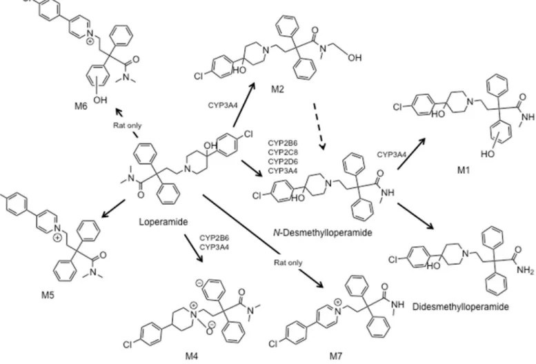
Figure 1.1. Furanocoumarin structures.. ... 37 Figure 1.2. Loperamide metabolites in rat and human liver
microsomes. ... 38 Chapter 2
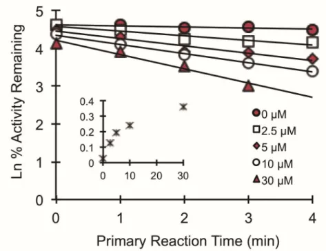
Figure 2.1. Clinical study design and procedures ... 78 Figure 2.3. Time- and concentration-dependent inhibition of
loperamide N-desmethylation by DHB in human
intestinal microsomes ... 80 Figure 2.4. Relationship between the predicted and observed
AUCGFJ/AUC for 15 test drug substrates of the
‘grapefruit juice effect’ due to inhibition of intestinal CYP3A4 ... 81 Figure 2.5. Relationship between the magnitude of a grapefruit juice-
drug interaction for varying enterocyte concentrations
of DHB and victim drug Fg. ... 82 Chapter 3
Figure 3.1. The general model structure of a physiologically-based
pharmacokinetic/ pharmacodynamic model ... 107 Figure 3.2. Observed geometric mean and model-predicted mean plasma
concentration-time profiles of loperamide taken with water or GFJ
and of DHB. ... 108 Figure 3.3. Observed and model predicted alfentanil plasma concentration-
and effect- time profiles after an intravenous or an oral dose. ... 109 Figure 3.4. Observed and model predicted methadone plasma concentration-
and effect- time profiles. ... 110 Figure 3.5. Model-predicted maximum decrease in pupil diameter with increasing
loperamide dose in the absence and presence of DHB. ... 111 Chapter 4
Figure 4.1. Study design and procedures ... 131 Figure 4.2. Concentration-time profiles for naloxone following intravenous,
xii
Figure 4.3. Mean pupil miosis-time profiles after administration
of oral alfentanil. ... 133 Figure 4.4. Area under the effect-time curve and Rmax following
oral administration of alfentanil. ... 134 Appendices
Figure A.1. HPLC-MS/MS separation and detection of DHB ... 165 Figure A.2. DHB stability following storage at -80C. ... 166 Figure A.3. DHB freeze-thaw stability in human plasma. ... 167 Figure B.1. Representative chromatograms following HPLC-MS/MS
analysis of plasma from two subjects. ... 171 Figure C.1. Michaelis-Menten plot for N-desmethylation of
loperamide by HIM. ... 176 Figure C.2. Parent disappearance of N-desmethylloperamide
by HIMs and HLMs. ... 177 Figure D.1 Rat P450 enzymes catalyzing the formation of
N-desmethylloperamide………182
Figure D.2. N-Desmethylloperamide formation at increasing loperamide
concentrations in HLMs, RLMs, HIMs and RIMs. ... 183 Figure E.3. Comparison of the effects of supplements labeled to DHB ... 189 Figure F.1. Loperamide PBPK model structure. ... 192 Figure F.2. Model simulated plasma concentration-time profiles
of loperamide and N-desmethylloperamide. ... 193 Figure G.1. Simulated loperamide hepatic fraction metabolized
xiii
LIST OF TABLES Chapter 1
Table 1.1. Phase I and II pathways and bioavailability (F)
of common opioids. ... 31 Table 1.2. Models to predict perpetrator or victim substance
specific parameters. ... 32 Table 1.3. Static models to assess drug interaction potential. ... 33 Table 1.4. Software packages for PBPK modeling. ... 36 Chapter 2
Table 2.1. Pharmacokinetic outcomes of loperamide and
N-desmethylloperamide in 16 healthy volunteers administered
loperamide with 240 ml of water or grapefruit juice. ... 76 Table 2.2. Victim drug, Fg, F, and clinical study information
for the IVIVE. ... 77 Chapter 3
Table 3.1. PBPK model input parameters. ... 104 Table 3.2. Pharmacodynamic model parameter input. ... 105 Table 3.3.Observed and model-predicted pharmacokinetic and
pharmacodynamic outcomes following oral opioid
administration. ... 106 Chapter 4
Table 4.1. Pharmacokinetics of naloxone after intravenous,
intramuscular, and intranasal naloxone ... 129 Table 4.2. Clinical study inclusion and exclusion criteria. ... 130 Appendices
Table A.1. Precision (%CV) and accuracy (RE) inter- and intra-day
variability of DHB in human plasma (n=3) ... 168 Table B.1. Individual DHB pharmacokinetic outcomes. ... 172 Table D.1. In vitro kinetic parameters for loperamide (Km and Vmax)
xiv
Table G.1. Pharmacokinetic outcomes following oral loperamide and DHB ... 203 Table H.1. Model-predicted pharmacokinetic and pharmacodynamic
outcomes following oral alfentanil administration in
xv
LIST OF ABREVIATIONS
ADAM Advanced Dissolution, Absorption and Metabolism AUC area under the plasma concentration-time curve AUCm AUC of the metabolite
AUCp AUC of the parent
AUEC area under the effect-time curve
BG bergamottin
B/P blood to plasma ratio bid two times daily
CL/F oral clearance
Clast last measured concentration Clint intrinsic clearance
Cmax maximum concentration
CTRC Clinical and Translational Research Center CV coefficient of variation
CYP cytochrome P450
DHB 6’,7’-dihydroxybergamottin DDI drug-drug interaction
DFB 3-[(3,4-difluorobenzyl)oxy]-5,5-dimethyl-4-[4-methylsulfonyl)phenyl] furan-2(5H)-one
DMSO dimethyl sulfoxide fa fraction absorbed
xvi fu fraction unbound
GFJ grapefruit juice
HIMs human intestinal microsomes HLMs human liver microsomes HTS high throughput screening
IC50 half-maximal inhibitory concentration IM intramuscular
IN intranasal
IV intravenous
IVIVE in vitro-to-in vivo extrapolation
ka first-order rate constant for absorption
kg kilogram
Ki reversible inhibitory potency
KI concentration to achieve half the maximal inactivate rate kinact maximal inactivation rate constant
Km concentration to achieve half the maximal metabolic rate LC/MS/MS liquid chromatography-tandem mass spectrometry MAT mean absorption time
MBI mechanism-based inhibition MDCK Madin-Darby canine kidney
mg milligram
mM millimolar
xvii
NADPH nicotinamide adenine dinucleotide phosphate ND not determined
nM nanomolar
OATP organic anion transporting polypeptide PBPK physiologically-based pharmacokinetic
PD pharmacodynamic
P-gp P-glycoprotein
pM picomolar
PK pharmacokinetics RAF Relative activity factor RIMs rat intestinal microsomes RLMs rat liver microsomes rCYP recombinant CYP SD standard deviation SE standard error
UDPGA uridine diphosphate glucuronic acid t1/2 terminal elimination half-life
tid three times daily tmax time to Cmax
Vmax maximal metabolic rate
λz terminal elimination rate constant λ first-order inactivation rate at a given I
xviii
pM picomolar
µM micromolar
1
CHAPTER 1 : INTRODUCTION
Dietary substance-drug interactions are a public health concern
The U.S. Environmental Protection Agency (EPA) implemented their ToxCast high throughput screening (HTS) initiative in 2007 (Dix et al., 2007). This project was designed to screen a bevy of failed pharmaceuticals, alternative plasticizers, pesticides, and food additives across 331 cell-free enzymatic and ligand-binding HTS assays (Haynes, 2010). These assays evaluated direct mechanisms of toxicity, such as those associated with carcinogenicity and neurotoxicity, and alterations in the activity of some cytochrome P450 (CYP) enzymes and transporters (Sipes et al., 2013). The ToxCast assays are demonstrating a potential to prioritize chemicals for more targeted risk assessment testing. The ToxCast approach parallels pharmaceutical discovery in certain ways; however, relative to drugs, little is understood about the quantitative influence of environmental exposures on the metabolism and transport of concurrent medications. Studies to determine the induction of certain CYPs by environmental agents have been reported. For example, heterocyclic aromatic amines,
2
between ‘victim’ drugs and ‘perpetrator’ herbs or foods when consumed concurrently (Won et al., 2012). Beverages, such as grapefruit juice (GFJ), are known to interact with >100 drugs through inhibition of intestinal CYP3A, P-glycoprotein (P-gp), or organic anion transporting polypeptide (OATP). Therefore, the result of consuming otherwise seemingly safe products with certain drugs may be detrimental and is concerning to public health.
The impact of certain dietary substances, including foods, nutritional
supplements, and exotic juices on public health is increasing due to an increase in consumer popularity resulting from the purported health benefits and the misperception that “natural” equates with “safe”. The ever-growing market appearance of these
substances raises concern when co-exposed with drugs, both prescription and non-prescription, potentially leading to untoward or toxic effects (Won et al., 2012). Both qualitative and quantitative assessment of dietary substance-enzyme/transporter interactions will provide more robust risk assessment of their combined intake.
Since dietary substances and many drugs are taken orally, mechanisms
involving the inhibition of first-pass (i.e., pre-systemic) elimination are most likely to be involved. Inhibition of the ‘first pass effect’ can result in substantial increases in victim drug systemic exposure, which may lead to untoward effects. The gut and the liver are the primary organs that mediate the first pass effect. CYP3A is a prominent enzyme subfamily in both the gut and liver and contributes to the metabolism of numerous
marketed drugs and some dietary substances (Shimada et al., 1994; Paine et al., 2006). The human CYP3A family includes four genes: CYP3A4, CYP3A5, CYP3A7 and
3
adults, whereas CYP3A7 is expressed in fetal and neonatal stages of life, and
CYP3A43 has a role in steroid and cholesterol metabolism (Fanni et al., 2014). Given that CYP3A4/5 are responsible for the metabolism of >30% of marketed drugs (Zanger and Schwab, 2013), the liver and intestine are major sites of an interaction.
While the liver remains a major contributor to first-pass metabolism, the gut is also sensitive to enzyme or transporter inhibition. The gut, primarily the small intestine, is a critical portal for the absorption of ingested xenobiotics. Xenobiotic oxidative
metabolism in the intestine is mediated primarily by CYP3A4/5 (Paine et al., 2006). The absorptive intestinal epithelial cells, enterocytes, provide active transport processes for the uptake and efflux of xenobiotics, including dietary substances and drugs. Organic anion transporting polypeptide (OATP) is one a family of uptake transporters expressed in the intestine, among other tissues, is and involved in the absorption of several drugs. A major apically located efflux transporter is P-glycoprotein (P-gp) and is expressed in a variety of other cell types, including endothelial cells in the liver and kidney and capillary cells constituting the blood-testis barrier and blood-brain barrier (BBB) (Ho and Kim, 2005). Perpetrator dietary substance constituents likely attain high concentrations in the enterocytes despite not achieving inhibitory concentrations in the liver.
4
requirements for botanicals than in the US, although safety assessment of these products frequently lags behind that of drugs (Health Canada, 2011; European Medicines Agency: Committee for Human Medical Products, 2012). The FDA
acknowledges the need for improved safety testing, and recent funding mechanisms offered by the National Center for Complementary and Alternative Medicines (division of the U.S. National Institute of Health) highlight this unmet need (de Lima Toccafondo Vieira and Huang, 2012).
Despite a change in the regulatory climate addressing the necessity for some standardization of products [European Medicines Agency (European Medicines Agency: Committee for Human Medical Products, 2012), Health Canada (Health Canada, 2011)] marketed as herbal products and dietary supplements, formal guidelines to assess the risk of dietary substance-drug interactions remain nonexistent. The study of dietary substances has certain challenges unique to those for drug development due to the highly variable biochemical makeup of dietary substances. A standard framework to assess these interactions without expensive clinical trials remains elusive. Since it would be extremely costly and time consuming to fully characterize all constituents of a given dietary substance, investigators have postulated that one or few constituents of the mixture, termed ‘marker constituents’, may be predictive of the effect of the whole mixture (Won et al., 2012; National Center for Complementary and Alternative Medicine, 2013; Ainslie et al., 2014). These marker constituents can be identified and used for testing. However, further work is needed using well-studied exemplar dietary
5 Common mechanisms of enzyme inhibition
Reversible inhibition involves the rapid association and disassociation of an enzyme-inhibitor complex. Reversible inhibition frequently occurs by one of three
specific mechanisms, including competitive, noncompetitive and uncompetitive (Shou et al., 2001; Walsh et al., 2011). Competitive inhibition is the result of a perpetrator
substance binding to the active site of the enzyme preventing access by the victim substrate. The result in victim drug enzyme kinetics is an increase in the concentration needed for half-maximal rate of metabolism (Km) with no change in maximal rate of metabolism (Vmax) (Lin and Lu, 1998; Venkatakrishnan et al., 2003). Noncompetitive inhibition occurs when a perpetrator substance associates to a site of the enzyme other than the active site to attenuate enzyme activity. Noncompetitive inhibition is revealed by a decrease in Vmax and no change in Km. Uncompetitive inhibition occurs with the perpetrator substance binding to the enzyme-substrate complex, resulting in a decrease in both Km and Vmax. In the case of reversible inhibition enzyme activity is restored with removal of the perpetrator substance (Lin and Lu, 1998; Venkatakrishnan et al., 2003).
6
In general irreversible inhibition results from covalent or tight binding (quasi-irreversible) of a chemically reactive intermediate resulting in a loss of enzyme function (Grimm et al., 2009). This specific mode involving enzyme inactivation is known as mechanism-based inhibition (MBI). The MBI associated constant (KI) and the rate of maximal enzyme inactivation (kinact) are the parameters associated with MBI (Kalgutkar et al., 2007; Venkatakrishnan et al., 2010). The effects of irreversible inhibition can be prolonged after the inhibitor is removed, since transcription of new enzyme or cell regeneration is required to recover enzyme activity.
Grapefruit juice as an exemplar dietary substance and 6’,7’-dihydroxybergamottin as a candidate marker constituent
Grapefruit juice (GFJ) is one of the most extensively studied dietary substances shown to perpetrateCYP3A-mediated inhibition of drug metabolism (Paine and
Oberlies, 2007; Bailey et al., 2013). Many of these drugs undergo extensive first-pass metabolism by CYP3A. An extensive list of these medications has been reported elsewhere (Bailey et al. 2013). When consumed in usual volumes, GFJ elevates
systemic concentrations of the victim drug by inhibiting enteric, but not hepatic, CYP3A. Despite that the juice inhibits only enteric CYP3A, the magnitude of the effect can be large enough to elicit untoward effects, such as severe muscle pain with some HMG-CoA reductase inhibitors (statins)(Dreier and Endres, 2004; Karch, 2004) and
hypotension/dizziness with some calcium channel antagonists (Bailey and Dresser, 2004).
7
irreversible inhibitors of CYP3A (Paine and Oberlies, 2007). Using a “furanocoumarin-free” GFJ suitable for human consumption and the CYP3A probe substrate felodipine, furanocoumarins, in aggregate, were demonstrated unequivocally as major CYP3A inhibitors in vivo. In addition to their ability to inhibit enteric CYP3A, furanocoumarins have been shown to inhibit the P-gp in vitro(Eagling et al., 1999; Paine and Oberlies, 2007). Moreover, using the aforementioned furanocoumarin-free juice,
GFJ/furanocoumarins were shown to inhibit the enteric P-gp-mediated translocation of the dual CYP3A/P-gp substrate cyclosporine in healthy volunteers, as well as in the human intestine-derived cell line Caco-2 (Paine et al., 2008). Parallel in vitro studies identified the furanocoumarins bergamottin (BG) and 6’,7’-dihydroxybergamottin (DHB) (Figure 1.1.) as mechanism based inhibitors of CYP3A (KI, 1.6 and 0.7 µM,
respectively) and P-gp activity (IC50, 0.74 and 0.33 µM, respectively) (Eagling et al., 1999; Paine et al., 2004).
Despite the potent inhibitory activity of BG towards CYP3A in vitro, BG has high tissue binding properties and is less readily absorbed than DHB, indicating it may contribute less to the GFJ effect in vivo (Paine et al., 2004; Paine et al., 2005). The clinical role of BG is evidenced largely by two studies designed to evaluate the
8
was not reported) prior to felodipine (10 mg). The lime juice was diluted such that the concentration of BG was the same as that in GFJ. Relative to water, GFJ increased felodipine area under the plasma concentration-time curve from time zero to infinite time (AUC0-inf) and maximum observed plasma concentration (Cmax) by 2- and 1.9-fold,
respectively. In contrast, lime juice had no effect on the pharmacokinetics of felodipine despite an extract having both reversible and irreversible inhibitory activity in a
microsomal CYP3A activity assay. In vitro activity assays were conducted using recombinant CYP3A4, increasing amounts of juice extracts, and testosterone 6β-hydroxylation as the index reaction. The observed in vitro-to-in vivo disconnect may be due to the slower absorption of BG compared to DHB in Caco-2 cells (Paine et al., 2005) and perhaps a delay in felodipine treatment following lime juice would have resulted in an interaction. Alternatively, there may have been components in the lime juice extract which did not attain high enough enteric concentrations.
9
and 12 mg increased felodipine AUC0-12h by 1.3-fold. The GFJ product containing 1.7 mg of BG resulted in a 1.5-fold increase, whereas 2 mg of BG had no significant effect. This study suggested that BG contributes to the GFJ effect but alone does not account for the effect of the mixture in vivo. Additionally, at the higher BG doses, DHB was detected in plasma, indicating that BG is biotransformed to DHB in vivo or the study product was contaminated with DHB.
In vitro assessment of other furanocoumarins, including dimers and trimers, have indicated that some of these constituents are highly potent as well (IC50, 0.003-1 µM) (Guo et al., 2000b; Guo et al., 2000a; Tassaneeyakul et al., 2000; Row et al., 2006; Oda et al., 2007). Despite these data, such constituents have unknown/poor stability (light, pH, and metabolic), and their clinical relevance is unknown (Morliere et al., 1990; Kent et al., 2006). Improved methods to quantify these constituents in both the juice and biologic matrices are required to further understand their role in the CYP3A-mediated GFJ effect.
10
that in healthy volunteers administered GFJ (Lown et al., 1997); and (4) the existence of commercially available authentic standards that are not cost prohibitive.
Grapefruit juice as a ‘pharmacokinetic boosting’ agent
The discovery of GFJ as an irreversible inhibitor of intestinal CYP3A4 has spurred the marketing of dietary supplements labeled to contain BG, DHB and/or GFJ extracts. These products have been marketed as ‘pharmacokinetic boosting’ agents aimed towards body builders to increase the bioavailability of oral hormones and supplements. Many hormones (e.g., androgens) are metabolized by CYP3A (Hara and Nishiyama, 2014), and GFJ has been shown to increase systemic exposure to the sex hormone, 17 β-estradiol (Schubert et al., 1994), providing loose justification for the products marketed use. Even in conventional pharmacotherapy, GFJ has been proposed as a means to increase the exposure to expensive and poorly bioavailable drugs as a cost-savings strategy, including some immunosuppressive and anticancer agents (Reif et al., 2002; Liu et al., 2009; Kimura et al., 2011; Cohen et al., 2012). Similarly, the role of GFJ as a pharmacokinetic booster has been implicated in
anecdotal reports as a means to increase the systemic exposure to certain opioids (e.g., methadone, morphine, oxycodone, hydrocodone and loperamide) (Bluelight, 2014b; Bluelight, 2014a).
Loperamide as a candidate GFJ victim drug with abuse potential
Zamek-11
Gliszczynski et al., 2012). The low bioavailability is in part due to the high first pass effect mediated primarily by CYP2C8 and CYP3A4 (Figure 1.2) (Kim et al., 2004). Anecdotal reports and case studies have implicated the potential for loperamide abuse when taken concomitantly with CYP3A4 or dual CYP3A/P-gp inhibitors (Daniulaityte et al., 2013). A limited number of case studies have also signified that high doses of loperamide can result in abuse or death (Langlitz et al., 2001; Sklerov et al., 2005). In contrast, clinical evaluations in healthy volunteers have shown that increases in
loperamide plasma exposure do not translate to a central opiate-like effect (Tayrouz et al., 2001; Skarke et al., 2003; Niemi et al., 2006). Although supratherapeutic doses of loperamide have been examined (up to 24 mg) in non-addict populations, these doses are much lower than those from anecdotal reports of abuse (70-200 mg) (Daniulaityte et al., 2013). Due to ethical and safety concerns, an alternative approach must be taken to evaluate the validity of these claims and to assess the abuse potential of loperamide. The in vivo impact of grapefruit juice on various clinically used opioids
Naturally-occurring opiates and synthetic opioids are metabolized predominately by CYP3A, CYP2B6, CYP2D6, and the UDP-glucuronosyltransferases (UGTs),
GFJ-12
opioid interaction studies is warranted. A common limitation of the following studies is lack of measured constituents in the study juice (Overholser and Foster, 2011). ALFENTANIL
Alfentanil is a short-acting (t1/2 = 1.5 h) (Egan et al., 1996) synthetic opioid used in anesthesia for surgical procedures (Scholz et al., 1996). Clinically, alfentanil is administered intravenously, although it has been administered both orally and intravenously as a ‘probe’ of intestinal and hepatic CYP3A (Kharasch et al., 2004a; Klees et al., 2005a; Klees et al., 2005b; Kharasch et al., 2007; Kharasch et al., 2011). A decade ago, GFJ was shown to increase oral alfentanil (23 µg/kg) plasma exposure (AUC) by 1.7-fold relative to water in healthy volunteers (Kharasch et al., 2004a). In this study, a pharmacodynamic endpoint (pupil diameter) was measured, resulting in a 1.4-fold increase in the area under the effect curve (AUEC).
METHADONE
Methadone is a long-acting opioid (t1/2 = 40 h) administered orally to treat opioid withdrawal symptoms in opioid-addicted patients and abusers (Nilsson et al., 1982). Since methadone is given both orally and chronically, a GFJ-methadone interaction would be concerning, as unforeseen accumulation of the drug would result in overdose. However, a clinical study conducted in 12 healthy volunteers with no history of drug abuse revealed a low risk of a GFJ-methadone interaction (1.2-fold increase in AUC) (Kharasch et al., 2004b). Pupil diameter also was measured in this study and was
13
study suggested that the role of intestinal CYP3A, in methadone metabolism, is minimal compared to that in the liver and of CYP2B6 (Kharasch et al., 2004b).
In a second GFJ-methadone interaction study, the racemic components were examined (Benmebarek et al., 2004). Subjects (n=8) undergoing methadone
maintenance therapy consumed water or GFJ (200 mL) 30 minutes prior to and in conjunction with their methadone dose for 5 days. Blood was collected for 24 hours following the methadone dose and quantified for total methadone and the individual enantiomers (R and S). A mean increase in AUC of 17% was observed for both
enantiomers, with a similar increase in Cmax. However, the maximal increase in plasma AUC of R-methadone, the most potent enantiomer, was 29%. This study in methadone patients confirmed that reported in healthy volunteers, which indicated a low magnitude of effect by GFJ.
MORPHINE
Morphine is perhaps the most historically significant opioid agonist. It was first used in opium-derived elixirs hundreds of years ago and later isolated and identified in 1804. The discovery of morphine spurred synthetic development of many of the opioids currently used in pain management. Morphine is primarily metabolized by hepatic UGT2B7, forming a 6-glucuronide conjugate that is a more potent µ-opioid receptor agonist than morphine, and the 3-glucuronide conjugate that is inactive and possibly antagonistic (Osborne et al., 1992; Handal et al., 2007; Klimas and Mikus, 2014).
14
UGT2B7-mediated glucuronide formation by hepatic UGT2B7 (Groer et al., 2014). Okura and colleagues studied the GFJ-morphine interaction in rats, by, demonstrating that a GFJ preparation could increase antinociception by 50% (Okura et al., 2008). This interaction has not been confirmed in humans.
OXYCODONE
Oxycodone is one of the most frequently prescribed oral opioids to treat pain. A clinical evaluation of the GFJ-oxycodone interaction detected a 70% increase in
oxycodone (10 mg dose) plasma AUC in the presence of GFJ (200 ml t.i.d. x 5 days). Effect was measured using the cold compression test, visual analog scale and the self-reported performance test of which only the self-self-reported performance test detected a significant albeit modest increase in response (20%) in the presence of GFJ (Nieminen et al., 2010). These findings may raise the concern of an alternative means of
oxycodone abuse. TILIDINE
Tilidine is an orally administered prodrug, given as a racemic mixture, which is believed to be primarily bioactivated by CYP3A viaN-desmethlyation in the gut to form the active metabolite nortidine (Grun et al., 2012; Wustrow et al., 2012; Eichbaum et al., 2014). Nortidine is further desmethylated and inactivated by the liver, forming
15
suggesting that intestinal CYP3A does not contribute to the first-pass metabolism of tilidine. The authors concluded that enzymes other than CYP3A might be involved in tilidine metabolism; however, the study juice was not quantified for furanocoumarin content, precluding definitive interpretation. The lack of measured marker constituents in study products is a common limitation of GFJ-drug interaction studies (Won et al., 2012).
IN VIVO OPIOID INTERACTION SUMMARY
Despite the limited number of GFJ-opioid interaction studies conducted to date, those existing provide important evidence to clinicians regarding the safety and efficacy of these dietary substance-drug combinations. While the interaction reported with alfentanil occurs, the clinical impact is likely minimal, whereas that with oxycodone may be concerning. The GFJ-oxycodone interaction may elicit accidental overdose or
promote intentional opioid abuse and deserves further investigation. Assessing the abuse and interaction potential of opioids clinically is accompanied by certain ethical concerns. (1) Opioids are often associated with abuse, dependence and overdose; therefore, studies investigating dietary substance-opioid interaction potential in opioid naïve subjects require more careful consideration than with relatively safer medications. (2) Despite the addictive properties of opioids, they remain important in pain
16
treatments for addicts, human studies have utilized opioid addict populations. Studies in these populations raise even more issues, which have been extensively reviewed, and include considerations of coercion and exploitation in subject recruit concerns (Smith, 2008; Timmermans and McKay, 2009). Finally, the Declaration of Helsinki provides a statement that, “In advance of a clinical trial, sponsors, researchers and host country governments should make provisions for post-trial access for all participants who still need an intervention identified as beneficial in the trial” (World Medical, 2013; Fletcher, 2014). As the ethical apprehensions are daunting, such clinical evaluation of a dietary substance-opioid interaction should be preceded with careful preclinical evaluation and considerations of risk. Preparation for such studies would be well-informed from
preclinical modeling and simulation approaches.
Preclinical models for assessing dietary substance-drug interaction potential In vitro-to-in vivo extrapolation (IVIVE) of dietary substance-drug interaction liability has been conducted using several techniques, which span a spectrum of cost, complexity and accuracy. Methods may be as straightforward as direct extrapolation of in vitro inhibition assay data to predict clinical interactions, to the application of simple or complex algebraic equations from in vitro data, to the use of more complex models comprised of differential equations.
17
of using live animals is that they can be administered a test beverage or extract in the presence of a victim drug with relative ease, by generally accepted oral administration routes (e.g. oral gavage, dietary supplementation), resulting in a moderate throughput assay. However, these models are limited by differences in metabolism and enzyme expression that is differentially distributed in humans. Despite limited reports of a reasonable assessment of CYP3A-mediated interactions in rats (Kosugi et al., 2012; Vuppugalla et al., 2012; Rioux et al., 2013), this approach has been generally
unsuccessful but remains in practice and may gain in popularity with the refinement of humanized rodent models (Jaiswal et al., 2014).
A second ‘direct extrapolation’ technique has been the use of human cell
fractions (microsomes, S9 or recombinantly expressed enzyme) as an enzyme source to determine inhibitory potency of dietary substances. This is a high throughput
technique involving the incubation of human microsomes (e.g. liver, intestine and
18
parameters of inhibitory potency, the Ki can be determined with a matrix of substrate and inhibitor concentration spanning, ideally, 5-fold below and above the estimated inhibitor Ki (e.g., based on IC50) and substrate Km (eq. 2). To assess MBI of an enzyme, a common approach analogous to the IC50 recovery is to determine the “IC50 shift”. The IC50 shift assay includes a pre-incubation of varying inhibitor concentrations in the presence of enzyme source (with and without) the appropriate cofactor, followed by enzyme activity measurements with the probe substrate of interest (Grimm et al., 2009). The fold change in the apparent IC50 is a widely used measure of time-dependent inhibition (TDI). MBI, a specific mechanism of TDI, can be further characterized by recovering the kinact and KI (Riley et al., 2007). Incubations containing crude extracts, a conventional approach, at times result in challenging data interpretation due to the unknown composition and often existence of fatty acids and lipids. These constituents have unknown clinical relevance due to poor cell membrane permeability of high
molecular weight lipids and extensive metabolism of fatty acids by gut microflora (Clarke et al., 2014; Trier et al., 2014). Single constituents have been postulated to be ideal if in vitro kinetic parameters are to be incorporated into static models or dynamic models (National Center for Complementary and Alternative Medicine, 2013).
eq. 1
where v denotes the measured reaction velocity; v0, initial reaction velocity; I, inhibitor concentration.
eq. 2
v= v0
1+ I
IC50
v= Vmax•S
Km• 1+ I
Ki
19
where Vmax denotes maximum reaction velocity; S, substrate concentration. eq. 3
where kinact,app denotes the apparent inactivation rate constant at each inhibitor concentration, determined by the slope of the monoexponential decline in activity.
Mathematical methods to extrapolate in vitro data to humans vary in complexity and can be categorized into two classes: those to predict parameters for victim drugs and inhibitors (e.g., inhibitor/drug plasma concentration; fraction of dose escaping intestinal extraction, Fg; or intrinsic clearance) (Table 1.2) and those to evaluate the interaction potential. The latter can be further divided into to three types of models: simple static, mechanistic static, and physiologically-based pharmacokinetic (PBPK). Simple static models (Table 1.3) allow for a quantifiable estimate of the interaction liability using as little as a single experimentally recovered parameter (e.g. Ki). Kinetic parameters for reversible (Ki) or irreversible (KI and kinact) inhibition can be used along with estimated inhibitor concentrations in humans to determine the likelihood of an interaction. These models are often used to justify further investigation or to declare the likelihood of an interaction to be low (US Food and Drug Administration, 2012a). Simple static models exist to predict both reversible and time-dependent inhibition; however, some lack the ability to predict the magnitude of interaction and do not account for the location of the interaction (i.e., intestine, liver, kidney). Lastly, these models are not typically robust enough to accurately predict the magnitude of a clinical interaction directly.
Mechanistic static models have been applied to account for interactions limited to specific tissues (e.g., gut or liver) and predict clinically important outcomes (e.g.,
kinact,app=kinact•I
20
changes in AUC, intrinsic clearance or Fg). Success by the pharmaceutical industry has been seen in many cases, justifying the continued use of mechanistic static models (Obach et al., 2006; Obach et al., 2007; Gertz et al., 2010). Popularity of mechanistic static models should grow in early assessment of dietary substance-drug interactions because the requisite data can be readily obtained from reversible inhibition or time dependent assay data (IC50 shift, KI/kinact ). One differentiating characteristic of simple versus mechanistic static models is that the latter requires more information (i.e., more experimentally derived parameters) about the victim drug, such as the Fg, which may not be known in early drug discovery, precluding dietary substance interaction risk at this early stage. Mechanistic static models (Table 1.3) may also be combined or refined to include additional modes of action or routes/modes of elimination/inhibition. The application of mechanistic static models to dietary substances as perpetrators is limited, due to the gap in human pharmacokinetic knowledge of perpetrator substances and/or the lack of isolated constituents for in vitro parameter recovery.
The use of either a simple or mechanistic static model remains limited with their inability to account for varying dose and dose frequency of test perpetrator substances. These limitations can be accounted for with PBPK models, which utilize physiological parameters to account for anatomy, enzyme expression and transporter activity among others.
21
al., 1977; Andersen et al., 1979; Clewell and Andersen, 1985). Ever since, these types of models have been used by pharmaceutical, cosmetic and chemical industries
(Charnick et al., 1995; Sinha et al., 2012; Rowland, 2013; Bachler et al., 2014; Chen et al., 2014; Cristofoletti and Dressman, 2014; Schuhmacher et al., 2014; Suemizu et al., 2014).
This growth has been catalyzed in part by the increased computing power of the modern era and available software packages. Programs available to construct and use PBPK models can be categorized as either ‘free form coding’ or ‘preassembled’. Free form coding programs such as Matlab®, Berkeley MadonnaTM, acslX, and ModelMaker® allow the user complete control of model structure and parameterization, limited only by the user’s ability and data availability. Preassembled programs include BioDMET, GastroplusTM, Simbiology® (Matlab), PK Sim and Simcyp® (Table 1.4) (Schmitt and Willmann, 2005; Graf et al., 2012). These packages comprise built-in model structures and physiological parameters. Some allow for modification and customization but are typically limited to the manufacturer’s specifications, which are ever evolving to meet the customer’s expectations and needs.
22
Physiologically based pharmacokinetic/pharmacodynamic models
Publications including simulated PK of drugs using PBPK models have exploded during the past several years. Clinical investigation of dietary substance-drug
interactions with both pharmacokinetic and effect endpoints has increased (Sugimoto et al., 2006; Misaka et al., 2013; Chakraborty et al., 2014; Ide et al., 2014). Following this trend, the next innovative step would be to incorporate pharmacodynamic endpoints into interaction simulations using PBPK/ PD models. As with the initial resurgence of PBPK models in the 1970s, contemporary environmental toxicologists have taken the lead on examining PBPK/PD models for exposure risk assessment. Models have been applied to assess the risk of arsenic, carbonyl, chlorpyrifos, parathion and methyl-diazion exposure (Ling and Liao, 2009; Foxenberg et al., 2011; Hinderliter et al., 2011; Tan et al., 2011; Knaak et al., 2012; Wason et al., 2012; Poet et al., 2014; Smith et al., 2014; Yoon et al., 2014). PBPK/PD modeling is entering pharmaceutical assessment as embodied in recent work aimed at predicting the bioequivalence of generic ibuprofen formulations (Cristofoletti and Dressman, 2014). In this study, the authors used the PBPK/PD modeling and simulation software, Simcyp®, and two reference studies where ibuprofen was given at 400 mg and 10 mg/kg. These outcomes were compared to test formulations at 280 mg and 7 mg/kg, where bioequivalence was archived based on effect but not on pharmacokinetic outcomes.
23
opioid effect lend toward the ability to construct such PBPK/PD models with this class of drugs. Namely, pupil diameter (miosis) offers a sensitive and relatively precise measure of effect of centrally-acting opioid agonists.
Clinical assessment of dietary substance-opioid interactions and reversal of opioid effect
Despite maturation of the abovementioned predictive models, clinical evaluation remains the only absolute measure of dietary substance-opioid interaction risk. Clinical studies can be used to validate predictive models or to gain mechanistic insight
following an in silico-to-in vivo disconnect. Either outcome may be informative. Once model simulations conducted in virtual healthy subjects are successfully validated with a healthy volunteer (i.e., ‘proof-of-concept’) clinical study, the PBPK or PBPK/PD model may be applied to special populations (pediatrics, geriatrics, patients). In a recent publication, Li et al. applied this PBPK modeling and simulation strategy to seven drugs (antipyrine, nisoldipine, repaglinide, glibenclamide, glimepiride, chlorzoxazone, and metformin) (Li et al., 2014). PBPK models of all seven drugs were individually
constructed, and model simulations were compared to outcomes observed in healthy volunteer studies. After confirming model robustness, the plasma concentration-time profiles for each drug were simulated in diabetic patients. The resulting model-predicted AUCs all lay within 50% of those observed in diabetic patients. Sensitivity analysis of key disease specific parameters was conducted, identifying gut transit time, altered hepatic enzyme activity and impaired renal function as disease specific
24
synergy between PBPK modeling and clinical evaluation may also be applied to dietary substance-opioid interactions.
In many cases, the primary obstacles to conducting clinical studies are time, cost, personnel and materials (The National Academies Collection: Reports funded by National Institutes of Health, 2010). Each of these barriers is interconnected with one another. While the time to obtain Institutional Review Board approval can vary greatly depending on the sponsoring institution and host country, the study duration is
dependent on the availability of personnel/facilities and speed of sample analysis (frequently quantification of drug in blood or plasma). Increased study complexity may benefit the scientific merit, yet detriments the cost-efficiency, as additional study phases or long collection time-periods accelerate expenditures.
25
considered. A short-acting and well-studied opioid would reduce the duration of a study day, and associated monetary burden of staff and subject compensation. A sensitive, noninvasive measurement of opioid effect would decrease analytical time and cost and at minimum help prioritize more thorough and complex studies.
The short-acting µ-opioid receptor agonist, alfentanil (t1/2, ~1.5 h), is a CYP3A specific substrate and candidate to assess dietary substance-opioid interactions mediated by CYP3A. Kharasch et al. have proposed alfentanil as a probe to assess CYP3A phenotypes in humans and has utility in assessing dietary substance-drug interactions mediated by CYP3A (Kharasch et al., 2004a; Kharasch et al., 2007; Kharasch et al., 2011). The promise of using this probe is that it can be administered both orally and intravenously in the absence and presence of a test dietary substance, and pupil diameter can be measured in lieu of plasma drug concentrations. This less invasive approach would require less medically trained staff, would decrease time and costs associated with plasma analysis and decrease the risk infectious disease
transmission.
As described earlier, the clinical application of opioids is challenged with the potential of adverse events, mainly respiratory depression. The definitive treatment for opioid overdose is the opioid antagonist, naloxone, approved for intravenous (IV) and intramuscular (IM) administration (Dowling et al., 2008). Oral delivery of naloxone results in poor absolute bioavailability (≤2%) due to extensive pre-systemic
glucuronidation in both the gut and liver (Gill et al., 2012; Smith et al., 2012). The high first-pass extraction of naloxone has been exploited in new tamper resistant
26
and morphine (Embeda® ; withdrawn from the market due to production issues) (Johnson et al., 2010; Stanos et al., 2012). When naloxone-containing combinatorial medications are used as intended, systemic naloxone concentrations to reverse the effects of the active opioid are not achieved. Conversely, if the formulation is altered (i.e., crushed) for intravenous use, naloxone is expected to attenuate opioid effects. A liability of these orally administered drugs is the potential for a complex dietary
substance-drug interaction. Naloxone is metabolized by primarily by UGT2B7 and therefore inhibition of these enzymes by perpetrator dietary substances could increase naloxone exposure sufficiently for it to act systemically and potentially elicit withdrawal symptoms (Gill et al., 2012; Gufford et al., 2014).
27
(4%) does not support widespread use or FDA approval (Barton et al., 2002; Dowling et al., 2008). Development of novel IN naloxone products will continue following a call by the FDA (US Food and Drug Administration, 2012b) and support by the Australian government (Lenton et al., 2014).
The recent development and market appearance of these new
naloxone-containing products is challenged with a lack of a human model to measure the efficacy (i.e., reversal of opioid effects) of naloxone formulations. Ideally, early stage clinical evaluation with such a model would facilitate the production of noninvasive naloxone products or help evaluate the dietary substance-drug interaction potential of naloxone-containing products. Taken together, there is an urgency for a noninvasive cost- and time- effective human model to (1) assess dietary substance-opioid interactions, (2) determine dietary substance-drug interaction potential of combinatorial naloxone products, and (3) develop novel antidotes to treat opioid overdose.
Summary and project aims
Certain dietary substances can perpetrate alterations in the absorption,
distribution, and elimination of victim drugs, resulting in altered systemic exposure and, potentially, untoward effects. Regulatory agencies are acknowledging this public health concern, yet there are no guidelines to assess dietary-substance-drug interaction risk. It has been postulated that, despite the complex mixtures of most dietary substances, one or a few marker constituents can be identified to serve as ‘marker constituent(s)’
reflective of the whole mixture. The ideal marker constituent would be readily measurable in the test product and in biological matrices and would not be
28
be evaluated using approaches taken from the pharmaceutical industry. Some of these methodologies include rodent models, in vitro testing and in silico predictions. While clinical evaluation is the definitive assessment of a dietary substance-drug interaction, certain measures should be taken to reduce the burden on sponsoring agencies to increase the throughput of dietary substance testing clinically. Despite the well-studied nature of GFJ in both the clinical and preclinical setting, there remain some gaps in current interaction risk knowledge and in methods to predict GFJ-drug interaction a priori. Recent restrictions on opioid prescribing may promote an alternate means of abuse, including pharmacokinetic boosting with GFJ. Methods to assess GFJ-opioid interaction potential are critical, since clinical studies with these drugs may be
particularly challenging due to ethical concerns. As such, in vitro studies and IVIVE techniques must be well developed to accurately inform clinical studies. Furthermore, the trends of opioid abuse have prompted the need for tamper resistant opioids and novel naloxone formulations for which a human model to assess the reversal of opioids is lacking. Since dietary substances may promote the abuse of opioids or interact with the antidote (i.e., naloxone), this dissertation will address these gaps in knowledge with the following specific aims:
Specific Aim 1: Develop robust in vitro methods to recover key kinetic parameters associated with DHB-mediated inhibition of loperamide metabolism.
Hypothesis: Human-derived in vitro systems will provide requisite kinetic parameters associated with the metabolism of loperamide in the absence and presence of DHB. 1a. Develop a rapid and sensitive LC/MS/MS method for the quantification of
29 DHB in biological matrices.
1b. Determine enzyme kinetic parameters (Km, Vmax) of loperamide N-demethylation using established human enzyme systems (human intestinal microsomes, recombinant CYP3A enzymes).
1c. Determine the reversible (Ki) and mechanism-based (KI, kinact) inhibition kinetics of DHB toward N-desmethylloperamide formation using human intestinal microsomes. Specific Aim 2: Predict the interaction risk of a GFJ with loperamide using a single marker constituent.
Hypothesis: A robust PBPK/PD model can be used to predict the likelihood and magnitude of an interaction between a GFJ and loperamide.
2a. Develop a PBPK/PD interaction model using kinetic parameters derived from Aim 1 and from the literature.
2b. Evaluate the PBPK model using existing in-house clinical data from a GFJ-loperamide study in which DHB was measured in the GFJ product.
2c. Simulate the PK and PD outcomes of a DHB supplement/GFJ-loperamide interaction under various DHB doses to emulate the rage found in dietary supplements, as well as with the therapeutic and maximum tolerated doses of loperamide.
Specific Aim 3: Evaluate the performance of a human model to assess the reversal of opioid effect.
Hypothesis: A human model using the model opioid, alfentanil, and the exemplar antagonist, naloxone can be used to test novel naloxone formulations.
30 absence and presence of grapefruit juice.
31
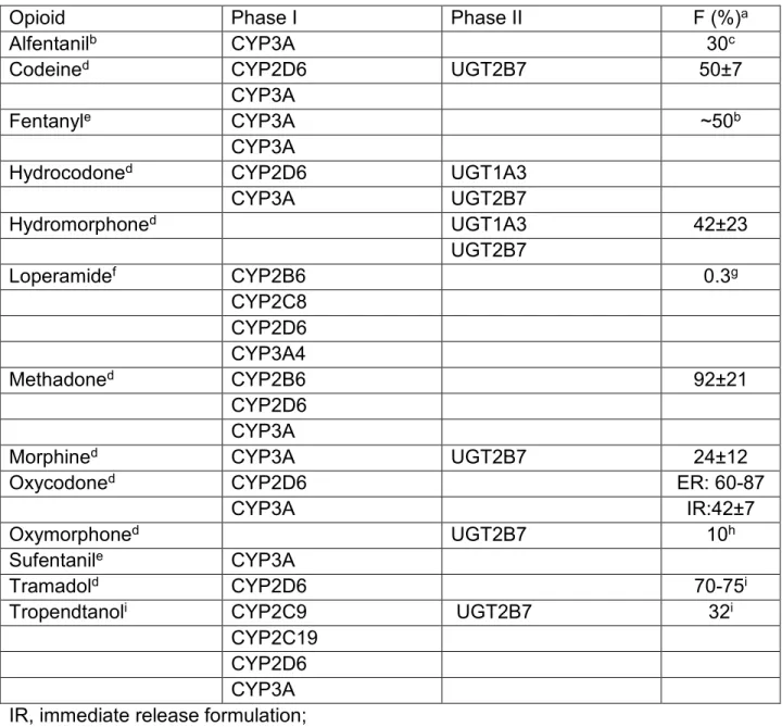
Table 1.1. Phase I and II pathways and bioavailability (F) of common opioids.
Opioid Phase I Phase II F (%)a
Alfentanilb CYP3A 30c
Codeined CYP2D6 UGT2B7 50±7
CYP3A
Fentanyle CYP3A ~50b
CYP3A
Hydrocodoned CYP2D6 UGT1A3
CYP3A UGT2B7
Hydromorphoned UGT1A3 42±23
UGT2B7
Loperamidef CYP2B6 0.3g
CYP2C8 CYP2D6 CYP3A4
Methadoned CYP2B6 92±21
CYP2D6
CYP3A
Morphined CYP3A UGT2B7 24±12
Oxycodoned CYP2D6 ER: 60-87
CYP3A IR:42±7
Oxymorphoned UGT2B7 10h
Sufentanile CYP3A
Tramadold CYP2D6 70-75i
Tropendtanoli CYP2C9 UGT2B7 32i
CYP2C19 CYP2D6
CYP3A
IR, immediate release formulation; ER, extended release formulation;
a Bioavailabilityreported in Goodman and Gilman’s unless otherwise denoted (Brunton et al., 2010).
b Transdermal administration
c Bioavailability reported by Klees et al. (2005a)
d Metabolic pathway reportedby Overholser and Foster (2011) e Metabolic pathway reported byGuitton et al. (1997)
32
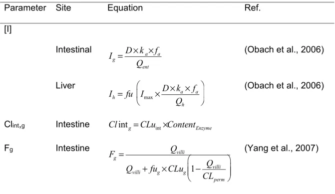
Table 1.2. Models to predict perpetrator or victim substance specific parameters.
Parameter Site Equation Ref.
[I]
Intestinal (Obach et al., 2006)
Liver (Obach et al., 2006)
Clint,g Intestine
Fg Intestine (Yang et al., 2007)
Ig, inhibitor concentration in the enterocyte; Ih, inhibitor concentration in the hepatocyte the hepatocyte; D, the oral dose of inhibitor; ka, the first-order oral absorption rate constant of inhibitor; fa, the fraction of dose of inhibitor absorbed into enterocytes; Qent, the enteric blood flow (may be interchanged with villous blood flow); fu, fraction unbound in plasma or tissue; Qh, hepatic blood flow; Fg, fraction of dose of drug/inhibitor escaping intestinal extraction; Qvilli, villous blood flow; Clintg, intrinsic metabolic clearance in the gut; Fug, fraction of drug unbound in the enterocyte; CLug, the net intrinsic metabolic clearance in the gut based on unbound drug concentration; CLperm, permeability clearance.
Ig =D×ka×fa
Qent
Ih = fu Imax×D×ka×fa
Qh
Clintg=CLuint×ContentEnzyme
Fg = Qvilli
Qvilli+ fug×CLug 1− Qvilli
CLperm
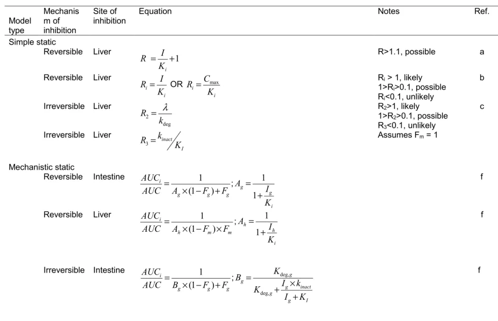
Table 1.3. Static models to assess drug interaction potential. Model type Mechanis m of inhibition Site of inhibition
Equation Notes Ref.
Simple static
Reversible Liver R>1.1, possible a
Reversible Liver
OR R1>Ri > 1, likely i>0.1, possible Ri<0.1, unlikely
b
Irreversible Liver R2>1, likely
1>R2>0.1, possible
R3<0.1, unlikely
c
Irreversible Liver Assumes Fm = 1
Mechanistic static
Reversible Intestine
; f
Reversible Liver
; f
Irreversible Intestine
; f
R = I Ki +1 Ri= I
Ki Ri = Cmax
Ki
R2 = λ kdeg R3=kinact
KI
AUCi AUC =
1
Ag×(1−Fg)+Fg Ag= 1
1+ Ig Ki AUCi
AUC =
1
Ah×(1−Fm)×Fm Ah= 1
1+ Ih Ki
AUCi AUC =
1
Bg×(1−Fg)+Fg Bg =
Kdeg,g Kdeg,g+Ig×kinact
Ig+KI
3
3
Irreversible Liver From IC50 shift data d
Irreversible Liver
;
Irreversible Intestine From IC50 shift data E
AUCi AUC =
1+ 1+
1+IC50
− IC50 +
kdeg×t
×Ln 2
1+IC5050
+
IC50−
× I IC50 −
1+ 1 IC50− AUCi
AUC =
1
Bh×(1−Fm)×Fm Bh=
Kdeg,h Kdeg,h+Ih×kinact
Ih+KI
AUCi AUC =
1
(1−Fm) 1+ Ig
tinc×IC50
+Fg
3
Irreversible Liver From IC50 shift data e
Combined Combined From IC50 shift data e
Combined Intestine f
Combined Liver F
Combined Combined F
R, value associated with drug interaction probability; I, inhibitor concentration; Cmax, maximal observed or predicted
inhibitor concentration; λ, first-order inactivation rate at a given MBI concentration; kdeg, degradation rate of enzyme; kinact,
maximal enzyme inactivation rate; KI, concentration to elicit half-the maximal rate of enzyme inactivation; AUCi/AUC, the
area under the victim drug plasma concentration-time curve in the presence of inhibitor over that in the absence of inhibitor; Fg, fraction of drug escaping intestinal extraction; fm, fraction of victim drug metabolized; Ki, the reversible
inhibition constant;IC50, inhibitor concentration to elicit 50% of the maximal inhibition; the subscript ‘h’ denotes the term
corresponds to the liver (hepatocyte); the subscript ‘g’ denotes the term refers to the gut (intestine); a US Food and Drug
Administration (2012a); b Bjornsson et al. (2003); c Fujioka et al. (2012); d Sekiguchi et al. (2009); e (Obach et al., 2005;
Obach et al., 2006; Obach et al., 2007); f Fahmi et al. (2009)
AUCi AUC =
1
Fm 1+ Ih
tinc×IC50
+(1−Fm)
AUCi AUC =
1
Fm 1+ Ih
tinc×IC50
+(1−Fm)
× 1
Fg× (1−Fg) 1+ Ig
tinc×IC50
AUCi
AUC =Cg =
1
[Ag×Bg]×(1−Fg)+Fg AUCi
AUC =Ch=
1
[Ah×Bh]×(1−Fm)×Fm
AUCi
AUC =Cg×Ch
3
36 Table 1.4. Software packages for PBPK modeling.
Program Provider Link Cost ($)
Free form
acslX AEgis Technologies Group
http://www.acslx.com 500-7,500
Berkeley Madonna www.berkeleymadonna.com 69-299
Matlab (Simulink) MathWorks® www.mathworks.com 3,250
ModelMaker ModelKinetix© www.modelkinetix.com 233-770
Powersim Studio PowerSim Software http://www.powersim.com 99+ Preassembled
BioDMET General Electric http://pdsl.research.ge.com Beta is free GastroplusTM SimulationPlus, Inc www.simulations-plus.com/Products a
PK Sim® Bayer Technology Services
www.systems-biology.com/products/pk-sim.html a
Simbiology® MathWorks®
http://www.mathworks.com/products
/simbiology 3,250
Simcyp® Certara http://www.simcyp.com a
37
Figure 1.1. Furanocoumarin structures. Bergamottin (A) and 6’,7’-dihydroxybergamottin (B).
38
39
REFERENCES
Abduljalil K, Jamei M, Rostami-Hodjegan A and Johnson TN (2014) Changes in individual drug-independent system parameters during virtual paediatric pharmacokinetic trials: introducing time-varying physiology into a paediatric PBPK model. The AAPS journal 16:568-576.
Ainslie GR, Wolf KK, Li Y, Connolly EA, Scarlett YV, Hull JH and Paine MF (2014) Assessment of a Candidate Marker Constituent Predictive of a Dietary Substance-Drug Interaction: Case Study with Grapefruit Juice and CYP3A4 Drug Substrates. J Pharmacol Exp Ther.
Al Saabi A, Allorge D, Sauvage FL, Tournel G, Gaulier JM, Marquet P and Picard N (2013) Involvement of UDP-glucuronosyltransferases UGT1A9 and UGT2B7 in ethanol glucuronidation, and interactions with common drugs of abuse. Drug Metab Dispos 41:568-574.
Andersen ME, Jones RA and Jenkins LJ, Jr. (1977) Enhancement of 1,1-dichloroethylene toxicity by pretreatment of fasted male rats with 2,3-epoxypropan-1-ol. Drug and chemical toxicology 1:63-74.
Andersen ME, French JE, Gargas ML, Jones RA and Jenkins LJ, Jr. (1979) Saturable metabolism and the acute toxicity of 1,1-dichloroethylene. Toxicol Appl Pharmacol 47:385-393.
Bachler G, von Goetz N and Hungerbuhler K (2014) Using physiologically based pharmacokinetic (PBPK) modeling for dietary risk assessment of titanium dioxide (TiO) nanoparticles. Nanotoxicology:1-8.
Bailey DG, Dresser GK and Bend JR (2003) Bergamottin, lime juice, and red wine as inhibitors of cytochrome P450 3A4 activity: comparison with grapefruit juice. Clin Pharmacol Ther 73:529-537.
Bailey DG and Dresser GK (2004) Interactions between grapefruit juice and cardiovascular drugs. Am J Cardiovasc Drugs 4:281-297.
Bailey DG, Dresser G and Arnold JM (2013) Grapefruit-medication interactions: Forbidden fruit or avoidable consequences? CMAJ 185:309-316.
Barton ED, Ramos J, Colwell C, Benson J, Baily J and Dunn W (2002) Intranasal administration of naloxone by paramedics. Prehospital emergency care : official journal of the National Association of EMS Physicians and the National Association of State EMS Directors 6:54-58.
40
Benmebarek M, Devaud C, Gex-Fabry M, Powell Golay K, Brogli C, Baumann P, Gravier B and Eap CB (2004) Effects of grapefruit juice on the pharmacokinetics of the enantiomers of methadone. Clin Pharmacol Ther 76:55-63.
Bjornsson TD, Callaghan JT, Einolf HJ, Fischer V, Gan L, Grimm S, Kao J, King SP, Miwa G, Ni L, Kumar G, McLeod J, Obach RS, Roberts S, Roe A, Shah A, Snikeris F, Sullivan JT, Tweedie D, Vega JM, Walsh J, Wrighton SA, Pharmaceutical R, Manufacturers of America Drug Metabolism/Clinical Pharmacology Technical Working G, Evaluation FDACfD and Research (2003) The conduct of in vitro and in vivo drug-drug interaction studies: a Pharmaceutical Research and Manufacturers of America (PhRMA) perspective. Drug Metab Dispos 31:815-832.
Bluelight (2014a) in, vBulletin.
Bluelight (2014b) What is it in grapfruit juice tht potentiates opioids?, in, vBulletin.
Brayfield A (2011) Martindale: The Complete Drug Reference, in Tapentadol hydrochloride: a novel analgesic (Brayfeild A ed), Pharmaceutical Press.
Brunton LL, Chabner BA and Knollmann BC (2010) Appendix II, in Goodman & Gilman's: The Pharmalogical Basis of Therapeutics (Brunton L ed), McGraw Hill Medical.
Chakraborty M, Kamath JV and Bhattacharjee A (2014) Pharmacodynamic Interaction of Green Tea Extract with Hydrochlorothiazide against Cyclophosphamide-Induced Myocardial Damage. Toxicology international 21:196-202.
Charnick SB, Kawai R, Nedelman JR, Lemaire M, Niederberger W and Sato H (1995) Perspectives in pharmacokinetics. Physiologically based pharmacokinetic modeling as a tool for drug development. J Pharmacokinet Biopharm 23:217-229.
Chen Y, Mao J and Hop CE (2014) Physiologically Based Pharmacokinetic Modeling to Predict Drug-Drug Interactions Involving Inhibitory Metabolite - A Case Study of Amiodarone. Drug Metab Dispos.
Cheng Y and Prusoff WH (1973) Relationship between the inhibition constant (K1) and the concentration of inhibitor which causes 50 per cent inhibition (I50) of an enzymatic reaction. Biochem Pharmacol 22:3099-3108.
Clarke G, Stilling RM, Kennedy PJ, Stanton C, Cryan JF and Dinan TG (2014) Minireview: Gut microbiota: the neglected endocrine organ. Molecular endocrinology 28:1221-1238.