Journal of Theoretical and Applied Information Technology 15thDecember 2016. Vol.94. No.1
© 2005 - 2016 JATIT & LLS. All rights reserved.
ISSN:1992-8645 www.jatit.org E-ISSN:1817-3195
AN ADAPTIVE CLUSTER BASED IMAGE SEARCH
AND RETRIEVE FOR INTERACTIVE ROI TO MRI IMAGE
FILTERING, SEGMENTATION, AND REGISTRATION
1PADMAJA GRANDHE,2DR. E. SREENIVASA REDDY,3DR.D.VASUMATHI 1Research Scholar, Department of CSE, JNTUK, Kakinada, A.P, India.
2Dean & Professor, Department of CSE, Acharya Nagarjuna University, Guntur, A.P, India. 3Professor, Department of CSE,JNTUH, Hyderabad, Telangana, India.
ABSTRACT
Recently, there has been an enormous development in compilation of diverse image databases in the appearance of digital. The majority of the user establishes it hard to investigate and recover necessary images in huge collection. In organize to supply an effectual and well-organized explore engine tool, to smooth the progress of high point examination of checkup image information in investigate and clinical environment the scheme has been put into practice. In image retrieval system, there is no methodologies contain been careful in a straight line to get back the images from databases. That featured images only have be measured for the retrieval process in order to retrieve exact desired images from the databases. This paper also highlights an thought of newly developed image clustering technique and their real time application such as Clustering based image linearization in ROI, The purpose of this effort is a scalable, immediate, illustration search engine for medical images, Preprocessing, feature extraction, Classification and retrieval steps in arrange to build an well-organized recovery tool. The main characteristic of this tool is used of CBISR of the extract feel pattern of the image and clustering algorithm for image categorization in arrange to get better retrieval efficiency. The future image retrieval scheme consists of three stages i.e., segmentation, texture feature extraction and clustering procedure. In the segmentation development, preprocessing step to section the image into block is carried out. A decrease in an image area to be process is approved out in the surface feature removal procedure and lastly, the extract image is clustered using K-means algorithm
Keywords:CBISR, MRI,K-Mean, Image Retrieval, Segmentation, Image Filter
1. INTRODUCTION
Three Dimensional display of part of person remains obtain by current analytical imaging method is even more normal. CBISR scheme is a technique for probing and retrieving of images base on their low level features (example texture, color, shape). It is a organization which discriminate the dissimilar region of an image based on their similarity and decide the likeness flanked by two images by devious the distance of these dissimilar region. In CBISR scheme, any type of imagery can be known as input image which depends upon the application supplies.
Brain Tumour is a frequent brain chaos that, according to an approximation of the affects almost 60 million people about the world. Approximately one in every 100 persons wills knowledge a Tumour at a number of times in their life [1]. Tumour is characterized by the
Tumour. In adding, some other linked issues, such as dataset and evaluation measures, are also discussed. Lastly, the appearance of algorithms is evaluated, and their capability and limits are described.[3]
Scheme inquiry consequences are a set of imagery sort by characteristic similarity with admiration to the query. However, images with high characteristic similarity to the query may be very dissimilar from the inquiry in terms of semantics. This is known as the semantic gap. We bring in a novel image retrieval scheme cluster based image search and retrieve for interactive ROI to MRI of images by unverified knowledge which tackles the semantic gap difficulty based on a theory: semantically images tend to be clustered in some feature space the analysis of brain tumour is consists of two phases: database building and query processing. MR images of the brain diseases store in the image database are pre processed by image de-noising and turning round alteration of askew images. Next, images are segmented automatically using clustering to identify the brain ROI region on MR images.
During MR image attainment, there can be misalignment of imagery due to group of the patient. The misalignment results in turning round or conversion of the image. The conversion will not cause problems in image analysis since the brain MRI can be segmented and analyzed irrespective of the site of brain area in the MR image. But image turning round limits the request of automatic tools for MR image analysis as it changes the shape and texture property of the tumor Skull removal of the brain in MR image is a significant beginning step in segmentation since it may cause misclassifications of pixels due to strength similarity with the brain regions.
The rest of the paper is organized as follows. The section 2 describes the literature survey of the related work and problem identification is described in section 3. The section 4 describes the proposed method and MRI medical image analysis and implementation are explained in section 5. The implementation procedure of the proposed method is explained in section 6 and analysis of the simulation results in section 7. Finally, the conclusions are given in section 8.
2. RELATED WORK
REswaraiah et.al [5] proposed a method used in telemedicine. It is an ordinary to switch medical descriptions flanked by hospitals situated at far-away places from side to side unsecure network like internet. During this move tamper may be introduce deliberately or by accident into medical images.Duke Aleut [7]proposed a method to describe the use of Mat lab in three-dimensional renovation of human brain MRI images. The programmed that were intended enables observe dissections of the gain 3D structure along three axes.
Panda Kr et.al [4]: proposed a method to Magnetic Resonance Imaging has turn out to be a extensively used method of high quality medical imaging. Magnetic resonance imaging (MRI) is a higher medical imaging method as long as rich in order about the human yielding tissue structure. Mathematical morphology provides a methodical move toward to analyze.
Vincent Chu[6] et.al proposed a method to make easy high level psychoanalysis of medical image data in investigate and scientific environments, a wrapper for the ITK toolkit is urbanized to allow ITK algorithms to be called in MATLAB. ITK is an influential open-source toolkit implement state of the art algorithms in medical image processing and analysis.[6]. Karen Simony et.al Proposed to the object of this labour is a scalable, real-time visual look for engine for medical images. In difference to obtainable system that gets back imagery that is internationally alike to a query image, we allow the user to select a inquiry area of Interest (ROI) and mechanically detect the matching region within all return images.
3. PROBLEM IDENTIFICATION
Existing methodology the medical image retrieval allows explore same images exterior with the dissimilar kind of analysis. It also allows the penetrating through large collections of disease-related illustration using the illustration attribute.
Journal of Theoretical and Applied Information Technology 15thDecember 2016. Vol.94. No.1
© 2005 - 2016 JATIT & LLS. All rights reserved.
ISSN:1992-8645 www.jatit.org E-ISSN:1817-3195
tumour discovery unit for offline and online monitor only on 2D dimensional image searching mechanism of epileptic patients. The unit is by as additional no of input information stream from MRI recording. The major drawback needs to analysis each database in both time and frequency and there is no proper combination factor for search medical image.[8].
4. RESEARCHER MOTIVATION IN PROPOSED METHODOLOGY:
Although more than a few studies are previously being conduct with respect to image search and retrieval, many challenging problems still exist: • Automatic description of ROI on the medical image without relying on the MRI. • A on its own characteristic vector will not do well in telling tumour since the features that are most capable in discriminating in the middle of descriptions from unlike classes may not be the most effectual for retrieval of images belong to the subclass within a class.
• Creation the CBISR system strong to misalignments of imagery that occur throughout MR image acquisition.
• As long as well-organized indexing arrangement for faster retrieval from cluster images from the database.
• Appropriate amalgamation of the dimensionality reduction techniques into the CBISR system so that the indexing structures can be advantageous.
Motivated by these requirements, in this paper, we propose a CBISR system for Cluster based image search and retrieve for interactive ROI to MRI image filtering, segmentation, and registration automatic extraction and analysis of the tumour region on MR images. The semantic gap between the high- and low-level features is abridged by raising a hierarchical framework that combine supervise and unsupervised categorization techniques with different set of tumor features at each level. Also, the scheme is made well-organized by apply modified K-means clustering on the characteristic set and adopt the indexing arrangement in low-dimensional feature space.
5 LIMITATIONS
The dynamics, location, and magnitude of the indication are extremely prejudiced by the more cluster group as it is sampled in each. If a happen to imprison large ROI effects, the magnitude of the image may be large, the time a bit more late than standard, and the location of the signal somewhat distal from the true region of activation. The problem of image filtering and registration coupling in MRI leftovers to some amount at all field strength and poses important limits on the depth and range of questions that can be addressed using MRI.
6. MAJOR FINDINGS:
Major and resemblance method only authorize MRI query image already in the database, not a new image. If using clustering approach, it first calculate distance to the first set, for the pertinent ones, then go additional into clusters to calculate distances, then rank the entire consequences. Now it only equipment to first level. It doesn’t work from Biren’s retrieval interface. Biren wrote some test program to do the experiment. Those test programs are not linked to the real system... files with names CbisrTest1, 2, 3 are the test programs, and the results in them are not direct as far as the major and resemblance method retrieval is anxious. The results of those examination programs are format and scheduled in a way to report them in his thesis, and those consequences can be used to get the main and resemblance way retrieval results. In his “Image Retrieval Process ROI”, there is some code for main method also along with first method, but it does not work. He tested but may not have removed that part of the code from the program.
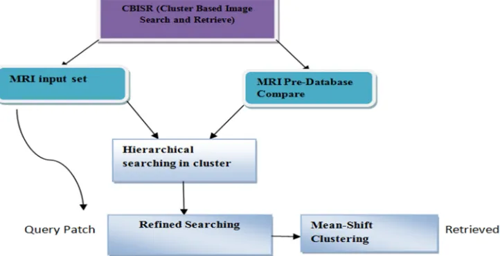
7. PROPOSED METHOD:CLUSTER BASED IMAGE SEARCH AND RETRIEVE-CBISR OVER MEDICAL ENVIRONMENT
image are careful for feature removal. For each image in the image database, characteristic vector value has been urbanized and which are store in feature database. When a database image is submit by the user, the same surface feature extraction and feature vector value building
[image:4.612.129.490.174.359.2]procedure has been practical to the query image in order to obtain the characteristic vector value to the query image.
Figure 1: Classification Flow Diagram
Improved the appearance of Brain Tumor detectors base wholly on MRI image. Two unlike approach were used to unite this extract in order. The primary move toward, recognized as clever characteristic fusion, involve combine features extract from normal and database Adaptive Brain Tumor Rate Variability (ABTRV) into a solitary characteristic vector previous to feed it to a classifier. The next move toward, called Support Vector machine,[3] is achieve by combine the self-governing decision of the ABTRV-based classifier, our ultimate aim is to proposed a real time valuable CBISR mechanism in three
different stages , but in previous work they not elaborate the working methodology but in this research we give more concentrate on
1. ROI
2 Image retrieve 3 Databases
Journal of Theoretical and Applied Information Technology 15thDecember 2016. Vol.94. No.1
© 2005 - 2016 JATIT & LLS. All rights reserved.
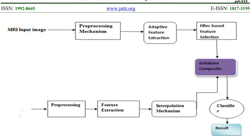
[image:5.612.89.522.67.303.2]ISSN:1992-8645 www.jatit.org E-ISSN:1817-3195
Figure 2:Functional Diagram of CBISR
Tumor obvious themselves in the signal as recurring that are absolutely unlike from the usual random-like background intellectual action. This independence has been downtrodden by a figure of at what time deceitful regular Tumor discovery methods. A figure of these technique are base on count this periodicity in (1) the occasion area using association purpose change in replica arrangement harmonization between channel and wave-sequence psychoanalysis the incidence area using power ethereal density and the time–incidence area by quadratic time– frequency The ultimate goal of proposed algorithm is to design and implement a novel technique for Cluster based medical image searching. The center of this procedure is the aptitude to calculate features that precisely and impartially explain the individuality of the imagery patch. [10]
8. ADAPTIVE REGION OF INTEREST IN MRI MEDICAL IMAGE ANALYSIS
A ROI technique for routine credit in ultrasound images which analysis infertility in patient, are obtainable using separate wavelet transform based K-means clustering [12]. A data-driven probabilistic arrangement perform atlas-guided segmentation of a varied set of brain MR images and clusters the images in homogeneous subgroups, [4] An effectual unverified move toward based on the joint dissimilarity image and k-means clustering is
Figure 3: Adaptive ROI image in exemplar image
The image which show the ROI of exemplar image which ensures the MRI parameters in different condition of segmented ROI MRI image it is usual to symbolize an representation depository as a fully-connected graph with vertices corresponding to images and edges weighted by the registration errors There are two significant choice to make: how to choose the exemplars and how to the function f, aggregate the transforms obtained using exemplars. In [14] the exemplars were selected randomly, and the aggregation was performed by taking a median. [15]
Table1: Analysis in Image Aggregation
Exemplar Aggregation Overlap ratio
Rand Mean
Median Single
K=1 (0.555) K=5 (0.532) K=7 (0.53)
Sum-min Mean
Median Single
K=1 (2.04) K=5(1.33) K=7(1.38)
8.1Real Time Parallel Execution
The parallel execution in our proposed CBISR which reduce the implementation time which means the overall performance parameters calculation is take place fast in manner dispensation large ensembles of imagery may still take a long occasion still when approved out on a high-end workplace. To speak to this computational confront, we have engineered an answer based on top of a master–worker parallelization plan for high-throughput dispensation of a set of images. We have selected
master–worker parallelization since resemblance computation on image tiles or whole image can be carried out separately. [17]
Wr(x) = {Dij| (δ,θ)}
Where, Dij (the co-occurrence probability between gray levels i and j)
Cij = Wij: ∑Wij
Where: Wij = Represents the number of occurrences of gray levels
i and j = Within the given image window, given a certain (δ, θ) Pair
G = the quantized number of gray levels
8.2 Pre Processing MRI
Journal of Theoretical and Applied Information Technology 15thDecember 2016. Vol.94. No.1
© 2005 - 2016 JATIT & LLS. All rights reserved.
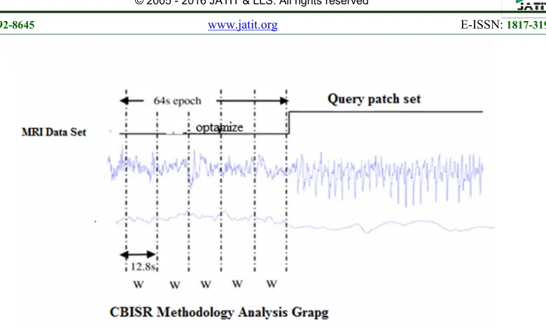
[image:7.612.125.515.64.298.2]ISSN:1992-8645 www.jatit.org E-ISSN:1817-3195
Figure 4: Algorithm Graph formation in MRI data set
9. STEPS IMPLEMENTED PROPOSED CLUSTER BASED IMAGE SEARCH AND RETRIEVE-CBISR
Step 1: Features
A sum of 96 skins is extract from the occasion and the TF domain for each AHRV era. A concise account of the extract skin is given below.
Time domain features: The denote, normal deviation, and limit (which give details the indication individuality in circumstances of action, mobility, and difficulty) were computed.
Time-Frequency features:Because AHAH is a non-stationary sign, we strong-minded to take out skin as of the time–incidence area in the direction of clarification for this. This procedure was not as easy as in the case of the occasion area skin. The time-frequency (TF) symbol was obtain by the Modified-B sharing (MBD) with its limit β set to 0.01The MBD has been selected to stand for the AHAH in the TF area as it is before set up to understand the best collaboration.
Step 2:
MRI and AHRV in order union in position to create the AHRV feature mixture likely, The signal frame rate is five times that of the, there is a disparity flanked by the skin of AHRV and MRI. To deal with this subject, we investigate three dissimilar solution allocate a stable value to all HRV windows, use linear exclamation, and use higher-order polynomials. The linear shout
was adopted as it realized a good trade off flanked by look and difficulty and resulted in a smooth change flanked by characteristic values. The flowchart of the proposed method is shown in figure 5 and the algorithm is represented in algorithm
1.Rational of sub-image retrieval functionality
Figure 5: Flow Chart for CBISR Proposed Algorithm
Algorithm 1: Cluster Based Image Search and Retrieve-CBISR Algorithm:
1: /*** MRI Data acquisition********/ 2: X=[x1,x2…xn]
3:/****** Query Patch Data acquisition *******/
4: Y= [y1,y2..yn]
5:/****** Feature extraction*******/ Z= [z1, z2,.zn]
6:To reduce Dimension Reduction A= BTZ = [a1,a2…..an]
7: The Advanced first classifier h1(A) = ATQ1A+V1TA+V01>0
Ifselect normal image ….
Else…
Select abnormal select vector D…. End
Result End…..
9.1 Real Time Wavelet Transform
Journal of Theoretical and Applied Information Technology 15thDecember 2016. Vol.94. No.1
© 2005 - 2016 JATIT & LLS. All rights reserved.
ISSN:1992-8645 www.jatit.org E-ISSN:1817-3195
Figure6: Circle flow of Brain cell detection
9.2 Proposed Mechanism Of Database Value Calculation
Show an example of an MRI signal including a Tumor era. It is obvious that there is dissimilarity flanked by Tumor and non-Tumor interval. As we are able to differentiate flanked by these interval visually, time area detection and forecast method attempt to differentiate flanked by them automatically, and assess the presentation using dissimilar metrics such as the sympathy, specificity, correctness, and false-positive value. These metrics are distinct as follows. [17]
In image database of real brain tumor MR images, along with their segmentations, may give the income to calculate the presentation of an algorithm by compare the results next to the unpredictability of the expert raters’ judgments. However, an purpose assessment to methodically contrast dissimilar methodologies also needs a ground truth with little or no unpredictability. An instance of such a ground truth is the artificial brain MRI database provided by the Montreal Neurological currently considered to be the common standard for evaluating the segmentations of healthy brain MR images.
The replicated brain tumor MR imagery can purpose as test data for any segmentation method and the ground truth can provide the means for object evaluation of segmentation performance. We do not aim to create a database of simulated brain tumor MR images that are impossible to differentiate from real brain tumor MR images.
At present, this meaning involve a large degree of instinct and cannot be formulate algorithmically. Our simulated data provide a standard for different tumor segmentation
methods that is currently not available to the community.
A) Set of Parameters in Real Time Sensitivity (i.e.) TP(True Positive)/TP(True Positive)+FN(False Negative) * 100 which ensembles the image database value calculation formula the positive and negative which indicate maximum and minimum pixel value of density image.
B) The valuable parameter set in one time
accuracy is belongs to
TP+TN/TN+FP+TP+FN*100 image pixel accuracy to be utilized in many different set of ROI MR Images. So our ultimate aim is to increases the pixel quality of original image to take the comparative result in simulation environment.
c) Final statement of Database value which ensures the 0 & 1 in binary values in data matrix format so we consider the prototype in manner of False Positive Value = TP/TP+FP*100
Where,
The awareness of tumor cells in particular area, we need to separate the particular area into cluster groups in ROI region to divide into particular segments of MRI datasets.
The image filter mechanism is only suitable for histogram from side to side tumor detection techniques i.e. so we considered a peak to peak ratio analyzer in different set of estimate variable of region of interest. The MRI image background and distance from cells to cells is expressed in terms of pre-define data set values. The author used to train a data set by use of Advanced Support Vector Machine (ASVM) classifier for this research task and achieve a standard sympathy of about 90% on self-recorded data.[15]
9.3 Medical Image Prediction Methods
The investigate work on the subject of time-domain Tumor guess is better-off than time-domain Tumor discovery due to the significance of the Tumor prediction difficulty.
We can believe of the Tumor forecast difficulty as a detection difficulty of the pre state on Tumor minutes. [6]
This requires a substantial long interstate for good forecast results. Alike figures to those used in Tumor discovery like the zero-crossing rate can be used for Tumor forecast used the zero crossing rate of MRI signal segment to develop a Patient-specific Tumor forecast method. A moving window analysis is used in this technique. The histograms of the dissimilar casement intervals are predictable,[8]
The first set of experiment was performing using the MATLAB completion of the CBISIR algorithm. MATLAB provide well-organized function and toolboxes that make easier to expand algorithms rapidly and professionally. However, MATLAB is not installing on many cluster systems. Hence, we urbanized a Java version of the hierarchical CBSIR algorithm and ported the parallel code to support the Java implementation. [6]
Figure7: CBISR working Methodology
Selected histogram bins are second-hand for categorization into pre-data and inter-data state base on contrast with orientation histograms. A difference Bayesian Gaussian combination model has be used for categorization. In this technique, a joint directory for the choice in use on chosen bins is compute and compare with a pre-defined patient-doorsill to lift a fear for awaiting Tumor this method has been tested on 561 h of scalp MRI hold 86 Tumor for 20 patients. It achieve a understanding of 88.34%, a false
prediction rate of 0.155 h−1, and a standard forecast time of 22.5 min. the formation algorithm is represented in algorithm 2.
Algorithm 2: Formation for Image searching
function [N]=filt(D); G=D+80;
imadjust(G);
BW = edge(G,'canny',.3); se = strel('disk',10);
Journal of Theoretical and Applied Information Technology 15thDecember 2016. Vol.94. No.1
© 2005 - 2016 JATIT & LLS. All rights reserved.
ISSN:1992-8645 www.jatit.org E-ISSN:1817-3195
[m,n]=size(BW2); M=min(min(D)); for j=1:n for i=1:m if BW2(i,j)== 0 D(i,j)=M; end; end; end; N=D;.
10. IMPLEMENTATION AND RESULT ANALYSIS
First the sign is in use and alienated into the blocks. Then denote is full of the exacting beat. Then that denote is subtracted from the unique signal. Thus we got the main mechanism. If the covariance is taken of the ensuing we with the help of which we can rebuild the original signal. The process is performing with the help of MATLAB Software and consequences are being display. The principal part analysis is performing for each of the cases. [18]
The key to obtain data density is signal representation, which concern the symbol of a given group of students of signals in a well-organized manner. If a separate signal comprises of n sample, then it can an n dimensional space. Each sample value is then a part of the data n vector x, that represent a discrete signal in this space. [19]
For a well-organized symbol of X, we secure an orthogonal change of X, which results in Y=TX where Y denotes the change vector and T represent the transformation matrix.
For data compression we will select a subset of m mechanism of Y, where m is substantially less than n. The balance of (n-m) components will be discarded without introducing any grave error when the signal is reconstructed using the m saved components of the vector Y. To Whom It May Concern quantify this error of approximation an error criterion is needed and that is mean square error. [20]
10.1 CBISR mathematical calculation
Approximate Reasoning steps of Binary Image Representation
I = X [f(0) + f(1)]
Pixels = Width (W) X Height (H) = 256 X 256 f (0) = white pixel (digit 0)
f(l) = black pixel (digit 1) No. of White Pixels :
P = Y f[(0)]
P = number of white pixels (width*height) 1 Pixel = 0.264 mm
Size of Tumor:
S = 0.264 √ ^2
P= no-of white pixels; W=width; H=height
10.2 Proposed CBISR Model Feature
1)The plan of a computer scheme able to notice the attendance of a tumor in the digital images of the brain, and to precisely describe its borderline.
2) The essential supposition is that dissimilar local feel in image can explain dissimilar corporeal individuality matching to dissimilar objects.
3) The supposition is that local feel of tumor cells is extremely dissimilar from restricted texture of other organic tissues. Thus, texture capacity in the picture could be part of an effectual bias technique amid healthy tissues and likely tumor areas.
4) A computer system has been intended and urbanized to know the typical facial appearance of the tumor in the digital form of images.
5) The textural features have been extracting using a co-occurrence move toward. The level of credit, among three likely types of image areas non-tumor, tumor and back ground. We are into tumor image segmentation.
Table 2: Comparison Of The Clustering Segmentation
Mean of the picture just point to the standard strength of the pixel and normal divergence is the ordinary way to explain the range of deviation. Pixel adds up and quantity extracted is abridged in segmentation with
pre-processing. This is due to the loss of surplus pixels like noise pixel, pixel which are there in the without pre processing segmentation. Pre-processing with segmentation gives correct edge or border detection and it protect the shape of the tumor.
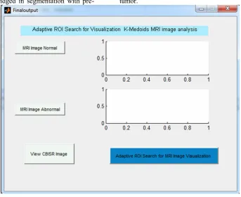
Figure 8 : Real Time Adaptive ROI Search For Visualization
[image:12.612.154.497.260.540.2]Journal of Theoretical and Applied Information Technology 15thDecember 2016. Vol.94. No.1
© 2005 - 2016 JATIT & LLS. All rights reserved.
ISSN:1992-8645 www.jatit.org E-ISSN:1817-3195
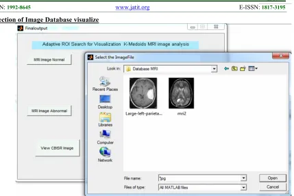
[image:13.612.105.534.71.358.2]Selection of Image Database visualize
Figure 9: Select The Database Image For Retrieval
We search and knowledgeable the influence of being image classifications and of performing a search for images with comparable certain features in a database using Region of Interest (ROI)and Advanced principal component analysis (PCA).
The selection of image data has a possible to be an efficient, complete, and without
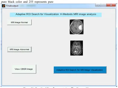
Figure 10: Graphical User Representations The Normal Image.
After selection of MRI normal image the pre-processing mechanism had been started in sense of image enhancement technique to ensure the quality of image to conversion of pixel region to 0 to 255...where 0 represents the pure black color and 255 represents pure
white in-between the pixel intensity varied simultaneously.
[image:14.612.111.502.419.711.2]Journal of Theoretical and Applied Information Technology 15thDecember 2016. Vol.94. No.1
© 2005 - 2016 JATIT & LLS. All rights reserved.
ISSN:1992-8645 www.jatit.org E-ISSN:1817-3195
[image:15.612.129.492.76.432.2]After selection of both normal and abnormal MRI database image the mathematical algorithm performance calculation take place in Mat lab command window the process in flow of minimum to maximum threshold value. The ROI of image segmentation is converting all pixel images into Regions...
Figure 12: CBISR ROI frame Analysis
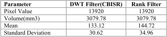
Table2 : Proposed Filter Analysis
Parameter DWT Filter(CBISR) Rank Filter
Pixel Value 13920 13920
Volume(mm3) 3079.78 3079.78
Mean 133.12 144.72
Standard Deviation 30.62 34.96
From the new results, DWT Filter (CBISR) filter gave better output and de-noising. Usually the noise is cause by bit errors that happen throughout data imprison or broadcast. Since only a little amount of pixels and be likely to live in the great gra deposition. While using DWT filter there is a dispersal of region and produce blurred the image. Also shape and edges are good conserved. DWT filter give better presentation than ranj filter and it is apt for
this application. The proposed CBISR algorithm plays a vital role in ROI image analysis the gray scale pixel image value converted into binary values to speed up the matrix process in back around algorithm mathematical formation and calculation.
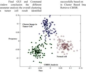
[image:15.612.154.431.493.555.2]Figure 13: Cluster Based Data Point’s classifier
Final GUI and Command window conclusion the different parameter analysis the overall clustering in tumor cell result identified
successfully based on K-MEDOIDS in Cluster Based Image Search and Retrieve-CBISR.
[image:16.612.130.458.422.701.2]Journal of Theoretical and Applied Information Technology 15thDecember 2016. Vol.94. No.1
© 2005 - 2016 JATIT & LLS. All rights reserved.
ISSN:1992-8645 www.jatit.org E-ISSN:1817-3195
The graphical which represent the different cluster cell groups to segregate the normal and abnormal cells. The X and Y axis which indicates frequency and time because the time is mandatory to analysis the final result...
11. CONCLUSION
In this research we have obtainable the design and implementation of cluster-based image search and retrieval (CBISR) and direction-findingstructure and its similar completion of ROI MRI.Segmentation of medical images can be alienated into twoclassifications: Pixel based local method and regionbased global method is called Region of Interest (ROI). Brain MR Image is a multifaceted system to be segmented with well-organizedmethod for having variable kind of tissues. MATLAB supports this function,but working with matrix this great would be very timeconsuming for home computers hierarchical searching and retrieve step is achieved enough that substantial reduction in resource usage can be used when a subset of images are selected and process, even when the cost of the hierarchical searching step is reduced and over all time efficiency is low.
12. FUTURE WORK
We will carry out experiment on different databases of images and performance indexing sets of images with different criteria.
13. ACKNOWLEDGMENT.
The authors also gratefully acknowledge the helpful comments and suggestions of the reviewers, which have improved the presentation.
REFERENCES:
[1] M. Natick, MATLAB Reference Guide, MathWorks, 1992.
[2] L. Obez and W. Shchroeder, ITK software guide : ITK 1.4 : the Insight segmentation and registration toolkit, Kitware Inc., 2003. [3] M. J. Ackerman, “Accessing the visible
human project,” D-Lib Magazine , 1995. [4] K. Friston, R. Dolan, and R. Frackowiak,
Statistical parametric mapping, Functional Neuroimaging: Technical Foundations, 1994.
[5] M. Wolforth, G. Ward, and S. Marrett, EMMA: Extensible MATLAB Medical Analysis, 1995.
[6] C. Hendriks, L. van Vliet, B. Rieger, and M. van Ginkel, DIPimage: a scientific image processing toolbox for MATLAB, Pattern Recognition Group, TU Delft, 2001. Proc. of SPIE Vol. 6144 61443T-7
[7] Rockinger, “Image fusion toolbox.” http://www.metapix.de/toolbox.htm
[Accessed Nov 20, 2005].
[8] P. D. Kovesi, “MATLAB and Octave functions for computer vision and image processing.” School of Computer Science & Software Engineering, The University of Western Australia.
[9] A. R.Kavitha, Dr.C.Chellamuthu, Ms.KavinRupa “An Efficient Approach for Brain TumourDetection Based on Modified Region Growing and Neural Network in MRI Images” International Conference on Computing, Electronics and Electrical Technologies [ICCEET] IEEE Xplorer2011, pp (1087 – 1096)
[10]Matlab, Help, sections: Visualizing MRI data: Volume Visualization Techniques (3-D Visualization); Image Processing Toolbox; Creating Graphical User Interfaces
[11] Dave Harvey, Dicom Objects User Manual, Medical Connections , pdf, 2001 Avni, U., Greenspan, H., Konen, E., Sharon, M., Goldberger, J.: X-ray categorization and retrieval on the organ and pathology level, using patch-based visual words. IEEE Trans. Med. Imag. 30(3), 733{746 (2011
[12] Burner, A., Donner, R., Mayerhoefer, M., Holzer, M., Kainberger, F., Langs, G.:Texture bags: Anomaly retrieval in medical images based on local 3d-texture similarity.In: M uller, H., Greenspan, H., Syeda-Mahmood, T.F. (eds.) MCBR-CDS. LNCS, vol. 7075, pp. 116{127. Springer, Heidelberg (2011)
[13] Cardoso, M.J., Clarkson, M.J., Ridgway, G.R., Modat, M., Fox, N.C., Ourselin,S.: LoAd: A locally adaptive cortical segmentation algorithm. NeuroImage 56(3),1386{1397 (2011)
[14] Cardoso, M., Modat, M., Ourselin, S., Keihaninejad, S., Cash, D.: Multi-STEPS:Multi-label similarity and truth estimation for propagated segmentations. In:Math. Meth.in Biomed. Im. Anal., IEEE Workshop on, pp. 153{158 (2012)
Knopman,D.S., Boeve, B.F., Ivnik, R.J., Smith, G.E., Cha, R.H., Tangalos, E.G., Petersen,R.C.: Comparison of di_erent MRI brain atrophy rate measures with clinical diseaseprogression in AD. Neurology 62(4), 591{600 (2004)
[16] Joachims, T.: Optimizing search engines using clickthrough data. In: ACMSIGKDD Int. Conf. on Knowl. Disc. and Data Mining, pp. 133{142. ACM Press,New York (2002) [17] G. Coatrieux, J. Montagner, H. Huang, and
Ch. Roux, “Mixed reversible and RONI watermarking for medical image reliability protection,” 29th IEEE International Conference of EMBS, Cite Internationale, Lyon, France, 2007.
[18]X. Luo, Q. Cheng, and J. Tan, "A lossless data embedding scheme for medical images in application of e-Diagnosis," in Proceedings of the 25" Annual International Conference of the IEEE EMBS, 2003, pp. 852-855.
[19] J.J. Eggers, R. Bauml, R. Tzschoppe, and B. Girod, “Scalar SOSTA scheme for information hiding,” IEEE Transactions on Signal Processing. 151(4) (2003), pp. 1003– 1019.
[20] N.A. Memon and S.A.M. Gilani, “NROI watermarking of medical images for content authentication,” Proceedings of 12th IEEE International Mutitopic Conference (INMIC’08), Karachi, Pakistan, 2008, pp. 106–110.
[21] C. Delong, “A semi-fragile watermarking for ROI image authentication,” MMIT, International Conference on Multimedia and Information Technology, 2008, pp. 302– 305.
[22] A. Giakoumaki, S. Pavlopulos, and D. Koutouris, “Multiple image watermaking applied to health information management,” IEEE Transactions on Information Technology and Biomedicine. 10(4) (2006), pp. 722–732.
[23] J. M. Zain and A. R. M. Fauzi, "Medical image watermarking with tamper detection and recovery," in Proceedings of the 28th IEEE EMBS Annual International Conference, 2006, pp. 3270-3273.