831
AN IMPROVED APPROACH FOR RETINAL VASCULAR
SKELETON EXTRACTION IN FUNDUS IMAGES
1JIANJUN SUN, 2LIANFEN HUANG 3YUE HUANG
1Assoc. Prof., Department of Radiology,the 476th clinic section of FUZHOU general Hospital of the PLA,
Fuzhou, Fujian, China
2 Assoc. Prof., School of Information Science and Engineering, Xiamen University, Xiamen, Fujian, China 3
Asstt. Prof., School of Information Science and Engineering, Xiamen University, Xiamen, Fujian, China E-mail: [email protected]
ABSTRACT
Retinal vascular imaging system offers a noninvasive research tool to probe the pathophysiology of the microvasculature. Retinal vascular skeleton extraction from fundus images is an essential step to analyze the retinal vascular branching pattern. An improved tracing based approach is presented in this work to extract the centerlines for retinal vasculatures in the fundus images automatically. Compared with existing work, the major contribution of the proposed algorithm is that it incorporates geometrical information and intensity distribution of the retinal vessels to define a new tracking updated direction scheme. In addition, the scale value in defining Hessian matrix is also dynamic and self-adaptive, as well as the size of searching window in identifying the boundary positions during the tracing process. Experiments results and validations have demonstrated that proposed approach has satisfied accuracy in vessel skeleton tracking.
Keywords: Retinal Vessel Image; Image Processing; Skeleton Extraction; Direct Exploratory; Hessian
Matrix Analysis
1.
INTRODUCTIONRetinal vessels are the only parts of the central circulation that can be observed and studied in vivo (1).Retinal vessel imaging systems, which take photographs of the back of the eyes, have significant potential as a noninvasive method for assessing retinal function (2). It has been reported that the studies on the morphology of retinal vascular play important roles in analyzing and diagnosing diseases such as hypertension, arteriosclerosis and diabetes, which affect the morphological pattern and functional characteristics of blood vessels (3, 4). Considered that manual labeling and analyzing are strongly time-consuming, fully automated image processing for retinal vessel image, especially vascular skeleton extraction is a prerequisite step in subsequent automatic vessel branching pattern analysis and automatic diagnosis systems(5).
Stacks of literatures have been devoted to the study on retinal vessel segmentation and extraction. Techniques such as matched filters, piece-wise threshold probing, region growing, probabilistic approach, scale multiplication, Gabor filters, wavelets, morphology edge operator and other supervised methods have been reported in the
existing work (6-12). Major purposes of these algorithms are segmenting retinal vessels from noisy and low contrast fundus images, and other processing steps such as skeleton extraction is always considered as a post-processing step. Another category of computational-aided medical research for retinal vessel images focuses on the extraction the vessel skeletons which are connected by centerline segments directly. The contribution of the latter category is that they can extract the skeleton line-structure vessel only while avoiding the challenges of dealing with other structure like optic disc and pathologies in the image. Skeleton extraction is very useful in assisting researchers in studying the tree structure of retinal vessel efficiently and effectively. Also, it has been reported as the first step in retinal vessel segmentation by reconstructing vessels from extracted centerlines (13).
832 approaches. The tracing algorithm models line-like structure, such as retinal vessel in this work, as piecewise linear generalized cylinder segments (3, 14). Seed points are automatically selected by rough grid searching at the beginning of tracing, and then the tracking process starts from the initial points and the tracking is updated recursively until certain pre-defined stop criterions are satisfied(15, 16). The coordinates and orientation of each
centerline point is determined by the corresponding boundary identification, which can be derived by applying templates and calculating correlation between patterns and original image(17). Approaches in this category are efficient since only pixels close to the ideal centerlines are processed; in addition, no linking step is required as post-processing.
[image:2.612.101.534.208.270.2](a) (b) (c)
Figure 1 Performances Comparison Between Proposed Approach And Traditional Exploratory Method In Vessel Overlapping. Original Image Is Shown In (A), Tracking Results With (B) The Proposed Method, And (C) Direct
Exploratory Approach.
[image:2.612.104.530.421.535.2](a) (b)
Figure 2 Performances Comparison Between (A) Direct Exploratory Approach And (B) Proposed Method With Vessel Bifurcation. (C) And (D) Are Close Observation Of A Crop In (A) And (C) Respectively.
50 100 150200 250300 350400 450500 550 50
100
150
200
250
300
350
400
450
500
550
50 100 150 200250 300 350 400450 500 550 50
100
150
200
250
300
350
400
450
500
550
(a) (b) (c) (d)
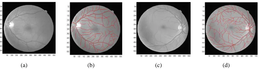
Figure 3 Tracking Results From Proposed Method In Retinal Vessel Images From DRIVE Database.
Algorithms in the second group are often called curvilinear structure detector, or ridge based approaches, which consider intensity image as 3D elevation maps in which intensity ridges or valleys approximating the skeleton of the line-structure.(18, 19) Eigen-analysis of Hessian matrix is always applied to estimate the orientation of each pixel in the image (20). The line structure skeletons can be derived by modeling the profile of the line-structure and examining its first and second derivatives with Gaussian kernel along the profiles (21). Hysteresis linking phrase is usually necessary in the post-processing to connect all the centerline points and detect the bifurcation markers (22). It has been
discussed that curvilinear structure detector always achieve high accuracy in centerline extraction; however, its performances are still limited especially on low contrast objects since the image is smoothen by the Gaussian filter(23). It also suffers from the limitation of high computational cost since it exams every pixel in the image.
833 approach is presented in section 4; finally we make a discussion and conclude the work.
2.
DATABASE DESCIPTIONSAll the images in this work come from the popular public retinal vessel image database DRIVE, which has been established in Utrech University. DRIVE consists of 40 images (7 with pathologies) captured by a Canon CR5 3CCD camera with 45 FOV. The slides were digitized to
700 605
×
pixels with 8 bits per color channel. The FOV in the images is approximately 650×550 pixels (18). Detailed description is available in http://www.isi.uu.nl/Research/Databases/DRIVE /.3.
METHODOLOGYSeed points, which are the original points for tracking, are labeled manually with the positions and their directions.
The core step of the direct exploratory approach is the tracking process, which is based on the adaptive tracking processing of the image(24). Traditional exploratory procedure sequentially detects the incremental sections along a retinal vessel segment.
Starting from a detected seed point pion or near the centerline, and an initial estimate of the direction
u
i, the line structure is traced recursively, estimating and refining successive points1 i
p+ ,pi+2,
…, along the centerline. The processing is repeated until a stopping criterion is satisfied. Typical edge based exploratory approach will be introduced at first, followed by description of proposal improved method.
As discussed above, the basic idea of exploratory algorithm contains two steps, estimation and correction. Before processing, it should be mentioned at first that all the directions in a two dimension digital image are quantized into 16 directions, each indicated by an index number from the set
u
i, where i=0,1, 2,...15.(1) Given the coordinate and orientation of current centerline pointpi, the next centerline point along the process
1 i
p+ can be predicted by the following update equation of the form:
~
1
k k k
p+ =p +du , (1)
Where
p
k is the x-y coordinate of current centerline point, ~1
k
p+ is the position of next vessel
skeleton point from prediction, ~ indicates approximation,
d
is the forward step size, andu
kis the orientation of the current pointpk.(2) Fine-turning step is applied to p~k+1 for extracting the precise direction and coordinate of centerline point
p
k+1 derived from prediction inEq.(1). pk~+1 can be achieved by Eq.(1), however, it may not exist exactly on the centerline. Therefore, the core issue in direct exploratory is the correction step. With the direction
u
k, it is possible to define two direction vector perpendiculars tou
k, which can be assigned asu
Rk andu
Lkrespectively. Theedge points corresponding to
~
1 k
p
+ , both in left side and right side can be determined by scanning alongthe direction
u
Rk andu
Lk from~ 1 k
p
+ . The two dimension 5 6× kernels are considered as a ‘template’ in searching the vessel boundary approximated positions. For convenience of exposition, the templates are grouped as left and right templates, which can be denoted asT
Li, andRi
T
, with subscriptsL
,R
,i
, indicating the left side, right side andi
th direction respectively. The algorithm motives an exhaustive search by scanning all the pixels within a pre-defined searching window along the directionu
Rkandu
Lk, with the templates rotated in different directions. Thetemplate response at a location
~
1 k
p
+ with coordinate (x y
,
) along the directionu
Rk andLk
u
is expressed as:,
( , , ) ( , )
i Rk Ri
x y
R x y u =
∑
I x+n y+m T(2)
,
( , , ) ( , )
i Lk Li
x y
L x y u =
∑
I x+n y+m T (3)Where
R
iandL
idenote the correlation betweenthe neighborhoods of
~
1
k
p+ in the image data
I
, and834 through the direction
u
Rk andu
Lk . The subscriptioni
indicates thei
th direction in the orientation setu
i . Location and rientations of boundary points are then estimated as those resulting in maximum template responses. Finally,~ 1 k
p+ is updated to pk+1 by averaging the left and
right boundary locations where the template response on each boundary is locally a maximum.
(3) Return to step (2) to obtain the subsequent skeleton chain until some stop criterions are satisfied.
As discussed in the introduction, direct exploratory approach and curvilinear structure detector are two major categories in detecting line-structure objects. Direct exploratory approach focuses on the geometrical topological feature of retinal vessels in the image only. Curvilinear structure detector always treats grayscale images as 3D elevation maps, in which the intensity ridges approximate the medial axes of the tubular objects such as vessels. The proposed method follows the basic idea of vector tracing, and then employs combination of two kinds of approaches in order to incorporate geometrical topological information and intensity distribution at the same time in order to achieve a better performance in the centerline point direction update scheme. A brief introduction of curvilinear structure detector will be introduced first, and then the contribution of proposal approach will be discussed later.
One of the key steps in curvilinear structure approaches is the construction and eigen-analysis of the Hessian matrix, which can be defined as follows:
( ), ( )
( ), ( )
xx xy
yx yy
I I
H
I I
σ
σ
σ
σ
=
(4)
Where Ixx,Ixy,Iyx and
yy
I are the normalized second order derivatives along the
x
,y
directions from the intensity image, which can be generated by convolving the image with a derivative of the Gaussian kernel,''( ) * ''( )
uv uv
I
σ
=I Gσ
(5)where Gdenotes a Gaussian kernel, subscripts
u
andv
representsx
ory
direction,σ
is the scale of Gaussian kernel(27, 28).A key parameter in the Hessian matrix construction is how to select the appropriate scale of Gaussian filter, the σ in Eq.(4) and (5). According to the literatures, the scale proportional to the width of current centerline point in retinal vessel. Existing methods determine
σ
from an ordered list of scale, in which the largest and smallest elements of the list are pre-defined by the users, as well as the step size in the ranges(22). Different scales generate different Hessian matrixes and then lead to different eigenvalues, therefore, the appropriate scale is the one which leads to the maximum absolute value among all the eigenvalues. However, in retinal vessels, vessel width varies from images in the stacks, so it is time-consuming for the users to measure and determine maximum and minimum value of the list for each image. As mentioned above, the proposal approach follows the basic idea of direct exploratory to trace the vessels and obtain the skeletons, so it is possible to derive the width of a given local point from a pair of boundary points. Based on the discussion, we utilize the width corresponding to centerlinepoint
p
k, calledw
k, to simulate the scale value inconstructing Hessian matrix for the next centerline
point
p
k+1, which avoids facing the difficulty indefining the scale range for each individual image. After eigen-analysis of Hessian matrix, two eigenvalues and their corresponding eigenvectors are obtained. One eigenvector points to the direction of the vessel, while the other eigenvector corresponds to the orthogonal direction of the vessel. In retinal vessel images, vessels are dark line-structures surrounded by the brighter background; therefore, skeletons of the vessels are the valley of the image and curvilinear structure detector method in this paper becomes valley based algorithm. Let
1 λand
2
λ denote the two eigenvalues of the Hessian matrix in Eq.(4),
v
1andv
2represent the corresponding eigenvectors, while it is also assumed that the two eigenvalues are ordered by1 2
|
λ
| |
<
λ
|
. Based on this, the directionu
for the local point in the vessel can be represented by normalized eigenvector v1, as shown in theexpression:
|| ||
u
2=
1
.In order to incorporate the geometrical information with the intensity information, the proposal algorithm defines the direction of each centerline point as(29):
(1
)
k ke kc
u
=
α
u
+ −
α
u
835 where
α
is the weighted factor which ranges from 0 to 1, subscriptionk
denotes thek
thiteration in the tracing process,
u
kerepresents the normalized direction from the average boundary orientations, which is similar in direct exploratory approach; whileu
kcis derived from eigen-analysis of Hessian matrix(27, 29). The definition in Eq.(6) considers both geometrical information and intensity distribution, which will lead to a better orientation estimation performance.ke
u
in Eq.(6) is determined by the geometrical topology of vessels, and can be derived from the directions of boundary points. Before direction estimation, the location of boundary points should be identified at first. As described before, two kinds of rotated templates are utilized in the correlation calculation, as shown in Eq.(2) and (3). It is necessary to start an exhaustive scan on each pixel
along the given orientation
u
Rk andLk
u
respectively, until it reaches the given maximum value of fixed iteration number M/2. It should be noticed that, in the searching process, correlation measurement has to be performed between image data and rotated pattern at different directions pixel by pixel along the given orientation. For example, the orientation space has been discredited to 16 values, yielding an angular precision of 22.5。and a total of 32 left and right templates; in addition, the maximum step size of scanning for each side is 5 pixels, therefore, 32*5=160 correlation calculations is processed in the searching for one centerline point. In order to accelerate the process, the proposed method employs the results of Hessian matrix to define the boundary points. While defining the Hessian matrix, second order derivatives of the image along
x
andy
direction are calculated; in addition,temporary local orientation is given as
u
k; it ispossible to derive the second order derivative of vessel profile with the direction perpendicular to
k
u
at local point by adopting steerable filters, asexpressed as:
'' 2 '' '' 2 ''
2
xx xy yy
uk xk xk yk yk
I =u I + u u I +u I (7)
Where
u
xk andu
yk indicate the normalizedelements of
u
k(30, 31).The partial derivatives inEq.(7) can be treated as the likelihood of a point
belonging to the centerline, and also can be called ‘strength value’ or ‘vessel confidence’(32). This value will change dramatically in the boundary position. In the correction step, given the orientation of centerline point pk and the
approximating position
~
1
k
p+ , we will still motive the
searching along the perpendicular direction of
k
u
from
~
1
k
p +
, which are uRkand
u
Lk. It is alsopossible to calculate strength value along the profile
of
~ 1 k
p+ . In the scanning process of each side, if the
detected strength value of a pixel is smaller than a predefine threshold, it will be considered as a boundary and the searching process stops. Since the scanning is applied along two perpendicular
direction of
u
k, there are two searching processesand the corresponding boundary position vector
B
Land BRcould be identified. It is also necessary to
estimate orientation of the boundaries; the proposal approach follows the idea in typical direct exploratory approaches which estimates the directions by calculating the correlation between rotated patterns and image data.
Moreover, the size of searching window is also adopted to be dynamic, which is assigned to be 1.5
w
k, wherew
kis the vessel width corresponding to previous centerline pointpk. This definition isbased on the assumption that the vessel width does not change abruptly in the tracing process.
The fully automated proposal algorithm can be finally concluded as following:
(1) Preprocessing is applied first to enhance the image contrast based on basic approaches, such as morphological operator and intensity adjustment. (2) Seed points are detected in order to be the starting point and ending points in the tracing process of following modified exploratory method.
Tracing framework in the work is similar as the traditional vector tracking, as described in 3.2.1. However, orientation updating scheme is modified by combining information from geometrical information and intensity information. Given a centerline point
k
p and its orientation uk , an
approximation position of the next point ~ 1
k p+ is
836 along the perpendicular direction of
u
k. Boundary identification can be finished by searching ~1
k p+ in
the strength value image, and their orientations are also estimated based on correlation measurement with rotated templates. After that, both of the position of
p
k+1and its orientation are derived. Finally, the estimated direction ofp
k+1is updated by Eq.(6).4.
RESULTSAs discussed above, each seed point is the first point of each tracing chain, and it may not exist exactly on the centerline of the retinal vessel segment. In addition, there is no prior direction estimation for a seed point; all we have is its coordinate. Therefore, traditional direct exploratory approach is applied here to refine its position and to estimate the direction. For the convenience in the implementation of Eq.(6), normalization is necessary. In the proposal algorithm, all the directions space in the image is quantized into 16 values, and then it is easy to get them normalized
with
||
u
k||
2=
1
. After the calculating in Eq.(6),k
u
is re-normalized to be an element in direction set
i
u
, and will be applied in the tracking process. In the proposed work, direction space is discrete into 16 values, and then the forward step in tracking is fixed 2 pixels so far. Another parameter in Eq.(6) is the weight valueαis setup to be 0.5, which takes account the geometrical information and intensity information equally.
Performances comparisons are tested especially in cases with complicated vessel distribution, such as overlapping and bifurcation, as shown in Fig.1 and Fig.2. Vessel skeletons identified from different methods are overlaid over the original images with red lines. It should be mentioned that the extracted centerlines in each case start from the same given seed point and have the same tracing length. Original image with vessel overlapping is shown in Fig.1(a), and tracking results achieved from proposed method and exploratory method without Hessian matrix analysis respectively, shown in Fig.1(b) and (c). It can be observed in Fig.1(c) that the centerline of one vessel segment appears to jump to another vessel in overlapping area, and then deviates outside the vessel. Typical case of vessel bifurcation is shown in Fig.2, Fig.2(c) and Fig.2 (d) are close investigation of Fig.2 (a) and Fig.2 (b) respectively. As in Fig.2(c), extracted centerline
points to the background instead of following either vessel in the bifurcation area. Performance from the proposed approach in Fig.2 (d) is satisfied. Tracking in Fig.1 and Fig.2 have demonstrated that the tracking results from the proposed method have higher accuracy. Tracing results of four retinal vessel images in DRIVE database are shown in Fig.3.
5.
DISCUSSION AND CONCLUSIONSThe method proposed in the paper has proved to be a valuable research tool for extract the retinal vascular skeleton network in retinal images. The main purpose of this work is to propose a novel approach to improve the performance of skeleton extraction of retinal vascular network, especially in some complicated situations, such as bifurcation and crossing area. Major contributions of the proposed approach can be organized into two parts: firstly, a improved seed point detection algorithm is presented to assign starting point and ending point for each tracing segment, which employs SNR measurement to remove redundant seed points from rough grid searching; secondly, a new tracking direction estimation scheme which incorporates geometrical and intensity features of the vascular structure is presented to yield higher estimation accuracy; in the application, the scale value of Gaussian model in defining Hessian matrix is self-adaptive, which is helpful to reduce the interactions from users and improve estimating performance; a dynamic searching window acknowledges the vessel width of previous traced centerline point is applied for detection of current boundary points. Quantitative analysis in validation section has demonstrated that performances from program have strong similarity with manual labeling.
837
ACKNOWLEDGEMENT
The work was supported by National Natural and Science Foundation of China No. 61172179, the Fundamental Research Funds for the Central Universities No. 2011121051 and Natural Science Foundation of Fujian Province of China 2012J05160.
REFRENCES:
[1] J. Lowell, A. Hunter, D. Steel, A. Basu, R. Ryder and R. L. Kennedy, "Measurement of retinal vessel widths from fundus images based on 2-D modeling," IEEE Trans Med Imaging
Vol.23, No.10, 2004, pp.1196-1204.
[2] N. Patton, T. Aslam, T. Macgillivray, A. Pattie, I. J. Deary and B. Dhillon, "Retinal vascular image analysis as a potential screening tool for cerebrovascular disease: a rationale based on homology between cerebral and retinal microvasculatures," J Anat Vol.206, No.4, 2005, pp. 319-348
[3] C. L. Tsai, B. Madore, M. J. Leotta, M. Sofka, G. Yang, A. Majerovics, H. L. Tanenbaum, C. V. Stewart and B. Roysam, "Automated retinal image analysis over the internet," IEEE Trans Inf Technol Biomed , Vol.12, No.4, 2008, pp.480-487
[4] H. Narasimha-Iyer, J. M. Beach, B. Khoobehi and B. Roysam, "Automatic identification of retinal arteries and veins from dual-wavelength images using structural and functional features," IEEE Trans Biomed Eng Vol.54, No.8, 2005, pp. 1427-1435
[5] C. Heneghan, J. Flynn, M. O'Keefe and M. Cahill, "Characterization of changes in blood vessel width and tortuosity in retinopathy of prematurity using image analysis," Med Image Anal, Vol.6 no.4, 2002, pp.407-429
[6] A. Hoover, V. Kouznetsova and M. Goldbaum, "Locating blood vessels in retinal images by piecewise threshold probing of a matched filter response," IEEE Trans Med Imaging Vol.19, no.3, 2000, pp. 203-210
[7] G. Lin, C. V. Stewart, B. Roysam, K. Fritzsche, G. Yang and H. L. Tanenbaum, "Predictive scheduling algorithms for real-time feature extraction and spatial referencing: application to retinal image sequences," IEEE Trans Biomed Eng Vol.51, No.1, 2004, 115-125 [8] E. Ricci and R. Perfetti, "Retinal blood vessel
segmentation using line operators and support vector classification," IEEE Trans Med
Imaging vol.26, No.10, 2007, 1357-1365 [9] Y. Sato, S. Nakajima, N. Shiraga, H. Atsumi, S.
Yoshida, T. Koller, G. Gerig and R. Kikinis, "Three-dimensional multi-scale line filter for segmentation and visualization of curvilinear structures in medical images," Med Image Anal
Vol.2, No.2, 1998, pp. 143-168
[10] K. A. Vermeer, F. M. Vos, H. G. Lemij and A. M. Vossepoel, "A model based method for retinal blood vessel detection," Comput Biol Med Vol.34, No.3, 2004, 209-219 (2004) [11] H. Narasimha-Iyer, V. Mahadevan, J. M. Beach
and B. Roysam, "Improved detection of the central reflex in retinal vessels using a generalized dual-gaussian model and robust hypothesis testing," IEEE Trans Inf Technol Biomed Vol.12, No.3, 2008, pp. 406-410
[12] F. Zana and J. C. Klein, "A multimodal registration algorithm of eye fundus images using vessels detection and hough transform,"
IEEE Trans Med Imaging Vol.18, No.5, 1999, pp. 419-428
[13] A. M. Mendonca and A. Campilho, "Segmentation of retinal blood vessels by combining the detection of centerlines and morphological reconstruction," IEEE Trans Med Imaging Vol.25, No.9, 2006, pp.1200-1213
[14] D. Wu, M. Zhang, J. C. Liu and W. Bauman, "On the adaptive detection of blood vessels in retinal images," IEEE Trans Biomed Eng
Vol.53, No.2, 2006, pp.341-343
[15] K. A. Al-Kofahi, A. Can, S. Lasek, D. H. Szarowski, N. Dowell-Mesfin, W. Shain, J. N. Turner and B. Roysam, "Median-based robust algorithms for tracing neurons from noisy confocal microscope images," IEEE Trans Inf Technol Biomed Vol.7, No.4, 2003, pp.302-317 [16] H. Shen, B. Roysam, C. V. Stewart, J. N.
Turner and H. L. Tanenbaum, "Optimal scheduling of tracing computations for real-time vascular landmark extraction from retinal fundus images," IEEE Trans Inf Technol Biomed Vol.5, No.1, 2001, pp.77-91
[17] M. A. Abdul-Karim, B. Roysam, N. M. Dowell-Mesfin, A. Jeromin, M. Yuksel and S. Kalyanaraman, "Automatic selection of parameters for vessel/neurite segmentation algorithms," IEEE Trans Image Process Vol.14, No.9, 2005, pp.1338-1350
838 retina," IEEE Trans Med Imaging Vol.23, No.4, 2004, pp.501-509
[19] L. Gang, O. Chutatape and S. M. Krishnan, "Detection and measurement of retinal vessels in fundus images using amplitude modified second-order Gaussian filter," IEEE Trans Biomed Eng Vol.49, No.2, 2002, pp.168-172 [20] B. Zhang, L. Zhang and F. Karray, "Retinal
vessel extraction by matched filter with first-order derivative of Gaussian," Comput Biol Med Vol.40, No.4, 2005, pp. 438-445
[21] C. Steger, "An Unbiased Detector of Curvilinear Structures," IEEE Trans Pattern Analysis and Machine Intelligence Vol.20, No.2, 1998, pp. 113-125
[22] G. Xiong, X. Zhou, A. Degterev, L. Ji and S. T. Wong, "Automated neurite labeling and analysis in fluorescence microscopy images,"
Cytometry A Vol.69, No.6, 2006, pp.494-505 [23] Y. Zhang, X. Zhou, A. Degterev, M. Lipinski,
D. Adjeroh, J. Yuan and S. T. Wong, "Automated neurite extraction using dynamic programming for high-throughput screening of neuron-based assays," Neuroimage Vol.35, No.4, 2007, pp.1502-1515
[24] A. Can, H. Shen, J. N. Turner, H. L. Tanenbaum and B. Roysam, "Rapid automated tracing and feature extraction from retinal fundus images using direct exploratory algorithms," IEEE Trans Inf Technol Biomed
Vol.3, No.2, 1999, pp.125-138
[25] Y. Zhang, X. Zhou, A. Degterev, M. Lipinski, D. Adjeroh, J. Yuan and S. T. Wong, "A novel tracing algorithm for high throughput imaging Screening of neuron-based assays," J Neurosci Methods Vol.160, No.1, 2007, pp.149-162 [26] N. Otsu, "A threshold selection method from
gray-level histograms," IEEE Trans. Systems, Man and Cybernetics Vol.9, No.1,1979, pp. 62-66
[27] Y. Xu, G. Hu, G. Jinzhao and S. Lihua, "Adaptive tracking extraction of vessel centerline using Hessian matrix in coronary arteriograms," J Tsinghua Univ(Sci&Tech)
Vol.47, No.6, 2007, pp. 889-892
[28] C. Lorenz, I. C. Carlsen and T. M. Buzug, "Multi-scale line segmentation with automatic estimation of width, contrast and tangential direction in 2D and 3D medical images," in
CVRMed-MRCAS, 1997, pp. 233-242.
[29] Y. Xu, H. Zhang, H. Li and G. Hu, "An improved algorithm for vessel centerline tracking in coronary angiograms," Comput Methods Programs Biomed Vol.88, No.2, 2007, pp. 131-143
[30] W. T. Freeman and E. H. Adelson, "The design and use of steerable filters," IEEE Trans Pattern Anal Mach Intell Vol.13, No.9, 1991, pp. 891-906.
[31] Y. Huang, X. Zhou, B. Miao, M. Lipinski, Y. Zhang, F. Li, A. Degterev, J. Yuan, G. Hu and S. T. Wong, "A computational framework for studying neuron morphology from in vitro high content neuron-based screening," J Neurosci Methods, Vol.190 No.2, 2010, pp.299-309