http://www.sciencepublishinggroup.com/j/bmb doi: 10.11648/j.bmb.20170206.13
ISSN: 2575-5064 (Print); ISSN: 2575-5048 (Online)
Antimicrobial and Physicochemical Evaluation of
Luffa
acutangular
Leaf Extracts
Alagbe Seyi Valerie
1, 2, Ibi Anna
1, *, Toge Christal
1, Amuzie Uzo Felicia
1, Ftepti Benson Jelani
3,
Raji Bamanga
31
Biotechnology Advanced Research Centre, Sheda Science and Technology Complex, Abuja, Nigeria
2Department of Biosciences, Salem University, Lokoja, Nigeria
3Department of Biotechnology, Modibbo Adama University of Technology, Yola, Nigeria
Email address:
[email protected] (I. Anna)
*Corresponding author
To cite this article:
Alagbe Seyi Valerie, Ibi Anna, Toge Christal, Amuzie Uzo Felicia, Ftepti Benson Jelani, Raji Bamanga. Antimicrobial and Physicochemical Evaluation of Luffa acutangular Leaf Extracts. Advances in Materials. Vol. 2, No. 6, 2017, pp. 80-85. doi: 10.11648/j.bmb.20170206.13
Received: September 8, 2017; Accepted: October 8, 2017; Published: November 11, 2017
Abstract:
Leaves of Luffa acutangular are consumed in some parts of Nigeria as part of folk medicine for the treatment of diseases and as a vegetable food source. This study was undertaken to evaluate the phytochemical constituents from ethanol extracts, antimicrobial resistance, proximate and mineral analysis of its leaf extracts. The phytochemical investigation revealed the presence of alkaloids, phenols, saponins, tannins, terpenoids, and triterpenoids. The elemental analysis of the dried leaf revealed the presence of Calcium (58.6 mg/g), Copper (0.6 mg/g), Magnesium (12.4 mg/g), Manganese (0.9 mg/g), Zinc (0.6 mg/g), Sodium (14.4 mg/g) and Potassium (143.6 mg/g) respectively. The samples were screened against Staphylococcus aureus, Staphylococcus pneumonia, Streptococcus pyrogens, Klebsciella pneumonia, Candida albicans and Candida tropicalisfor their anti-microbial activity using Ciprofloxacin, Streptomycin and Fluconazole as control. Minimum inhibitory concentration (MIC), minimum bactericidal concentration (MBC) and minimum fungicidal concentration (MFC) values were determined. Proximate analysis revealed moisture content of 10.6%, ash 6.3%, crude protein 2.6%, crude fiber 4.0%, fat 5.1% and carbohydrate content of 71.4%. The study showed that the leaf extract of L. acutangular may be used to manage some common diseases caused by the tested organisms. The major antimicrobial activity is tailored to the phyto-constituents. This confirms the folkloric use of the plants in the management of various diseases.
Keywords:
Luffa acutangular, Phytochemical Constituents, Antimicrobial Activities, AAS, Proximate Analysis1. Introduction
In the past decade, clinical drug research programs have turned towards plants as a reservoir of untapped therapeutic agents, primarily used in orthodox medical practices by over 75% of global populace [1]. Such plants contain varied
secondary metabolites; phyto-compounds including
alkaloids, flavonoids, glycosides, phenols, saponins,
terpernoids, minerals, etc which exhibit unique activities that are of pharmaceutical interest [2-4]. These metabolites generally do not participate in plant development and growth, rather in plant defensive mechanisms to combat onslaughts of abiotic and biotic stresses [5-6].
The curative interest in these compounds emanates from
their unique mechanisms of resistance against a horde of disease causing organisms by circumventing the host-pathogen interaction which would have otherwise led to the propagation of the vector, ultimately producing a diseased state in the host [7]. It can then be inferred that the unique chemical diversity derived from natural products; phyto-compounds, is considered to be superior whereby the active compounds’ framework is absent in its synthetic variants, a factor which could be associated with certain side effects via consumption of the latter.
prophylactic, thereby authenticating their use in folk medicine [8]. As a raw material in biopharming, nutraceuticals, etc, several studies have been conducted in many parts of the world to validate their use as antimicrobial agents or biological control agents [9-12]. However, some plants belonging to known families with therapeutic potential have either been neglected nor experienced satisfactory attention. Agricultural expansion strategies in developing countries necessitates exploring the full potential of indigenous or wild food plants as an essential part of our diet [13]. Reports indicate that food crops grown in the wild possess an array of minerals, micronutrients and therapeutic properties that are crucial towards combating nutritional deficiencies especially during periods of food scarcity, diseases, etc [14-15]. One of such crops is Luffa acutangular
(L.) Roxb var. amara (sponge guard) from the Cucurbitaceae family which is widely distributed across China, Korea, India, Japan and Central America and grows wildly in the western and north central parts of Nigeria where it is consumed as a vegetable [16]. There is however a limited amount of scientific information on the nutritional, mineral and phytochemical composition of some indigenous crops, like the diversity of Luffa acutangular (L.) Roxb var. amara
(sponge guard) grown in Nigeria. There is therefore a need to evaluate this in order to fill the knowledge gap and raise the awareness towards cultivation and consumption. This study was therefore aimed at identifying the phytochemical, antimicrobial, nutritional and mineral composition of indigenous Luffa acutangular (L.) Roxb var. amara (sponge guard) obtained in Nigeria.
2. Materials and Methods
Leaves of Luffa acutangular were collected in fresh condition at Sheda and Abakiliki regions of Kogi state, Nigeria. The plants were identified, air-dried and kept in airtight containers until required for further laboratory analysis. Crispy plant leaf sample (100g) was placed and soaked with ethanol in a Soxhlet apparatus for 6-8 hours. The crude extracts were later concentrated using rotary evaporator. Phytochemical screening was then performed using standard procedures [17].
2.1. Mineral Analysis
The metal analysis was determined using an Atomic Absorption spectrometer (iCE 3000, Thermo fisher). Five grams of oven dried samples were weighed into a crucible and transferred to a furnace at 600°C and left to ash for 3 hours. The furnace was cooled to about 120°C and then placed in a desiccator for an hour to cool before weighing. This process was repeated until a constant weight was obtained. The ashed samples (0.5g) were weighed and transferred into the digestion tube. Distilled water, concentrated HNO3 and perchloric acid (5mL of each) were
added and the content mixed. The tubes were placed into the digestion block inside a fume cupboard and the temperature was set at 150°C for 90 minutes. The temperature was then
adjusted to 230°C and incubated for another 30 minutes to obtain white fumes. The temperature was then reduced to 150°C, followed by the addition of 1 mL of hydrochloric acid to the tubes within a few minutes. Water was added to the tube to make up to the mark, mixed and filtered.
Elemental analysis of the solution obtained was then performed using an atomic absorption spectrophotometer (AAS) at an appropriate wavelength, temperature and lamp-current for the different elements. The following elements
were determined, calcium (Ca), magnesium (Mg),
manganese (Mn), iron (Fe), copper (Cu), zinc (Zn), potassium (K), sodium (Na).
2.2. Proximate Analysis
The moisture, protein, fat, ash, crude fibre and carbohydrate content of the dried leaves were determined. For moisture content, 5g of dried leaf samples was weighed and dried in an oven at 105°C to a constant weight. The percentage weight loss was determined. Fat content was determined by extracting 5g of dried leaf sample with hexane or petroleum ether in a Soxhlet apparatus for 8 hours. The ash content was estimated by incinerating 5g of dried sample in a muffle furnace (Carbolite-RHF 1600) at 550°C for 4 hours, and then the percentage ash content was determined. The micro-Kjedahl method was employed for estimation of crude protein. All experiments were done in triplicate and results were expressed as the averages on dry weight basis.
2.3. Preparation of Inoculum
The bacterial isolates were collected from Medical Microbiology Department of Specialist Teaching Hospital, University of Abuja, F. C. T., Nigeria on a slant Nutrient agar. The isolates were restored on Nutrient broth and confirmed using standard biochemical tests according to the Bergey’s manual of Bacteriology [18].
2.4. Biological Screening of Extracts
The crude extract was screened for antimicrobial activity using agar well diffusion technique with little modification.
Clinical isolates utilised were Staphylococcus aureus,
Staphylococcus pneumonia, Streptococcus pyrogens, Klebsciella pneumonia, Candida albicans and Candida tropicalis. The minimum inhibitory concentration (MIC) was determined on the test organisms that were sensitive to the extracts and was done by broth dilution method [19]. Mueller Hinton broth was prepared, dispersed into test tubes and the broth was sterilized at 121°C for 15 minutes, the broth was allowed to cool. Normal saline was prepared, 10mls was dispersed into sterile test tube and the test microbes was inoculated and incubated at 39°C for 6hrs. Dilution of the test microbes was done in the normal saline until the turbidity marched that of the McFarland’s standard scale by visual comparison at this point, this test microbes has a
concentration of about 1.5 x 10⁸ CFU/ml. Standard
sulfide (DMSO) used as negative control. Minimum bactericidal and fungal concentrations (MBC/MFC) were evaluated by plating the bacterial suspensions from individual well at the beginning and at the end of the experiments on Mueller Hinton agar medium for estimation of MBC. The culture from MIC well was taken and streaked on the surface of fresh Mueller Hinton agar in a 90-mm plate with division and incubated at 37°C for 24 hours (bacteria) after which the plates of the medium was observed for colony growth, the MBC was the plates with lowest concentration of the extract without colony growth.
3. Results
Table 1 represents the phytochemical composition of ethanol extracts from the leaves L. acutangular. The powdered leaves tested positive for the presence of certain phyto-compounds such as alkaloids, phenolic acid, saponins, tannins and terpenoids (table 1).
The mineral content analysis of the leaves of Luffa acutangular revealed that the most predominant micronutrients were potassium (143.6 mg/g) and calcium (58.6 mg/g) whereas the least concentrations of 0.6 (mg/g) each was determined for zinc and copper (Table 2).
Table 1. Phytochemical Analysis of leaf sample.
Phyto-Constituents Ethanol
Alkaloids +
Balsams -
Cardenolides -
Cardiac glycosides -
Flavonoids -
Glycosides -
Phenolic acid +
Saponins +
Steroids -
Tannins +
Terpenoids +
Triterpenoids +
Key: -= Negative/Absent, += Positive/Present
Nutritional analysis revealed that the leaves possessed a high moisture to ash and fat content at 10.6%, 6.3% and 5.2% respectively (Table 3). The carbohydrate content was highest (71.4%).
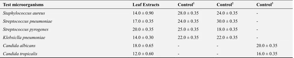
The highest zone of inhibition recorded for the alcoholic
extracts of Luffa acutangular leaves was against
Streptococcus pyrogens (20.0±0.35 mm), followed by
18.0±0.65 (mm) against Candida albicans (Table 4).
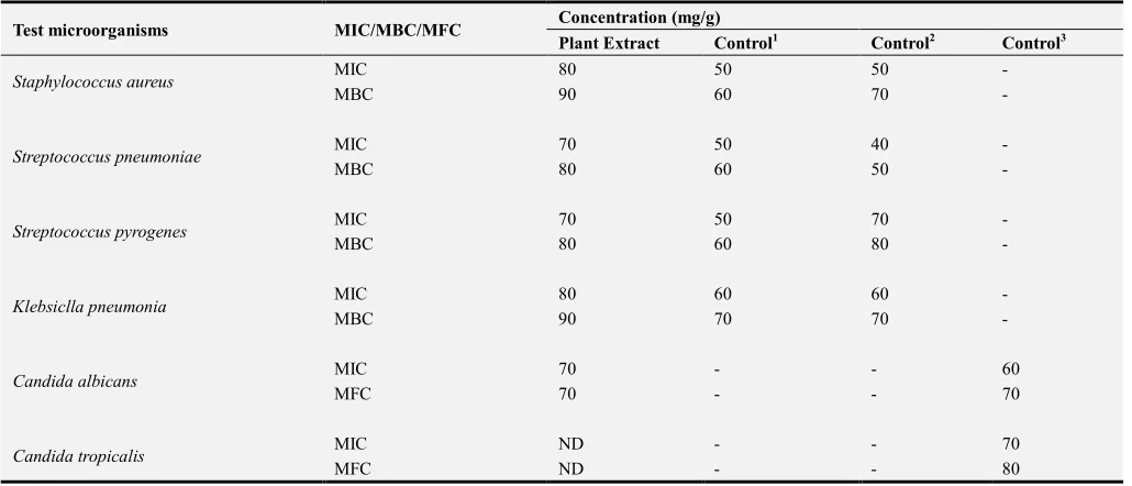
The lowest combined MIC and MBC values obtained (70
and 80 mg/ml) was against Streptococcus pneumonia and
Streptococcus pyrogens (Table 5).
The lowest combined MIC and MFC values obtained (70 and 70 mg/ml) was against Candida albicans (Table 5).
Table 2. Mineral Analysis.
Calcium (Ca) (mg/g)
Iron (Fe) (mg/g)
Magnesium (Mg) (mg/g)
Manganese (Mn) (mg/g)
Zinc (Zn) (mg/g)
Copper (Cu) (mg/g)
Sodium (Na) (mg/g)
Potassium (K) (mg/g)
58.6 8.1 12.4 0.9 0.6 0.6 14.4 143.6
Table 3. Proximate Composition of leaves.
Ash (%) Fat (%) Fibre (%) Protein (%) Moisture (%) Carbohydrate (%)
6.3 5.1 4.0 2.6 10.6 71.4
Table 4. Antimicrobial activity of Luffa acutangular leaf extracts against test organisms Zone of inhibition (mm).
Test microorganisms Leaf Extracts Control1 Control2 Control3
Staphylococcus aureus 14.0 ± 0.90 28.0 ± 0.35 24.0 ± 0.35 -
Streptococcus pneumoniae 17.0 ± 0.35 24.0 ± 0.35 30.0 ± 0.35 -
Streptococcus pyrogenes 20.0 ± 0.35 25.0 ± 0.35 18.0 ± 0.35 -
Klebsiclla pneumoniae 14.0 ± 0.30 22.0 ± 0.35 22.0 ± 0.35 -
Candida albicans 18.0 ± 0.65 - - 20.0 ± 0.35
Candida tropicalis 12.0 ± 0.60 - - 16.0 ± 0.35
Table 5. Minimum Inhibitory Concentration (mg/ml ± SD) of L. acutangular leaf extracts against test organisms.
Test microorganisms MIC/MBC/MFC Concentration (mg/g)
Plant Extract Control1 Control2 Control3
Staphylococcus aureus MIC 80 50 50 -
MBC 90 60 70 -
Streptococcus pneumoniae MIC 70 50 40 -
MBC 80 60 50 -
Streptococcus pyrogenes MIC 70 50 70 -
MBC 80 60 80 -
Klebsiclla pneumonia MIC 80 60 60 -
MBC 90 70 70 -
Candida albicans MIC 70 - - 60
MFC 70 - - 70
Candida tropicalis MIC ND - - 70
MFC ND - - 80
Control1= Ciproflaxin (100 µg/g), Control2= Streptomycin (100 µg/g), Control3= Fluconazole (100 µg/g). Each value represents mean (n = 3). ND= Not Detected
4. Discussion
Whole plants, herbs and vegetables provide established launch-pads through the field of phyto-chemistry and pharmacognosy for the development of new drugs and their intermediates that have adequate therapeutic uses [20-21]. A plethora of essential molecules in plants is responsible for both their nutritional, energy giving and prophylactic effects [21]. The mode of action of different complex plant products bear a resemblance to ligands, hormones, signal transduction molecules, etc thus have beneficial medicinal effects on humans due to their potential target sites similarities. In this study, the presence of alkaloids, phenolic acid, saponins, tannins, terpenoids and triterpenoids (table 1) suggests that the plant possess a unique antimicrobial range as these individual compounds exert action that thwarts the growth or development of disease causing pathogens [23]. Recent studies have shown that alkaloids, obtained from different plant extracts like Callistemon citrinus and Vernonia adoensis
successfully diminished the growth of Staphylococcus aureus
when compared to β-lactam antibiotics [24]. Bioactive compounds like tannins has also been studied and found to be active against Staphylococcus aureus alongside a host of bacterial pathogens linked with a variety of infections [25-26]. The presence of alkaloids suggests that the leaves of Luffa acutangular could be used as neuro-stimulants or in new drug therapies [27]. As a result of the saponin content, ingestion of these leaves may aid in lowering cholesterol metabolism thereby boosting liver function in the body [28-29]. Saponins would also deter Na+ efflux and trigger the Na+- Ca2+ antiporter in cardiac muscle thereby elevating the cytosolic Ca2+ via the influx of calcium, thus strengthening the contraction of cardiac muscles towards abating congestive heart failure [30]. The presence of phenols, flavonoids and
tannins indicate that these leaves possess antioxidant potentials that may be applied towards end-products that aid the prevention of stroke, cardiovascular diseases, cancer and other neurological disorders [31-33]. Steroids play a vital role in enhancing the well-being of animals and humans as they function in resisting infiltration by many pathogens owing to their antimicrobial properties [34].
such water rich plants keeps the body hydrated and aids in digestion [38]. This study showed that the leaves extract of
L. acutangula had antimicrobial and anti-fungal activity. The ethanolic extract of L. acutangula leaves exhibited a stronger anti-microbial activity on streptococcus pyrogens
compared to the controls suggesting a new drug with little or no side effect for the treatment of diseases caused by
streptococcus pyrogens. The effect of the ethanolic leaf extracts on the other microorganisms and fungi compared to
the control was less than the control. Therefore L.
acutangula leaves can be used as a preventive drug to upper respiratory and urinary tract organisms (table 4).
The antimicrobial screen for the studied plant revealed that
Luffa acutangular leaves confer a higher degree of resistance to Streptococcus pyrogenes (20mm), Candida albicans (18
mm) followed by Streptococcus pneumonia (17mm), thereby
hinting at the possibility of new drug candidates from the plant (table 4). The findings from table 4 compliments the results given in tables 1 and 2 whereby other studies have shown that bioactive compounds detected as well as the presence of some minerals triggers resistance against a range of disease causing pathogens linked with respiratory infections [25, 26]. In particular, interference of bacterial enzyme activity is commonly observed in the presence of tannins also detected in this study [40]. Table 5 data shows the MIC, MBC and MFC of the crude leaf extracts of L. acutangular. The data confirms that the crude leaf extracts
were more active against Candida albicans and both
Streptococcus species. The general observation from the displayed activity against the selected microorganisms and detected minerals suggests that the studied plant part could be employed towards alleviating infections as a plant for food and medicine.
5. Conclusion
This study explored the potential of an indigenous medicinal plant in the treatment of infections and its nutritional content towards encouraging the cultivation and consumption of the plant as food and to boost commercialization. The study has provided additional scientific knowledge of the ethnomedicinal ability of the plant, thereby supporting the folkloric use of the plant in the management of infections. It was observed that the plant has a high amount of potassium and calcium, showing that the plant could be nutritional supplement in diet. The anti-microbial activity of the plant extract suggested that the plant could be used as a prophylactic for the management of infections caused by the tested micro-organisms. The plant could be a possible drug candidate for the management of infections caused by Streptococcus pyrogens, due to its high anti-microbial activity against the organism compared to streptomycin which was used as a control. Since this plant is one of the forgotten indigenous plants that grows mainly in the wild in Nigeria, cultivation and consumption could be encouraged based on the possibilities obtained from this study.
References
[1] Kirbag, S., Zengin, F., and Kursat, M. (2009): Antimicrobial Activities of Extracts of some Plants. Pakistan J. Bot., 41: 2067-2070.
[2] Shakeri A., Hazeri N., Vlizadeh J., Ghasemi A., and Tavallaei F., (2012): Phytochemical Screening, Antimicrobial and Antioxidant Activity of Anabasis aphylla L. Extracts. Kragujevac J. Sci., 34: 71-78.
[3] Uttu A. J., Sallau M. S., Hamisu I., Mubarak B. D., and Abdullahi Y. I., (2015): Phytochemical and antimicrobial screening of stem bark extracts from Glossonema boveanum
(Decne). Journal Pharma. Phytochem., 4: 86-88.
[4] Madhuri, S., and Pandey, G. (2009): Some anticancer medicinal plants of foreign origin. Curr. Sci., 96: 25.
[5] Edvera, A., Velikova, V., Tsonev, T., Dagnon, S., Gurel, A., Aktas, L., and Gesheva, V. (2008): Stress protective role of secondary metabolites: Diversity of functions and mechanisms. Gen. Appl. plant Phys., 34: 67-78.
[6] Achakzai, A. K., Achakzai P., Masood, A., Kayani, S. A., and Tareen, R. B. (2009): Response of plants parts and age on the distribution of secondary metabolites on plants found in Quetta. Pakistan J. Bot., 41: 2129-2135.
[7] Shakeri, A., Hazeri, N., Vlizadeh, J., Ghasemi, A., and Tavallaei, F. (2012): Phytochemical Screening, Antimicrobial and Antioxidant Activity of Anabasis aphylla L. Extracts. Kragujevac J. Sci.,34: 71-78.
[8] Ngwu N. W., Effa E. B., Ftepti B. J., Gali A. I., Useh M. U., and Samuel C. J., (2016): Biochemical Studies of Ocimum sanctum and Olax subscorpioidea Leaf Extracts. British J. Pharm. Res., 12:1-9.
[9] Okwute S. K., Olajide O. O., Etuk-Udo G., and Orishadipe A. T., (2016):Phytochemical Screening, In-vitro Antimicrobial Activity and Antioxidant Characteristics of Tetrapleura tetraptera
Extracts. European J. Med. Plants, 17:1-10.
[10] Jong, H. J., Ji, W. L., Kyoung, S. K., Ju-Sung, K., Sang, N. H., Chang, Y. Y., Ju, K. L., Yong, S. K., and Myong, J. K. (2010): Antioxidant and antimicrobial activities of extracts from a medicinal plant, Sea Buckthorn. J. Kor. Soc. Appl. Bio. Chem., 53: 33-38.
[11] Bhalodia, N. R., and Shukla, V. J. (2011): Antibacterial and antifungal activities from leaf extracts of Cassia fistula l.: An ethnomedicinal plant. J. Adv. Pharm. Tech. Res., 2: 104-109.
[12] Sanjay, M. G., Atul, K. G., Zakwan, A., and Anil, K. (2011): Antibacterial and antifungal activity in leaf, seed extract and seed oil of Seabuckthorn (Hippophae salicifolia D. Don) Plant. J. Plant. Path. Microbio., 2: 1-4.
[13] Murray, S. S., Schoeninger, M. J., Bunn, H. T., Pickering, T. R., and Marlett, J. A. (2001): Nutritional Composition of Some Wild Plant Foods and Honey Used by Hadza Foragers of Tanzania. J. Food Comp. Anal.,14:3-13.
[15] Aline, L. M., Lamien, C. E., Compaoré, M. M. Y., Meda, R. N. T., Kiendrebeogo, M., Jeanne, B. Z., Millogo, and Nacoulma, O. G. (2008): Polyphenol Content and Antioxidant Activity of Fourteen Wild Edible Fruits from Burkina Faso. Mol.,13:581-594.
[16] Bal, K. E., Bal, Y., and Lallam, A., (2004): Gross morphology and absorption capacity of cell-fibers from the fibrous vascular system of Loofah (Luffa cylindrica). Text Res. J., 74: 241-247.
[17] Trease, G., and Evans, W. (2002): A textbook of pharmacognosy (fifth edition). E. Elsevier ltd. Edinburgh, 20-23.
[18] Cheesbrough, M. (2002). Reaction Isolates on Tropical Diseases: The Effects. Cambridge University press, London, 2: 76-100.
[19] Suffredini, I. B., Sander, H. S., Goncalves, A. G., Reis, A. O., Gales, A. C., Varella, A. D., and Younes, R. N. (2004): Screening of anti-bacterial extracts from plants native to Brazilian Amazon Rain Forest and Atlantic forest. Braz. J. Med. Bio. Res., 37: 379-384.
[20] Tanaka, J. C. A., da Silva, C. C., de Oliveira, A. J. B., Nakamura, C. V., and Dias-Filho, B. P. (2006): Antibacterial activity of indole alkaloids from Aspidosperma ramiflorum.
Braz. J. Med. Bio. Res.,39: 387-391.
[21] Adebiyi, A., Effa, E. B., Ayo, R., Bello, I., Habila, J., and Gali, A. I. (2016): Anti-mycobacterial, Antimicrobial and Phytochemical Evaluation of Pulicaria crispa and Scoparia dulcis Plant Extracts. J. Adv. Med. Pharm. Sci.,7:1-11.
[22] Zampini, I. C., Cuello, S., Alberto, M. R., Ordonez, R. M., Almeida, R. D., Solorzano, E., and Isla, M. I. (2009): Antimicrobial activity of selected plant species from the Argentine Puna against sensitive and multi-resistant bacteria. J. Ethanopharm., 124: 499-505.
[23] Omojate, G. C., Enwa, F. O., Jewo, A. O., and Eze, C. O. (2014): Mechanisms of Antimicrobial Actions of Phytochemicals against Enteric Pathogens – A Review. J. Pharm. Chem. Bio. Sci., 2: 77-85.
[24] Mabhiza, D., Chitemerere, T., and Mukanganyama, S. (2016): Antibacterial Properties of Alkaloid Extracts from
Callistemon citrinus and Vernonia adoensis against
Staphylococcus aureus and Pseudomonas aeruginosa. Int. J. Med. Chem., 1: 1-7.
[25] Newton, S. M., Lau, C., Gurch, S. S., Besra, G. S., and Wright, C. W. (2002): The evaluation of forty-three plant species for in vitro anti-mycobacterial activities; isolation of active constituents from Psoralea corylifolia and Sanguinaria canadensis. J. Ethnopharm., 79: 57–67.
[26] Dahiya, P., and Purkayastha, S. (2012): Phytochemical screening and antimicrobial activity of some medicinal plants against multi-drug resistant bacteria from clinical isolates. Indian. J. Pharm. Sci., 74: 443–450.
[27] Zenk, M. H., and Juenger, M. (2007): Evolution and current status of the phytochemistry of nitrogenous compounds. Phytochem., 68: 2757–2772.
[28] Shi, J., Arunasalam, K., Yeung, D., Kakuda, Y., Mittal, G., and Jiang, Y. M. (2004): Saponins from edible legumes: chemistry, processing, and health benefits. J. Med. Food., 7: 67–78.
[29] Sparg, S. G., Light, M. E., and van Staden, J. (2004): Biological activities and distribution of plant saponins. J. Ethnopharm., 94: 219–243.
[30] Schneider, G., and Wolfling, j. (2004): Synthetic Cardenolides and related compounds. Curr. Org. Chem., 8.
[31] Commenges, D., Scotet, V., Renaud, S., Jacqmin-Gadda, H., Barberger-Gateau, P., and Dartigues, J. F. (2000): Intake of flavonoids and risk of dementia. Euro. J. Epidem., 16: 357– 363.
[32] Ng, T. P., Feng, L., Niti, M., Kua, E. H., and Yap, K. B. (2008). Tea consumption and cognitive impairment and decline in older Chinese adults. American J. Clin. Nut.,88: 224–231.
[33] Nurk, E., Refsum, H., Drevon, C. A., Tell, G. S., Nygaard, H. A., Engedal, K., and Smith, A. D. (2009): Intake of flavonoid-rich wine, tea, and chocolate by elderly men and women is associated with better cognitive test performance. J. Nutr. 139: 120–127.
[34] Hassan, M. A., Oyewale, A. O., Amupitan, J. O., Abdullahi, M. S., and Okonkwo, E. M. (2004): Preliminary phytochemical and antimicrobial investigation of crude extract of root bark of Deterium Microcarpum.Nig. J. Chem. Sci., 29: 36-49.
[35] Mutch, D. M., Wahli, W., and Williamson G. (2005): Nutrigenomics and nutrigenetics: the emerging faces of nutrition. FASEB, 19: 1602–1616.
[36] Mathers, J. C., (2006). Plant foods for human health: research challenges. Proc., Nutr. Soc., 65: 198–203.
[37] Vance, C. P., Uhde-Stone, C., and Allan, D. L. (2003): Phosphorus acquisition and use: critical adaptations by plants for securing a non-renewable resource. New. Phytol., 157: 432–449.
[38] Raghavendra, N., Sneha, D. B., Acharya, R. N., and Shukla, V. J. (2015): Evaluation of phytochemical content, nutritional value and antioxidant activity of Olax scandens (Roxb) leaves. Int. Ayurv. Med. J.,3: 702-709.
[39] Jianyou, G., Tingjun, L., Linna, H., and Yongmei, L. (2009): The effect of Corn silk on glyceamic metabolism. Nut. Met., 6: 47.
[40] Sallau, A. B., Njoki, G. C., Olokisi, A. R., Wurochekke, A. U., Abdukadir, A. A., Isah, S., Abubakar, M. S., and Ibrahim, S. (2005): Effects of Guiera senegalinsis leaf extracts on some