INTERNATIONAL JOURNAL OF PHARMACEUTICAL, CHEMICAL AND BIOLOGICAL SCIENCES
Available online atwww.ijpcbs.com
EFFECTIVENESS OF ALPHA LIPOIC ACID AS
HEPATOPROTECTIVE AND ANTIOXIDANT
Eswaran Maheswari
1*, Ganesan Raja Lekshmi Saraswathy
2and Thaakur Santhranii
31Department of Pharmacy Practice, M.S. Ramaiah College of Pharmacy,
Rajiv Gandhi University of Health Sciences, Bangalore, Karnataka, India. 2Department of Pharmacology, M.S. Ramaiah College of Pharmacy,
Rajiv Gandhi University of Health Sciences, Bangalore, Karnataka, India. 3Division of Pharmacology, Institute of Pharmaceutical Technology,
Sri Padmavathi Mahila Visvavidyalayam (Women’s university), Tirupati, Andhra Pradesh, India.
INTRODUCTION
Carbamazepine (CBZ) is used for the treatment of seizure, as well as affective and behavioral disorders. The common adverse effects of CBZ include hepatitis and hepatotoxicity1,2. CBZ
produce serious hepatotoxicity and manifest as acute granulomatous hepatitis which is self-limiting if the drug is withdrawn. Administration of CBZ has produced primary biliary cirrhosis, primary sclerosing cholangitis, heavy portal liver fibrosis (precirrhotic process), acute or chronic hepatotoxicity, cholestatic3,
hepatocellular injury4 and even granuloma
formation5 in the liver. Oxidative stress occurs
due to an imbalance in the oxidants and
antioxidants system causing potential cellular damage6. Oxidative stress plays an vital role in
the pathogenesis of liver injuries, hepatotoxicity7 and liver diseases8. The clinical
utilization of aromatic antiepileptic drug is limited by its diverse adverse effects and hepatotoxicity9. The aromatic antiepileptic drug
provoked hepatotoxicity is attributed due to defective detoxification by the epoxide hydrolase and accumulation of arene oxides10.
There is a growing body of evidence suggesting that idiosyncratic drug-induced hepatotoxicity may be mediated by oxidative stress, characterized by enhanced levels of ROS such as hydroxyl radical, superoxide anion and
Research Article
ABSTRACT
The present study investigates the hepatoprotective and antioxidant activity of alpha lipoic acid (ALA) on carbamazepine (CBZ) administered rats. 30 animals were divided into 5 groups. Animals in group 1 received drinking water orally and served as control, group 2 received CBZ 50 mg/Kg dissolved in water daily by oral gavage, group 3, 4 and 5 received (50, 100 and 200 mg/Kg) of ALA in 0.2% carboxy methyl cellulose respectively 1 hr prior to administration of 50 mg/Kg CBZ for a duration of 45 days. The levels of serum glutamate oxaloacetate transaminase (SGOT), serum glutamate pyruvate transaminase (SGPT), alkaline phosphatase, bilirubin, albumin and total protein were estimated to determine the hepatoprotective activity. The levels of superoxide dismutase (SOD), glutathione (GSH), catalase and lipid peroxidation levels were estimated to determine the antioxidant activity. Histopathological examination was also done. CBZ increased the level of SGOT, GSH, catalase and increased lipid peroxidation. Administration of ALA reversed the CBZ induced hepatotoxicity. Histopathological examination revealed preservation of liver integrity in ALA administered rats compared to CBZ alone treated rats. The hepatoprotective effect of ALA was due to its antioxidant activity by scavenging free radicals.
hydrogen peroxide, due to reduced elimination and increased production of ROS11. It has been
recently demonstrated that aromatic antiepileptic drug mediated hepatotoxicity is associated with mitochondrial dysfunction and genetic or acquired mitochondrial defects8. In
the present study, we have addressed the oxidative stress as a potential mechanism responsible for CBZ-induced hepatotoxicity.
MATERIALS AND METHODS Animals
The pathogen free adult male albino rats weighing 150-250 g were used. The rats were housed in polypropylene cages at room temperature (25 ± 3oC) with 12/12 hours light
and dark cycle and the animals were fed with a balanced diet and tap water ad libitum. The study protocol was approved by the Institutional Animal Ethical Committee of M.S. Ramaiah College of Pharmacy, Bangalore, Karnataka (Ref. No. 220/abc/CPCSEA).
Study Protocol
The rats were divided into five groups with six animals in each group. First group served as control and received drinking water orally daily by gavage for 45 days. Second group received 50 mg/Kg CBZ dissolved in water daily by oral gavage for 45 days between 11.00 hrs and 12.00 hrs. Third, fourth and fifth group received 50, 100 and 200 mg/Kg (p.o.) of ALA respectively 1 hr prior to administration of 50 mg/Kg CBZ for 45 days between 11.00 and 12.00 hrs.
On 45th day of drug administration, the animals
were anaesthetized under light ether anaesthesia and the blood samples were collected from retro orbital plexus for estimation of biochemical parameters such as SGOT, SGPT, ALP, total bilirubin, total protein and albumin. Serum was separated by centrifuging blood at 2500 rpm for 10 minutes and the levels of SGOT, SGPT, ALP, bilirubin, albumin and total protein were analyzed by using a commercially available enzymatic kit (AGAPPE, India) and an autoanalyser (Chemistry Analyser (CA 2005), B4B Diagnostic Division, China). The animals were then sacrificed, liver tissues were isolated and rinsed with cold phosphate buffer (PB, 100 mM, pH 7.4), weighed, sliced for histopathological studies and stored at -40º C. The stored tissues were homogenized and the homogenate was centrifuged at 10,000 x g for 10 minutes at 4º C. The supernatant was stored at -40º C for further biochemical estimations of endogenous activities of antioxidants such as SOD, GSH12,
catalase and lipid peroxidation13.
Histopathological Studies
The histopathological study in liver tissue was conducted according to Li et al14. Rats were
anesthetized under ether anaesthesia and sacrificed. The liver was fixed in 4% paraformaldehyde overnight. A block was prepared in block preparation unit (Shandon Histocenter-2) and coronal sections (10 µm) were cut with the help of a microtome (Leica RM 2255, Lab India) and picked up on poly-l-lysine coated slides and were stained with hematoxylin and eosin (HE).
Statistical analysis
The results are expressed as mean±SEM. Statistical analysis was performed using one way analysis of variance (ANOVA) with Tukey's post hoc statistical tests. P < 0.001 was considered significant.
RESULTS
The CBZ treated group significantly elevated the levels of SGOT, SGPT, ALP and total bilirubin, whereas reduced the levels of total protein and albumin (P<0.001) as compared to the control group. Administration of CBZ along with ALA showed significant reduction in the levels of SGOT, SGPT, ALP and total bilirubin and increased the levels of total protein and albumin (P < 0.001) (Table 1).
The carbamazepine treated group significantly increased the liver lipid peroxidation as compared to the control group. CBZ plus ALA 50, 100 and 200 mg/kg showed dose dependent reduction (P < 0.001) in the levels of CBZ induced lipid peroxidation (Table 1).
Table 2 shows the effect of chronic treatment of CBZ and CBZ + ALA on SOD, GSH and catalase. Chronic CBZ treatment significantly decreased the reduced SOD, GSH and catalase levels when compared to control animals. ALA at the dose of 50, 100 and 200 mg/Kg significantly increased the SOD, GSH and catalase when compared to CBZ treated animals (Table 2).
At the end of 45th day of treatment with CBZ,
there was a statistically significant decrease in bodyweight and an increase in the absolute and relative liver weights when compared to the control group (P < 0.001). ALA at the dose of 50, 100 and 200 mg/kg showed increase in body weight and decrease in absolute and relative liver weights compared with CBZ group (P < 0.001) (Table 3).
Histopathological Studies
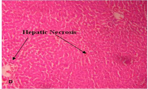
sinusoidal congestion with hepatic damage. (C) CBZ + ALA at a dose of 50 mg/ kg showed fatty degeneration and hepatic necrosis. (D) CBZ + 100 mg/kg ALA showed hepatic necrosis. (E) CBZ + 200 mg/kg ALA revealed normal hepatocytes with central vein and appeared similar to the control liver.
DISCUSSION
The imbalance in the serum oxidant/antioxidant status of epileptic children under antiepileptic monotherapy with CBZ was demonstrated and it was suggested that the hepatotoxic effects might be associated with the side effects of these drugs15. Our study suggests that the toxicity of
CBZ might be secondary to the oxidative stress induced by their metabolites, probably the intermediate arene oxides, formed after biotransformation of aromatic antiepileptic drug and due to disturbance in the GSH metabolism16.
In the present study, we observed the depletion of GSH in liver. Oxidative stress might have occurred as a consequence of disruption of thiol redox circuits, which are controlled by GSH. Liver is the main detoxifying organ for many toxic agents and drugs that contribute to oxidative stress17. The increased ROS
production due to depletion of GSH might lead to oxidative damage and a critical role in the development of hepatic damage18. ALA is
effective in preventing the development of
hepatic damage19,20. ALA is found to be a
potential therapeutic agent in the treatment and prevention of different pathologies that are related to an imbalance of the oxidoreductive cellular status, which occurs in the case of hepatic disorder status21. In addition, several
researchers have recently reported the protective effects of ALA on the liver which is induced by oxidative agents.
CONCLUSION
In the present study, supplementation with ALA decreased the markers of hepatotoxicity such as SGOT, SGPT, bilirubin and increased the levels of albumin and total protein. ALA (50 mg/Kg) showed mild hepatoprotective activity, fatty degeneration and hepatic necrosis. But, ALA (200 mg/Kg) improved the histopathological changes and hepatic damage induced by CBZ. ALA exerts significant protection against CBZ induced toxicity by its ability to decrease the lipid peroxidation and thus oxidative stress through its free radical scavenging activity, which improved the levels of antioxidant defense system.
ACKNOWLEDGEMENT
We wish to thank Dr. V. Madhavan, Principal, M.S. Ramaiah College of Pharmacy for encouraging us to successfully carryout this work.
Table 1: Effect of chronic treatment of carbamazepine and carbamazepine + ALA on liver enzymes, bilirubin, albumin, total protein and liver lipid peroxidation
Values are expressed as mean± SEM of 6 animals. ***( p< 0.001), **(p< 0.01), *(p< 0.05) Vs Control group. +++( p< 0.001), ++(p< 0.01), +(p< 0.05) Vs CBZ group.
SGOT-Serum Glutamate Oxaloacetete Transaminase SGPT-Serum Glutamate Pyruvate Transaminase ALP- Alkaline Phosphatase
LLP-Liver Lipid Peroxidation
Parameters Control Carbamazepine Carbamazepine + ALA 50mg/kg + ALA 100mg/kg Carbamazepine + ALA 200mg/kg Carbamazepine
SGOT 260.5±4.8+++ 378.66±3.59*** 318.66±2.201***,
+++ 295.83±5.61***,+++ 266±1.46+++ SGPT 67.05±1.42+++ 91.47±1.11*** 75.01±0.669***,+++ 72±0.84*,+++ 68.5±0.61+++ ALP 149.5±3.35+++ 252.6±3.44*** 215.66±2.39***,+++ 180.16±6.00***,+++ 157.66±2.04+++
Bilirubin 1.22±0.00 +++ 2.63±0.06*** 1.98±0.118***,+++ 1.72±0.05**,+++ 1.405±0.06+++
Albumin 4.6±0.13+++ 3.02±0.05*** 3.785±0.14***,+++ 3.98±0.15*,+++ 4.36±0.14+++ Total Protein 7.965±0.22+++ 5.15±0.17*** 6.35±0.22***,+++ 6.785±0.058***,+++ 7.5±0.0568+++
Table 2: Effect of CBZ and CBZ + NAC on Superoxide dismutase, catalase and glutathione
Values are expressed as mean± SEM of 6 animals. ***( p< 0.001), **(p< 0.01), *(p< 0.05) Vs Control group. +++( p< 0.001), ++(p< 0.01), +(p< 0.05) Vs Carbamazepine group.
Table 3: Effect of CBZ and CBZ + Alpha Lipoic Acid on body weight, absolute and relative liver weight
Group Body weight in gram Absolute liver weight (g)
Relative liver weight (g) Initial (g) Final (g) % Change
Control 225 268.3±2.1 ↑19.2±0.93+++ 12.45±0.14 +++ 4.64±0.05 +++ Carbamazepine 221.16±2.3 201.6±1.0 ↓8.7±1.03*** 14.16±0.09 *** 7.02±0.04 *** Carbamazepine +ALA
50mg/Kg 226.6±1.0 215±1.52 ↓4.73±0.7 ***,+++ 13.58±0.07***,+++ 6.29±0.06***,+++ Carbamazepine +ALA
100mg/Kg 209.16±4.3 206.6±4.0 ↓1.16±0.52 ***,+++ 12.85±0.13*,+++ 6.22±0.15***,+++ Carbamazepine +ALA
200mg/Kg 225±2.5 221±3.3 ↓1.5±0.47 ***,+++ 12.7±0.21***,+++ 5.81±0.12***,+++ Values are expressed as mean± SEM of 6 animals. ***( p< 0.001), **(p< 0.01), *(p< 0.05) Vs
Control group. +++( p< 0.001), ++(p< 0.01), +(p< 0.05) Vs Carbamazepine group.
Fig. a): control
Groups
Superoxide Dismutase (superoxide anion reduced/mg
protein/min)
Catalase (µmolH2O2degraded/ mg
protein/min)
Glutathione (g/dl)
Control 6.35±0.2455+++ 60.033±0.88+++ 17.43±0.61+++
Carbamazepine
(50 mg/Kg) 2.023±0.197*** 37.47±0.58*** 10.585±0.42***
Carbamazepine
+ ALA 50 mg/Kg 3.88±0.168***+++ 42.6±0.35***+++ 12.69±0.20***+++
Carbamazepine
+ ALA 100mg/Kg 4.6±0.202***+++ 52.83±1.09***+++ 13.67±0.32***+++
Carbamazepine
+ ALA 200 mg/Kg 5.58±0.096+++ 58.24±0.702+++ 14.64±0.25***+++
Normal hepatic
architecture
Fig. b): Carbamazepine (50 mg/Kg)
Effect of ALA on CBZ induced alterations in liver histopathology
Fig. c): Carbamazepine + ALA (50 mg/Kg)
Fig. d): Carbamazepine + ALA (100 mg/Kg)
Sinusoidal
congestion
Haemorrhage
Centrilobular
congestion
Fatty Degeneration and Hepatic
Necrosis
B
Fig. 1: Micrograph showing effect of CBZ on hepatocytes. (A) Control liver showed normal hepatic architecture.
(B) carbamazepine treated group showed haemorrhage, centrilobular and sinusoidal congestion. (C) Carbamazepine + ALA at a dose of 50 mg/ kg showed fatty degeneration and hepatic necrosis.
(D) Carbamazepine + 100 mg/kg ALA showed hepatic necrosis.
(E) Carbamazepine + 200 mg/kg ALA showed normal hepatocytes with central vein.
REFERENCES
1. Ahmed SN and Siddiqi ZA. Antiepileptic drugs and liver disease. Seizure. 2006;15:156.
2. Walia KS, Khan EA, Ko DH, Raza SS and Khan YN. Side effects of antiepileptics - a review. Pain prac. 2004;4:194. 3. Forbes GM, Jeffrey GP, Shilkin KB and
Reed WD. Carbamazepine hepatotoxicity: another cause of the vanishing bile duct syndrome. Gastroenterol. 1992;102:1385.
4. Zucker P, Daum F and Cohen MI. Fatal carbamazepine hepatitis. J Pediatr. 1977;91:667.
5. Mitchell MC, Boitnott JK, Arregui A and Maddrey WC. Granulomatous hepatitis associated with carbamazepine therapy. Am J Med. 1981;71:733.
6. Azzi KJA and Kelly DF. Free radical biology - terminology and critical thinking. FEBS Lett. 2004;558:3. 7. Nitti M, Pronzato MA, Marinari UM and
Domenicotti C. PKC signaling in oxidative hepatic damage. Mol Aspects Med. 2008;29:36.
8. Ravikumar S and Gnanadesigan M. Hepatoprotective and antioxidant activity of a mangrove plant Luminitzera racemosa. Asian Pac J Trop Med. 2011;1:348.
9. Santos NAG, Medina WSG, Martins NM, Rodrigues MAC, Curti C and Santos AC. Involvement of oxidative stress in the hepatotoxicity induced by aromatic antiepileptic drugs. Toxicology In Vitro. 2008;22:1820.
10. Bavdekar SB, Muranjan MN, Gogtay NJ, Kantharia V and Kshirsagar NA. Anticonvulsant hypersensitivity syndrome: lymphocyte toxicity assay for the confirmation of diagnosis and risk assessment. Ann Pharmacother. 2004;38:1648.
11. Chang TKH and Abbott FS. Oxidative stress as a mechanism of valproic acid-associated hepatotoxicity. Drug Metab Rev. 2006;38:627.
12. Sedlak J and Lindsay RH. Estimation of total, protein bound & non-protein SH groups in tissue with Ellman’s reagent. Anal Biochem. 1968;25:192.
13. Mihara M and Uchiyama M. Determination of malonaldehyde precursors in tissues by thiobarbituric acid test. Anal Biochem. 1978;86:271. 14. Li Y, Powers C, Jiang N and Chopp M.
Intact, injured, necrotic and apoptotic cells after focal cerebral ischemia in the rat. J Neurol Sci. 1998;156:119.
antioxidative balance in epileptic children. Eur Neurol. 2007;57:65. 16. Kalapos MP. Carbamazepine-provoked
hepatotoxicity and possible aetiopathological role of glutathione in the events. Retrospective review of old data and call for new investigation. Adverse Drug React Toxicol Rev. 2002;21:123.
17. Stevens MJ, Obrosova I, Coa X, Huysen CV and Greene DA. Effects of DL-α-lipoic acid on peripheral nerve conduction, blood flow, energy metabolism and oxidative stress in experimental diabetic neuropathy. Diabetes. 2000;49:1006.
18. Gorąca A, Huk-Kolega H, Piechota A, Kleniewska P, Ciejka E and Skibska B. Lipoic acid- biological activity and therapeutic potential. Pharmacol Rep. 63:2011;849.
19. Min AK, Kim MK, Seo HY, Kim HS, Jang BK, Hwang JS, Choi HS, Lee KU, Park KG and Lee IK. Alpha-lipoic acid inhibits hepatic PAI-1 expression and fibrosis by inhibiting the TGF- signaling pathway. Biochem Biophys Res Commun. 2010;393:536.
20. Park KG, Min AK, Koh EH, Kim HS, Kim MO, Park HS, Kim YD, Yoon TS, Jang BK, Hwang JS, Kim JB, Choi HS, Park JY, Lee IK and Lee KU. Alpha-lipoic acid decreases hepatic lipogenesis through adenosine monophosphate-activated protein kinase (AMPK)-dependent and AMPK-independent pathways. Hepatology. 2008;48:1477.