1
"This is an un-copyedited authored manuscript copyrighted by the
1
American Association for Clinical Chemistry (AACC). This may not be
2duplicated or reproduced, other than for personal use or within the rule of
3'Fair Use of Copyrighted Materials' (section 107, Title 17, U.S. Code)
4without permission of the copyright owner, AACC. The AACC disclaims
5any responsibility or liability for errors or omissions in this version of the
6manuscript or in any version derived from it by the National Institutes of
7Health or other parties. The final publisher-authenticated version of the
8article is available at
http://www.clinchem.org
.”
910
Ultra-sensitive mutation detection and genome-wide DNA copy
11number reconstruction by error corrected circulating tumor DNA
12sequencing
1314
Sonia Mansukhani1*, Louise J. Barber1*, Dimitrios Kleftogiannis1, Sing Yu 15
Moorcraft2, Michael Davidson2, Andrew Woolston1, Paula Zuzanna Proszek3,
16
Beatrice Griffiths1, Kerry Fenwick4, Bram Herman5, Nik Matthews4, Ben O’Leary6,
17
Sanna Hulkki3, David Gonzalez De Castro7, Anisha Patel8, Andrew Wotherspoon9,
18
Aleruchi Okachi2, Isma Rana2, Ruwaida Begum2, Matthew N. Davies1,10, Thomas
19
Powles11, Katharina von Loga1, Michael Hubank3, Nick Turner6,12, David Watkins2,
20
Ian Chau2, David Cunningham2, Stefano Lise1, Naureen Starling2 and Marco 21
Gerlinger1,2
22
23
Running title: Error corrected circulating tumor DNA sequencing 24
25
2 1Centre for Evolution and Cancer, Division of Molecular Pathology, The Institute of 27
Cancer Research, London, United Kingdom.
28
2Gastrointestinal Cancer Unit, The Royal Marsden NHS Foundation Trust, London 29
and Sutton, United Kingdom.
30
3Centre for Molecular Pathology, The Royal Marsden NHS Foundation Trust, Sutton, 31
United Kingdom.
32
4Tumour Profiling Unit, The Institute of Cancer Research, London, United Kingdom. 33
5Diagnostics and Genomics Group, Agilent Technologies Inc., Santa Clara, USA 34
6Breast Cancer Now Research Centre, The Institute of Cancer Research, London, 35
United Kingdom.7Centre for Cancer Research and Cell Biology, Belfast, United
36
Kingdom.
37
8Department for Radiology, The Royal Marsden NHS Foundation Trust, London and 38
Sutton, United Kingdom.
39
9Department of Histopathology, The Royal Marsden NHS Foundation Trust, London 40
and Sutton, United Kingdom.
41
10Current address: Achilles Therapeutics, Francis Crick Institute, London, United 42
Kingdom.
43
11Barts Cancer Institute, Queen Mary University of London, London, United 44
Kingdom.
45
12Breast Cancer Unit, The Royal Marsden NHS Foundation Trust 46
*These authors contributed equally to this work
47 48
Corresponding Author: 49
Dr Marco Gerlinger
50
Centre for Evolution and Cancer, The Institute of Cancer Research
3
237 Fulham Road, London SW3 6JB, United Kingdom
52
Tel: +44 207 153 5234
53
email: [email protected]
54 55
Keywords: cancer genomics, circulating tumor DNA, liquid biopsy, molecular
56
barcodes, sequencing error correction.
57 58
Abstract 59
Background: Circulating free DNA sequencing (cfDNA-Seq) can portray cancer
60
genome landscapes but highly sensitive and specific technologies are necessary to
61
accurately detect mutations with often low variant frequencies.
62
Methods: We developed a customizable hybrid-capture cfDNA-Seq technology
63
using off-the-shelf molecular barcodes and a novel duplex DNA-molecule
64
identification tool for enhanced error correction.
65
Results: Modelling based on cfDNA-yields from 58 patients showed this technology,
66
requiring 25 ng cfDNA, could be applied to >95% of patients with metastatic
67
colorectal cancer (mCRC). cfDNA-Seq of a 32-gene/163.3kbp target region detected
68
100% of single nucleotide variants with 0.15% variant frequency in spike-in
69
experiments. Molecular barcode error correction reduced false positive mutation
70
calls by 97.5%. In 28 consecutively analyzed patients with mCRC, 80 out of 91
71
mutations previously detected by tumor tissue sequencing were called in the cfDNA.
72
Call rates were similar for point mutations and indels. cfDNA-Seq identified typical
73
mCRC driver mutations in patients where biopsy sequencing had failed or did not
74
include key mCRC driver genes. Mutations only called in cfDNA but undetectable in
75
matched biopsies included a subclonal resistance driver mutation to anti-EGFR
4
antibodies in KRAS, parallel evolution of multiple PIK3CA mutations in two cases,
77
and TP53 mutations originating from clonal hematopoiesis. Furthermore, cfDNA-Seq
78
off-target read analysis allowed simultaneous genome-wide copy number profile
79
reconstruction in 20 of 28 cases. Copy number profiles were validated by
low-80
coverage whole genome sequencing.
81
Conclusions: This error-corrected ultra-deep cfDNA-Seq technology with a
82
customizable target region and publicly available bioinformatics tools enables broad
83
insights into cancer genomes and evolution.
5 Introduction
85
Many tumors release cell free DNA (cfDNA) into the circulation, allowing the
86
analysis of cancer genetic aberrations from blood samples [1-6]. Such ‘liquid
87
biopsies’ can inform tailored therapies [7] or predict recurrences after surgery [8, 9].
88
cfDNA analysis also permits subclonal mutation detection that is often missed by
89
biopsies due to spatial intratumor heterogeneity [10, 11]. Genetic techniques with
90
high analytical sensitivity and low false positive error rates are crucial for accurate
91
cfDNA-Seq due to low tumor-derived cfDNA fractions and low abundances of
92
subclonal mutations. Digital droplet PCR (ddPCR) and BEAMing assays can
93
accurately detect point mutations present at frequencies ≤0.1% but are restricted to
94
the analysis of a small number of genomic loci [8, 12]. Targeted next generation
95
sequencing (NGS) can interrogate larger regions such as gene panels but the error
96
rate of NGS complicates the calling of mutations with variant allele frequencies
97
(VAFs) <5% [13]. Error correction through random molecular barcodes (MBC) has
98
been incorporated into NGS cfDNA assays to reduce this error rate [14, 15] and has
99
enabled mutation calling with VAFs ≤0.1%. However, these methods have often
100
used amplicon sequencing, which can hamper coverage of entire genes due to
101
primer design restrictions. Some methods have employed solution hybrid-capture,
102
which is ideal to target entire genes, but used bespoke or proprietary rather than
off-103
the-shelf reagents and publicly available bioinformatics tools, limiting their broad
104
application for clinical or research purposes.
105
Here we assessed how novel, commercially available off-the-shelf MBC
106
reagents combined with customized capture-based target enrichment technology
107
could be optimized for ultra-deep error-corrected cfDNA-Seq. We developed a
6
duplex-DNA molecule calling tool to improve the calling accuracy and assessed
109
concordance of mutation calls from cfDNA with clinical grade tumor tissue
110
sequencing in patients with metastatic colorectal cancer (mCRC).
111
METHODS 112
Patients and samples 113
Plasma samples and clinical data were available from the FOrMAT trial
114
(Feasibility Of Molecular characterization Approach to Treatment [16], Chief
115
Investigator: N Starling ClinicalTrials.gov NCT02112357). Healthy donor (HD) cfDNA
116
was obtained through the Tissue Collection Framework to Improve Outcomes in
117
Solid Tumours (Chief Investigator: T Powles). Both trials were approved by UK
118
ethics committees and all patients provided written informed consent. Details of
119
clinical trials, patients, samples, sample processing and experimental techniques are
120
provided in the online Supplemental Methods file.
121 122
cfDNA sequencing 123
SureSelectXT-HS (Agilent) was used to prepare sequencing libraries using our
124
optimized protocol (online Supplemental Methods file) and a custom designed
125
SureSelect bait-library (online Supplemental Table 1). Sequencing libraries were
126
clustered using the cBot and sequenced with paired-end 75 reads on an Illumina
127
HiSeq2500 in rapid mode.
128
SureCall software (version 4.0.1.45, Agilent) was used to trim and align fastq
129
reads to the hg19 reference genome with default parameters and for MBC
de-130
duplication, permitting one base mismatch within each MBC. Consensus families
7
comprising of single reads were removed, on-target depths were assessed and
132
variants were called with SureCall SNPPET.
133
To identify variants supported by duplexes we developed the freely available
134
duplexCaller bioinformatics tool [17].
135
All variant positions identified in patient cfDNA were assessed in six HD
136
samples using bam-readcount [18]. Most called variants were absent in HD samples
137
(online Supplemental Table 2) but mutations with VAF less than double that of an
138
identical variant in HD were removed as false positives.
139
BAM files resulting from MBC de-duplication before removal of single-read
140
consensus families were used to generate genome-wide DNA copy number profiles
141
with CNVkit [19], with Antitarget average size set to 30 kb. HD samples were used
142
as the normal reference pooled dataset.
143
144
Low coverage whole genome sequencing (lcWGS) 145
Genomic libraries were constructed from 10 ng cfDNA with the NEBNext Ultra
146
II kit and sequenced with 100bp single-end reads on HiSeq2500 in rapid mode
147
(0.42x median coverage). Data was aligned (hg19 reference genome) with Bowtie
148
(v0.12.9), and processed as described [20]. logRatios were normalised against a
149
gender-matched pooled dataset from HD cfDNA (9 male, 8 female) before
150
segmentation and median centering.
151
152
8
ddPCR was performed in case 8 (BRAF V600E) and to validate discordant
154
variants between cfDNA and tumor tissue. 4 of 11 such cases had sufficient
155
remaining cfDNA to validate subclonal variants (online Supplemental Methods file).
156
157
cfDNA sequencing with a commercial kit 158
25 ng, 17 ng and 25 ng cfDNA (cases 3, 15, and 23, respectively) were
159
processed using the Roche AVENIO Expanded kit as per the manufacturer’s
160
protocol. Libraries were sequenced with 151 bp paired-end reads on Illumina
161
NextSeq500 to 2,689-6,420x depth after de-duplication. Data was analyzed using
162
the Roche AVENIO ctDNA Analysis Software v1.0.0 with default parameters.
163 164
RESULTS 165
cfDNA sequencing optimization 166
Modelling based on cfDNA yields from 58 patients with mCRC showed that 25
167
ng of cfDNA could be extracted from 20-30 ml blood from >95% of cases (online
168
Supplemental Figure 2C). 25 ng was therefore chosen as our standard cfDNA input
169
quantity. We designed a solution hybrid-capture panel targeting 32 genes including
170
all major CRC driver genes, (163.3 kb, online Supplemental Table 1) and used
171
Agilent SureSelectXT-HS kit, which tags each DNA strand with a random 10-base
172
MBC, for sequencing library preparation. The SureSelectXT-HS protocol was optimized
173
to perform reliably with 25 ng cfDNA input (online Supplemental Methods file). The
174
fraction of on-target reads is usually low when using small targeted sequencing
175
panels and low input DNA, so we first assessed how the on-target fraction could be
176
optimized by varying the stringency of the post-capture wash. Two library
9
preparations were started in parallel from each of four cfDNA samples, using the 1.5
178
h fast-hybridization protocol. Then, post-capture washes were performed at 65°C in 179
one library and at 70°C in the other. Sequencing generated similar read numbers 180
(65°C: 92,820,887; 70°C: 102,582,694 median reads/sample) and the on-target
181
fraction significantly (p=0.0011) increased from 30-35% to 71-74% with the 70°C 182
protocol (Figure 1A). Hence, the more stringent conditions were chosen for our
183
standard protocol. Target exon coverage was even with this solution hybrid-capture
184
technique and was not subject to the gaps commonly seen with commercial
185
amplicon sequencing designs (online Supplemental Figure 3). This would be
186
particularly advantageous for the analysis of tumor suppressor genes where driver
187
mutations often spread across large parts of the gene.
188
We next used MBCs to de-duplicate sequencing data and perform error
189
correction. SureCall creates families of reads with matched MBC that also align to
190
the same genomic position and then identifies the most likely consensus sequence
191
for each family (Figure 1B). This reduces random errors arising during PCR and
192
sequencing, as these are not common to all reads of a family. Consensus families
193
contained a median of 8 to 15 supporting reads in samples sequenced with the
194
optimized protocol (online Supplemental Figure 4), which was within the optimal
195
range for barcode error correction [21]. After MBC de-duplication, the median
on-196
target depth with the 70oC protocol was 1,782x. This was theoretically sufficient to
197
achieve a detection limit as low as 1 mutated DNA fragment in 1,782 molecules
198
(0.056%). However, the analytical sensitivity for de novo mutation detection is lower
199
in practice since more than one read is required to support robust bioinformatics
200
calling. Thus, we designed a mixing experiment to test the ability to detect and
201
bioinformatically call mutations with low VAFs.
10 Assay sensitivity and specificity
203
cfDNA from two donors that differed in 16 homozygous single nucleotide
204
polymorphisms (SNPs) within the targeted region were used to prepare a dilution
205
series with 0.15%, 0.075% and 0.0375% cfDNA from donor A spiked into cfDNA
206
from donor B. Sequencing a median of 74,030,118 reads/sample generated a
207
median on-target depth of 21,651x before de-duplication. Data from each sample
208
was then processed in two ways: first, we used MBCs for de-duplication and calling
209
of consensus sequences; second, we performed standard de-duplication using only
210
the genomic position of each read pair. The median on-target depth was higher after
211
MBC de-duplication (MBC 2,420x versus 1,587x with standard de-duplication; Figure
212
1C). This was anticipated as different MBCs tag distinct DNA fragments that would
213
otherwise be counted as duplicates. For example, the forward and reverse strands of
214
each original ‘duplex’ dsDNA molecule were separately tagged by MBC and so were
215
retained as independent consensus families (Figure 1D). Standard de-duplication
216
cannot distinguish these reads from PCR duplicates.
217
We first investigated whether the spiked-in SNPs could be re-identified in the
218
MBC de-duplicated BAM files using the Integrative Genomics Viewer (IGV) [22] and
219
tried to understand patterns associated with true positive variants. All 16 SNPs were
220
detected in the 0.15% mix, 14/16 at 0.075% and 11/16 at 0.0375% mixing ratios
221
(Figure 1E). Thus, our ultra-deep cfDNA-Seq assay allowed robust detection of
222
variants at 0.15% and retained a high detection capability at 0.075%. We then
223
assessed if MBC error correction improved the bioinformatics calling accuracy of
224
ultra-low frequency variants, which is more challenging than re-identification of
225
known variants. While interrogating sequencing data manually in IGV, we had
226
observed that all true variants were at least supported by two consensus families
11
mapping to the same genomic position but differing in whether the variant was seen
228
in read 1or read 2 in paired-end sequencing (Figure 1D). These were highly likely to
229
represent the forward and reverse strand of the double-stranded input cfDNA
230
molecule as observed previously [15]. Based on this observation, we developed the
231
duplexCaller bioinformatics tool that identified variants supported by duplex reads
232
(online Supplemental Methods file) and added the requirement for such a
‘duplex-233
configuration’ to be present to accept a mutation as genuine. The presence of a
234
variant in at least one additional family with a different alignment position was also
235
added to the post-call filters to assure high specificity. Thus, a variant had to be
236
present in ≥3 consensus DNA families in order to be accepted as a mutation call in 237
the MBC de-duplicated data. For a meaningful comparison, mutations in the
238
standard de-duplicated data were also required to be present in ≥3 reads. 239
We then compared SureCall calls for the mixing experiment on standard-
240
versus MBC-deduplicated data and quantified how many of the homozygous SNPs
241
from sample A that were present at 0.15% in the cfDNA mixture were called.
242
Although samples A and B differed at 16 homozygous SNP positions, only the 9
243
variant SNPs in spiked-in sample A could be assessed for capability to call at low
244
frequency against the reference genome. The other 7 SNPs were reference
wild-245
type in spiked-in sample A and so could not be called. Mutation calling after standard
246
de-duplication with low stringency caller settings (variant call quality threshold
247
[VCQT]=40) detected 5/9 homozygous SNPs (Figure 1F) but also generated 156
248
additional calls. These additional variants were likely false positives, since they had
249
not been identified by deep sequencing of the individual cfDNA samples used in the
250
mixing experiment. Stepwise increase of the VCQT reduced false positives but this
251
was accompanied by a loss of analytical sensitivity. When the same data were called
12
using MBCs and a low stringency VCQT=40 (Figure 1F), 4 of the spiked-in SNPs
253
were called with only 2 likely false positive variants. We assessed why calling with
254
MBC error correction failed to identify the 5 other SNPs. Each of these had VAFs
255
<0.1% when visualized in IGV [23], which was below the minimum VAF of 0.1% that
256
can be called by SureCall. We also assessed the number of false positive calls in
257
standard de-duplicated data at the maximum VCQT that identified the same four true
258
positive variants detected with MBC: 81 likely false positives were called compared
259
to just 2 using MBC. Hence at the same analytical sensitivity, de-duplication using
260
the MBCs dramatically decreased false positives by 97.5%. Mutation calling in 6 HD
261
samples subjected to cfDNA-Seq only identified heterozygous and homozygous
262
SNPs but no mutations with lower frequency (online Supplemental Table 3), further
263
supporting the high analytical specificity of this MBC technology.
264
Concordance of cfDNA- and tumor-sequencing in mCRC patients 265
cfDNA from 28 patients with mCRC were consecutively analyzed. Seven were
266
sequenced with the 65°C protocol and 21 with the 70°C protocol. The median 267
sequencing depth was higher with 70°C (2,087x) than 65°C (1,205x) (Figure 2A). 268
We then analyzed the concordance and discordance of mutation calls within
269
the target regions common to the tumor biopsy sequencing assay and our
cfDNA-270
Seq panel. Biopsies of 23 cases had been sequenced with the FOrMAT NGS panel
271
(online Supplemental Table 4) and four biopsies had been subjected to routine
272
clinical amplicon sequencing of 5 genes (BRAF, KRAS, NRAS, PIK3CA and TP53).
273
One case had failed tissue sequencing.
274
88% (80/91) of all mutations that had been found by tumor sequencing were
275
called in the cfDNA (Figure 2A). All 11 mutations not called in cfDNA were from only
13
3 cases. Inspection of the sequencing data on IGV revealed that 5/11 mutations
277
were present in cfDNA at VAFs below the SureCall detection limit (Figure 2B).
278
Sufficient cfDNA remained from case 8 for orthogonal analysis by ddPCR. Using
279
manufacturer-validated ddPCR-probes for the BRAF V600E mutation we identified
280
2,830 wild type DNA fragments but no mutated fragments (data not shown). This
281
confirmed that the absence of sufficiently abundant tumor-derived cfDNA molecules,
282
rather than technical failure, explained the inability to detect mutations.
283
We next assessed mutations called by cfDNA-Seq in genes that had not been
284
sequenced in corresponding tumor tissue. APC mutations were detected in each of 4
285
cases whose tumors had only been analyzed with the 5-gene amplicon panel (Figure
286
2A). Furthermore, one mutation was found in each of FBXW7, CTNNB1, TCF7L2, 287
ATM and SMAD4. We also detected mutations in APC, TP53 and KRAS in case 28
288
that had failed prior tumor tissue sequencing attempts. In total, 11 of these 13
289
mutations (85%) encoded protein changes previously reported in the COSMIC
290
cancer mutation database [24] and all variants in the tumour suppressor genes APC 291
and FBXW7 were truncating and hence likely driver mutations. This demonstrated
292
that our assay could detect biologically and clinically important cancer mutations
293
directly from cfDNA.
294
We then investigated mutations that had been called in cfDNA but were
295
absent when the same gene had been analyzed in tumor tissue: 7 in TP53, 7 in
296
ATM, 3 in PIK3CA, 2 in SMAD4 and one each in KRAS, FBXW7 and TCF7L2. All
297
four mutations called in the oncogenes KRAS and PIK3CA were canonical cancer
298
driver mutations. 8/18 mutations (44%) located in tumor suppressor genes were
299
nonsense mutations or encoded for amino acid changes found recurrently in cancer
300
[24], suggesting that these were also driver mutations. Together, 54.5% (12/22) of
14
variants detected only in cfDNA were likely cancer driver mutations. The VAFs of
302
mutations that were only detected in cfDNA but not in tumor tissue were a mean
303
105-fold lower than the VAF of the most abundant mutation detected in the same
304
cfDNA sample (online Supplemental Figure 1); these variants likely originated from
305
small cancer subclones. However, two TP53 mutations present in cfDNA but not in
306
matched tumor tissue (Cases 9, 13) were also detected with similar VAF in DNA
307
from blood cells (online Supplemental Table 5). These TP53 mutations hence
308
originated from a clonal expansion of blood cells [9], termed clonal hematopoiesis
309
[25, 26].
310
An activating mutation in KRAS (Q61H) was detected with a VAF of 0.37% in
311
cfDNA but not in the matched tumor (case 10). This was the only patient that had
312
received treatment with the anti-EGFR antibody cetuximab prior to blood collection
313
and the KRAS mutation was likely a driver of acquired resistance that evolved during
314
therapy [27]. ddPCR testing of cfDNA provided orthogonal validation (Figure 2C),
315
showing that our technology is suitable for the detection of subclonal resistance
316
driver mutations. Suspected driver mutations in PIK3CA were frequently discordant
317
with 3/7 mutations only detectable in cfDNA (E545K, H1046R, R1023*). Two cases
318
(17,26) harbored parallel evolution events, as further activating PIK3CA mutations
319
were present in the tumors and the cfDNA. These results are consistent with studies
320
showing that intratumor heterogeneity of PIK3CA mutations is common in mCRCs
321
whereas heterogeneity is rare for mutations in APC and, in tumors not previously
322
treated with anti-EGFR antibodies, for KRAS, NRAS and BRAF mutations [28].
323
Mutations in ATM tumor suppressor gene were called in 8/28 cfDNA samples.
324
Sequencing of matched tumor showed wild-type sequence in seven of these and
325
one tumor had only been sequenced with the 5-gene panel. All ATM mutations had
15
low VAFs (median: 0.17%) and only 2/8 encode protein changes previously
327
catalogued in cancer [24], making it difficult to interpret their functional relevance. No
328
ATM mutations were called in 6 healthy donors, indicating that the mutation calls in
329
cfDNA from mCRC patients are unlikely the result of a high false positive call rate in
330
this gene.
331
Next, we used ddPCR to validate further subclonal mutations called in cfDNA
332
but not in tumor tissue. All subclonal variants with VAF <2% from samples where
333
sufficient cfDNA material was available and where a custom ddPCR-assay could be
334
designed were assessed (online Supplemental Methods file). ddPCR validated all 6
335
tested mutations and VAFs were similar to those found by our error-corrected cfDNA
336
technology (Figure 2D, online Supplemental Table 6).
337
Additionally, we re-sequenced three cfDNA samples containing low VAF
338
(<2%) mutations (cases 3, 15, 23) with the commercially available AVENIO ctDNA
339
kit. 9/10 point mutations in genes targeted by both panels were concordant (online
340
Supplemental Table 7). The low frequency TP53 R175H variant in case 3 was not
341
called by AVENIO software but was seen to be present upon manual review of the
342
BAM file. Three indels in APC (cases 3,23) were not called by AVENIO analysis.
343
This comparison further confirmed the reliable performance of our customizable
344
cfDNA assay.
345
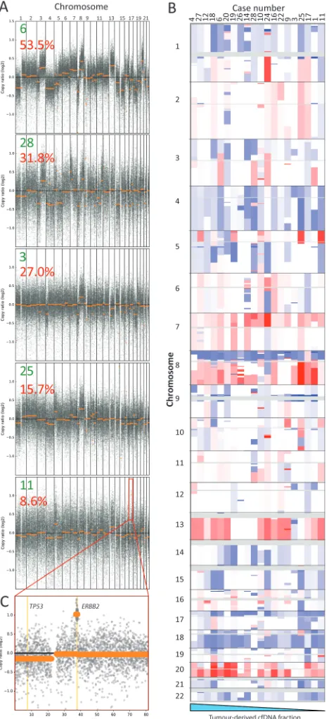
Genome wide DNA copy number aberration analysis 346
We finally assessed if we could maximise the information gain from a targeted
347
cfDNA assay through simultaneous reconstruction of genome-wide copy number
348
aberration (CNA) profiles. Applying the CNVkit-package [19] that uses off-target
349
reads to infer copy number changes, we generated genome-wide CNA profiles for
16
20/28 cases (71%) (Figure 3A-B). Chromosome arm losses (Chr17p and 18q) and
351
gains (Chr1q, 7, 8q, 13 and 20), which are typical for mCRC, were observed [29]. All
352
8 samples with a flat CNA profile had very low maximum VAFs ≤5.6%. A high-level
353
targetable amplification involving the ERBB2 oncogene was detected despite a low
354
tumor-derived cfDNA fraction (8.6% VAF) in case 11 (Figure 3C). This amplification
355
had also been detected in the matched tumor, validating the ability to profile CNAs
356
with our cfDNA-Seq technology. No other amplifications had been detected in tumor
357
biopsies with the FOrMAT NGS panel. Low-coverage whole genome sequencing is
358
an established approach for genome wide copy number profiling and we applied this
359
to 18 samples with sufficient cfDNA. This independent validation showed a median
360
weighted Spearman correlation of 0.886 with the profiles generated from cfDNA-Seq
361
using CNVkit (online Supplemental Figure 5).
362
DISCUSSION 363
Our ultra-deep and error-corrected cfDNA-Seq protocol that uses off-the-shelf
364
MBCs in combination with a custom-designed solution hybrid capture panel detected
365
100% of the known variants with VAFs of 0.15% in a mixing experiment. The use of
366
MBC error correction and the requirement for variants to be supported by a
duplex-367
pair of consensus families reduced false positive mutation calls by 97.5% while
368
maintaining true positives. We developed the DuplexCaller bioinformatics tool, which
369
can be run directly after MBC de-duplication to facilitate mutation calling; all
370
bioinformatics tools for the analysis of data generated with this technology are hence
371
freely available. Our approach did not rely on background error correction models
372
that are constructed from large numbers of healthy donor samples and are therefore
373
impractical for applications requiring frequently changing custom gene panels,
374
includingclinical assay development.
17
Importantly, the 1.5 h fast-hybridization step (standard protocol: 16h) used in
376
our assay dramatically reduces library preparation time which is advantageous when
377
fast turnaround is critical. Increasing the wash temperature after capture dramatically
378
reduced off target reads. The higher temperature likely relaxes the target/bait-bond
379
in hybridised molecules with a higher number of mismatches, reducing the
non-380
specific carry over of DNA fragments into the library.
381
cfDNA-Seq of 28 mCRC patients demonstrated that 88% of mutations
382
detected by clinical grade tumor tissue sequencing were also called in cfDNA. This
383
detection capability is similar to that reported for MBC-error corrected cfDNA-Seq
384
with a 5-gene assay using amplicons (87.2%) [1] and a 54-gene assay using
target-385
capture (85%) [14, 30]. Furthermore, indels are more difficult to call than point
386
mutations. Yet, our cfDNA assay called 23/26 indels (88.5%) that were known based
387
on tumor sequencing, showing a similar performance to point mutation detection
388
(87.7% called).
389
cfDNA-Seq detected several additional driver mutations not reported by tumor
390
sequencing. Seven were in TP53. Two were also observed in the matched blood
391
cells, indicating that they originated from clonal hematopoiesis. The discovery of
392
clonal hematopoiesis in 7% of our cohort demonstrates the importance of
393
sequencing DNA extracted from blood cells to avoid misinterpreting such variants as
394
cancer-associated mutations. In one patient who received cetuximab therapy, we
395
detected a KRAS Q61H variant that was absent from the matched tumor and likely
396
represents the evolution of a drug resistant subclone. Multiple PIK3CA activating
397
mutations detected in two anti-EGFR therapy naive patients represent parallel
398
evolution events. These examples show that our cfDNA assay can provide insights
399
into cancer evolution. Because the minimally invasive nature of cfDNA-Seq allows
18
application at multiple time-points, this could be used to monitor the evolution of
401
subclonal drug resistance driver mutations without prior knowledge of specific loci
402
where resistance mutations will occur. We finally demonstrate that cfDNA-Seq allows
403
genome-wide CNA reconstruction and validate this against low-coverage genome
404
sequencing. As the number of targeted therapies increases, custom target
405
enrichment panels that can be readily adapted and scaled for the tumor type and
406
therapeutic agent in question could be used to investigate the full tumor genomic
407
landscape of point mutations, indels and CNAs. This would facilitate the identification
408
of novel resistance mechanisms. Importantly, this ultra-sensitive cfDNA-Seq
409
technology can also address the subset of 20% of patients with mCRC who cannot
410
be molecularly profiled due to unobtainable or inadequate biopsy tissues [16, 31].
411
In conclusion, this cfDNA-Seq approach with customizable and off-the-shelf
412
reagents showed a similar performance to published techniques that use bespoke
413
reagents and more complex analyses.
414 415 416 417
418
419
420
19 Data access
422
Sequencing fastq files have been deposited into the NCBI Sequence Read
423
Archive (SRA submission code SUB3510375).
424 425
Acknowledgements 426
We would like to thank all patients participating in the FOrMAT clinical trial
427
and the clinical research team members at the Royal Marsden Hospital who
428
supported the sample collection. The study was supported by charitable donations
429
from Tim Morgan to the Institute of Cancer Research, from Philip Moodie to The
430
Royal Marsden Cancer Charity and by a Clive and Ann Smith Fellowship. The study
431
received funding by Cancer Research UK, a Wellcome Trust Strategic Grant
432
(105104/Z/14/Z), the Royal Marsden Hospital/Institute of Cancer Research National
433
Institute for Health Research Biomedical Research Centre for Cancer and by a
434
Cancer Research UK Clinical PhD Studentship.
435 436
Disclosure Declaration 437
The authors had pre-marketing access to Agilent SureSelectXT-HS reagents. BH is an
438
employee of Agilent. The other authors received no financial support or
439
compensation from Agilent.
440 441
Statement of Author Contributions 442
SM, LJB and MG conceived the study and wrote the manuscript; SM, LJB and BG
443
processed samples; SM, LJB and BH developed the cfDNA-Seq assay; DK and SL
444
developed the DuplexCaller tool; SM, LJB, DK, AW, MND and MG analyzed the
445
data; SYM, MD, AP, AO, IR, RB, DW, AWoth, KvL, IC, DC, NS and TP provided
20
clinical data and samples. KF and NM sequenced the cfDNA libraries, PZP, DGDC,
447
SH and MH provided tumor biopsy sequencing data from the FORMAT panel and
448
ran the Avenio analysis, NT and BOL provided support for ddPCR.
449 450
Sonia Mansukhani1*, Louise J. Barber1*, Dimitrios Kleftogiannis1, Sing Yu
451
Moorcraft2, Michael Davidson2, Andrew Woolston1, Paula Zuzanna Proszek3, 452
Beatrice Griffiths1, Kerry Fenwick4, Bram Herman5, Nik Matthews4, Ben O’Leary6,
453
Sanna Hulkki3, David Gonzalez De Castro7, Anisha Patel8, Andrew Wotherspoon9,
454
Aleruchi Okachi2, Isma Rana2, Ruwaida Begum2, Matthew N. Davies1,10, Thomas 455
Powles11, Katharina von Loga1, Michael Hubank3, Nick Turner6,12, David Watkins2,
456
Ian Chau2, David Cunningham2, Stefano Lise1, Naureen Starling2 and Marco 457
Gerlinger1,2
21 References
1. Bettegowda C, Sausen M, Leary RJ, Kinde I, Wang Y, Agrawal N, et al. Detection of circulating tumor DNA in early- and late-stage human malignancies. Sci Transl Med 2014, 6(224):224ra224.
2. Siravegna G, Marsoni S, Siena S, Bardelli A: Integrating liquid biopsies into the management of cancer. Nat Rev Clin Oncol 2017, 14(9):531-548.
3. Heitzer E, Ulz P, Belic J, Gutschi S, Quehenberger F, Fischereder K, et al. Tumor-associated copy number changes in the circulation of patients with prostate cancer identified through whole-genome sequencing. Genome Med 2013, 5(4):30.
4. Haber DA, Velculescu VE: Blood-Based Analyses of Cancer: Circulating Tumor Cells and Circulating Tumor DNA. Cancer Discov 2014, 4(6):650-661.
5. Leary RJ, Sausen M, Kinde I, Papadopoulos N, Carpten JD, Craig D, et al. Detection of chromosomal alterations in the circulation of cancer patients with whole-genome sequencing. Sci Transl Med 2012, 4(162):162ra154.
6. Dawson SJ, Tsui DW, Murtaza M, Biggs H, Rueda OM, Chin SF, et al. Analysis of circulating tumor DNA to monitor metastatic breast cancer. N Engl J Med 2013, 368(13):1199-1209. 7. Mok T, Wu YL, Lee JS, Yu CJ, Sriuranpong V, Sandoval-Tan J, et al. Detection and Dynamic
Changes of EGFR Mutations from Circulating Tumor DNA as a Predictor of Survival Outcomes in NSCLC Patients Treated with First-line Intercalated Erlotinib and Chemotherapy. Clin Cancer Res 2015, 21(14):3196-3203.
8. Garcia-Murillas I, Schiavon G, Weigelt B, Ng C, Hrebien S, Cutts RJ, et al. Mutation tracking in circulating tumor DNA predicts relapse in early breast cancer. Sci Transl Med 2015, 7(302):302ra133.
9. Phallen J, Sausen M, Adleff V, Leal A, Hruban C, White J, et al. Direct detection of early-stage cancers using circulating tumor DNA. Sci Transl Med 2017, 9(403).
10. Abbosh C, Birkbak NJ, Wilson GA, Jamal-Hanjani M, Constantin T, Salari R, et al. Phylogenetic ctDNA analysis depicts early-stage lung cancer evolution. Nature 2017, 545(7655):446-451. 11. Gerlinger M, Horswell S, Larkin J, Rowan AJ, Salm MP, Varela I, et al. Genomic architecture
and evolution of clear cell renal cell carcinomas defined by multiregion sequencing. Nat Genet 2014, 46(3):225-233.
12. Diehl F, Li M, He Y, Kinzler KW, Vogelstein B, Dressman D: BEAMing: single-molecule PCR on microparticles in water-in-oil emulsions. Nat Methods 2006, 3(7):551-559.
13. Perakis S, Speicher MR: Emerging concepts in liquid biopsies. BMC Med 2017, 15(1):75. 14. Lanman RB, Mortimer SA, Zill OA, Sebisanovic D, Lopez R, Blau S, et al. Analytical and Clinical
Validation of a Digital Sequencing Panel for Quantitative, Highly Accurate Evaluation of Cell-Free Circulating Tumor DNA. PLoS One 2015, 10(10):e0140712.
15. Newman AM, Lovejoy AF, Klass DM, Kurtz DM, Chabon JJ, Scherer F, et al. Integrated digital error suppression for improved detection of circulating tumor DNA. Nat Biotechnol 2016, 34(5):547-555.
16. Moorcraft SY, Gonzalez de Castro D, Cunningham D, Jones T, Walker BA, Peckitt C, et al. Investigating the feasibility of tumour molecular profiling in gastrointestinal malignancies in routine clinical practice. Ann Oncol 2018, 29(1):230-236.
17. GitHub. duplexCaller.
https://github.com/dkleftogi/duplexFiltering/blob/master/duplexCaller.py. (Accessed January 2018).
18. GitHub. bam-readcount. https://github.com/genome/bam-readcount (Accessed October 2017)
22 20. Baslan T, Kendall J, Rodgers L, Cox H, Riggs M, Stepansky A, et al. Genome-wide copy
number analysis of single cells. Nat Protoc 2012, 7(6):1024-1041.
21. Kennedy SR, Schmitt MW, Fox EJ, Kohrn BF, Salk JJ, Ahn EH, et al. Detecting ultralow-frequency mutations by Duplex Sequencing. Nat Protoc 2014, 9(11):2586-2606.
22. Robinson JT, Thorvaldsdottir H, Winckler W, Guttman M, Lander ES, Getz G, Mesirov JP: Integrative genomics viewer. Nat Biotechnol 2011, 29(1):24-26.
23. James T. Robinson HT, Wendy Winckler, Mitchell Guttman, Eric S. Lander, Gad Getz, Jill P. Mesirov: Integrative Genomics Viewer. Nat Biotechnol 2011, 29:24-26.
24. Forbes SA, Beare D, Bindal N, Bamford S, Ward S, Cole CG, et al: COSMIC: High-Resolution Cancer Genetics Using the Catalogue of Somatic Mutations in Cancer. Curr Protoc Hum Genet 2016, 91:10 11 11-10 11 37.
25. Xie M, Lu C, Wang J, McLellan MD, Johnson KJ, Wendl MC, et al: Age-related mutations associated with clonal hematopoietic expansion and malignancies. Nat Med 2014, 20(12):1472-1478.
26. Coombs CC, Zehir A, Devlin SM, Kishtagari A, Syed A, Jonsson P, et al. Therapy-Related Clonal Hematopoiesis in Patients with Non-hematologic Cancers Is Common and Associated with Adverse Clinical Outcomes. Cell Stem Cell 2017, 21(3):374-382.e374.
27. Misale S, Yaeger R, Hobor S, Scala E, Janakiraman M, Liska D, et al: Emergence of KRAS mutations and acquired resistance to anti EGFR therapy in colorectal cancer. Nature 2012, 486(7404):532-536.
28. Brannon AR, Vakiani E, Sylvester BE, Scott SN, McDermott G, Shah RH, et al. Comparative sequencing analysis reveals high genomic concordance between matched primary and metastatic colorectal cancer lesions. Genome Biol 2014, 15(8):454.
29. TCGA: Comprehensive molecular characterization of human colon and rectal cancer. Nature 2012, 487(7407):330-337.
30. Kim ST, Lee WS, Lanman RB, Mortimer S, Zill OA, Kim K-M, et al. Prospective blinded study of somatic mutation detection in cell-free DNA utilizing a targeted 54-gene next generation sequencing panel in metastatic solid tumor patients. Oncotarget 2015, 6(37):40360-40369. 31. Khakoo S, Georgiou A, Gerlinger M, Cunningham D, Starling N: Circulating tumour DNA, a
23 Figures
24
experiment. (F) Impact of MBC error correction on true positive and false positive calls. The top panels show the number of true positive variants (expected SNPs) that were bioinformatically called in the mixing experiment with standard de-duplication (left) and MBC de-duplication (right) using different variant call quality thresholds. The lower panel shows the number of likely false positive variant calls (not observed in the deep sequencing of either cfDNA sample used in the mix) for standard de-duplication (left) and MBC de-de-duplication (right).
26
28