R E S E A R C H A R T I C L E
Open Access
Goldmann applanation tonometry compared
with corneal-compensated intraocular pressure in
the evaluation of primary open-angle Glaucoma
Joshua R Ehrlich
1, Nathan M Radcliffe
1*and Mitsugu Shimmyo
2Abstract
Background:To better understand the role of corneal properties and intraocular pressure (IOP) in the evaluation of primary open-angle glaucoma (POAG); and to determine the feasibility of identifying glaucomatous optic
neuropathy (GON) using IOP corrected and uncorrected for corneal biomechanics.
Methods:Records from 1,875 eyes of consecutively evaluated new patients were reviewed. Eyes were excluded if central corneal thickness (CCT) or Ocular Response Analyzer (ORA) measurements were unavailable. Presence or absence of GON was determined based on morphology of the optic disc, rim and retinal nerve fiber layer at the time of clinical examination, fundus photography and Heidelberg Retinal Tomography. Goldmann-applanation tonometry (GAT) in the untreated state was recorded and Goldmann-correlated (IOPg) and corneal-compensated IOP (IOPcc) were obtained using the ORA. Glaucomatous eyes were classified as normal or high-tension (NTG, HTG) using the conventional cutoff of 21 mm Hg. One eligible eye was randomly selected from each patient for
inclusion.
Results:A total of 357 normal, 155 HTG and 102 NTG eyes were included. Among NTG eyes, IOPcc was greater than GAT (19.8 and 14.4 mm Hg; p < 0.001) and the difference between IOPcc and GAT was greatest for this subgroup of patients with NTG (p≤0.01). The maximum combined sensitivity and specificity for detection of GON occurred at 20.9 mm Hg for GAT (59%, 90%) and 18.4 mm Hg for IOPcc (85%, 85%) and the area under the curve was greater for IOPcc (0.93 vs. 0.78; p < 0.001).
Conclusions:IOPcc may account for measurement error induced by corneal biomechanics. Compared to GAT, IOPcc may be a superior test in the evaluation of glaucoma but is unlikely to represent an effective diagnostic test.
Keywords:Open-angle glaucoma, Low tension glaucoma, Intraocular pressure, Intraocular pressure, Ocular tonometry
Background
Glaucoma is an optic neuropathy for which intraocular pressure (IOP) is the only known modifiable risk factor. Historically, elevated IOP was essential to the diagnosis of glaucoma, however the current American Academy of Ophthalmology definition does not include elevated IOP as a requirement [1,2]. IOP as measured by Goldmann applanation tonometry (GAT) does not correlate well with glaucomatous optic neuropathy (GON) as there are
entities such as ocular hypertensive eyes with elevated GAT readings without GON as well as normal tension glaucoma (NTG) eyes with progressive GON despite normal or low GAT measurements [3-6].
Notwithstanding, data from clinical trials support a role for IOP reduction in patients with ocular hyperten-sion, early or advanced primary open-angle glaucoma (POAG), and NTG [7-11]. As IOP remains the only modifiable risk factor for eyes with GON and IOP re-duction remains the mainstay of treatment, greater ac-curacy and less confounding in IOP measurement should have value in the management of most glauco-mas [7-11]. It is not entirely clear whether the * Correspondence:[email protected]
1
Department of Ophthalmology, Weill Cornell Medical College, 1305 York Avenue, New York, NY 10021, USA
Full list of author information is available at the end of the article
inconsistent association between IOP and glaucoma is related to shortcomings of office based IOP measument, or whether IOP-independent risk factors are re-sponsible for the pathological process in some glaucomas. While both may be operative, there is evi-dence that in applanation tonometry various factors can cause systematic measurement errors. In fact, Goldmann warned that extremely thick corneas would be measured inaccurately [12]. Ehlers measured IOP by in vivo can-nulation of human eyes and introduced a table to cor-rect the GAT errors caused by variations in central corneal thickness (CCT) measured by optical pachyme-try [13]. Liu and Roberts quantitatively analyzed the the-oretically larger effect of biomechanical properties of the cornea in addition to corneal curvature and thickness [14]. Also, recent studies have shown that GAT calibra-tion error is common [15].
The Ocular Response Analyzer (ORA; Reichert Oph-thalmic Instruments, Buffalo, NY) functions as a non-contact tonometer and was designed to provide IOP assessments that are independent of CCT. The instru-ment provides a Goldmann-correlated measure of IOP (IOPg) and a corneal compensated IOP (IOPcc) that is not correlated with CCT [16]. A number of investigators have illustrated that the ORA may avoid some of the aforementioned drawbacks of GAT, including confound-ing by corneal thickness, calibration error, and concerns regarding contamination and sanitation [17-21]. It has been demonstrated that IOPg serves as a good approxi-mation of GAT and may offer the clinician a tool to transition toward a corneal compensated IOP without fully abandoning GAT [17,22]. Ultimately, transitioning to a corneal compensated measure of IOP might provide the clinician with a better approximation of anterior chamber pressure, independent of corneal biomechanical properties [18,21,23,24].
It is unclear whether an IOP estimation less affected by confounding corneal properties could more effect-ively detect and/or aid in the evaluation and manage-ment of glaucoma. In the present study we sought to compare the sensitivity and specificity of measurements corrected and uncorrected for corneal biomechanics for the detection of GON in a population of patients under evaluation in a comprehensive ophthalmology office.
Methods
Data collection for this cross-sectional retrospective study was performed within the private office of one of the authors (MS). Approval for this cross-sectional retrospective study was obtained from the institutional review board at the New York Eye and Ear Infirmary and this investigation adhered to the tenets of the Dec-laration of Helsinki.
We reviewed IOP measurements for 1,875 consecutive eyes of new patients evaluated from January 2, 2004 to December 31, 2007 in a comprehensive ophthalmology practice in New York City. Consecutive patients with or without glaucoma undergoing initial eye examination were included in the study. Eyes were classified as nor-mal or glaucomatous based on the absence or presence of morphological GON ascertained by clinical examin-ation, fundus photography and Heidelberg Retinal Tom-ography II (Heidelberg Engineering, Heidelberg, Germany) of the optic disc and neuroretinal rim using previously defined criteria [25] that include changes such as: optic disc asymmetry, vertical/horizontal disparity, rim area and rim volume asymmetry, notching of the rim, violation of the ISNT rule and disc hemorrhages, with or without visual field defects, regardless of IOP level.
Eyes of patients without clinically evident GON were designated as a normal group, including patients under evaluation for glaucoma who were generally referred on the basis on family history or physiologic cupping.
All included patients had undergone CCT measure-ments with the DGH 550 pachette ultrasonic pachy-meter (DGH Technologies, Pensylvania) and evaluation with the ORA (Reichert Inc, Depew, NY). Eyes without
documented CCT or ORA measurements were
excluded. All patients in this practice underwent routine CCT and ORA assessment regardless of diagnosis or reason for referral; however these were not always per-formed on the same visit and patients were only included when these data were acquired on the same date. Other exclusion criteria included acute or chronic angle closure glaucoma; secondary glaucoma; pseudoex-foliation syndrome; eyes with previous medical, laser or surgical treatment for glaucoma or corneal conditions; and lens and vitreous opacities that prevented optic nerve and retinal nerve fiber examinations. In addition, eyes were excluded if there was a history or diagnosis of ischemic optic neuropathy, vascular occlusive diseases, cerebrovascular accident, or other pertinent neurological disease. Data were extracted for analysis by randomly selecting one eye of each subject.
Using the conventional delineating GAT value of 21 mm Hg, we divided the population of all glaucomat-ous eyes into 2 groups: eyes with GAT greater than 21 mm Hg were designated as high-tension glaucoma (HTG); and eyes with GAT of 21 mm Hg or less as NTG. NTG eyes were then subdivided by IOPcc values; IOPcc greater than 21 mm Hg (high IOPcc NTG) and IOPcc less than or equal to 21 mm Hg (low IOPcc NTG). In each category and subgroup, age, GAT, IOPg and IOPcc were compared.
Differences in age, GAT, IOPg and IOPcc were determined among normal, HTG and NTG eyes. Sen-sitivity and specificity of GAT and IOPcc for the detec-tion of GON at different thresholds of IOP were determined from the entire study population of normal and glaucomatous eyes; presence or absence of GON, as previously described, was designated as the gold standard for comparison. Receiver operating character-istic (ROC) curves of GAT and IOPcc were then con-structed. The optimum IOP cutoff was defined as the value corresponding to the point on the ROC curve closest to the upper-left corner. A multiple ANOVA and post-hoc analyses were also performed and Benfer-roni corrections were applied when indicated. All stat-istical tests were two-sided with a 0.05 level of significance.
Results
A total of 1,261 normal eyes were excluded due to absence of CCT or ORA measurements. Demographic and IOP data for normal, HTG and NTG eyes are
shown in Table 1 for the 614 patients included in the study (357 normal, 155 POAG HTG, 102 POAG NTG).
Frequency distributions of IOP measurements in nor-mal and glaucomatous eyes are shown in Figure 1. GAT was chosen to represent IOP in normal eyes since all pressure measurements (GAT, IOPg and IOPcc) were statistically equivalent in this group (p > 0.05). The fre-quency distribution of both GAT and IOPcc is illustrated for all glaucomatous eyes (Figure 1).
Among NTG eyes, mean IOPcc was significantly higher than mean GAT (19.8 vs. 14.4 mm Hg; p < 0.001). Furthermore, the difference between GAT and IOPcc (expressed as [IOPcc –GAT] ) was significantly greater in NTG (5.4 ± 1.1 mm Hg) than in either normal (0.5 ± 1.7 mm Hg; p < 0.001) or HTG eyes (1.2 ± 1.2 mm Hg; p < 0.001). CCT was significantly correlated with IOPcc in NTG (p = 0.01) but not normal (p = 0.48) or HTG eyes (p = 0.93).
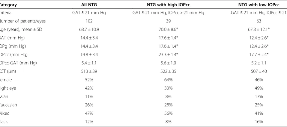
In this study, NTG eyes were defined as eyes with GON and a maximum recorded GAT less than or equal to 21 mm Hg. However, 39 (38%) of the 102 GAT defined NTG eyes had an IOPcc higher than 21 mm Hg; the mean IOPcc of these eyes was 23.3 mm Hg (Table 2). Of the same cohort of NTG eyes, 73 (72%) had an IOPcc greater than 18 mm Hg, 93 (91%) had an IOPcc greater than 15 mm Hg, and only 9 eyes (9%) had an IOPcc of 15 mm Hg or less.
Sensitivities and specificities for identifying glaucoma using GAT (Figure 2a) and IOPcc (Figure 2b) were determined from the total study population of normal
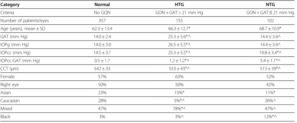
Table 1 Patient characteristics
Category Normal HTG NTG
Criteria No GON GON + GAT > 21 mm Hg GON + GAT≤21 mm Hg
Number of patients/eyes 357 155 102
Age (years), mean ± SD 62.3 ± 13.4 66.3 ± 12.7* 68.7 ± 10.9*
GAT (mm Hg) 14.0 ± 2.4 25.3 ± 5.6*^ 14.4 ± 3.4^
IOPg (mm Hg) 14.0 ± 3.0 26.5 ± 5.5*^ 14.4 ± 3.4^
IOPcc (mm Hg) 14.5 ± 3.1 25.3 ± 5.5*^ 19.8 ± 3.4*^
IOPcc-GAT (mm Hg) 0.5 ± 1.7 1.2 ± 1.2*^ 5.4 ± 1.1*^
CCT (μm) 542 ± 33 553 ± 43*^ 513 ± 39*^
Female 57% 63% 52%
Right eye 50% 50% 42%
Asian 23% 15%* 11%*
Caucasian 28% 5%*^ 26%^
Mixed 47% 78%*^ 47%^
Black 3% 3%^ 12%*^
*p≤0.05 for comparison with normal group.
^p≤0.05 for comparison between NTG and HTG groups.
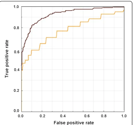
and glaucomatous eyes, using assessment of GON as the gold standard reference. The traditional GAT threshold of 21 mm Hg had a sensitivity of 46% (specificity 99%) for detecting glaucoma. The optimal GAT threshold to maximize combined sensitivity and specificity was 20.9 mm Hg (sensitivity 59%, specificity 90%). Likewise, an IOPcc cutoff of 18.4 mm Hg resulted in the highest combination of sensitivity (85%) and specificity (85%) for detecting glaucoma. The areas under the curve (AUC) of ROC curves were compared and demonstrated a greater AUC for IOPcc (0.93) compared to GAT (0.78) in the detection of GON (test for difference of AUCs: p < 0.001) Figure 3.
Discussion
In this comparison of several different assessments of IOP for the detection of GON, we found evidence that an IOP assessment that compensates for corneal
Table 2 Normal-tension glaucoma patient characteristics
Category All NTG NTG with high IOPcc NTG with low IOPcc
Criteria GAT≤21 mm Hg GAT≤21 mm Hg, IOPcc > 21 mm Hg GAT≤21 mm Hg, IOPcc≤21
Number of patients/eyes 102 39 63
Age (years), mean ± SD 68.7 ± 10.9 70.0 ± 8.6* 67.8 ± 12.1*
GAT (mm Hg) 14.4 ± 3.4 17.6 ± 1.4* 12.4 ± 2.6*
IOPg (mm Hg) 14.4 ± 3.4 17.6 ± 1.4* 12.4 ± 2.6*
IOPcc (mm Hg) 19.8 ± 3.4 23.3 ± 1.4* 17.7 ± 2.4*
IOPcc-GAT (mm Hg) 5.4 ± 1.1 5.6 ± 1.0 5.2 ± 1.1
CCT (μm) 513 ± 39 522 ± 35 507 ± 40
Female 52% 64% 46%
Right eye 42% 33% 49%
Asian 11% 8% 13%
Caucasian 26% 28% 25%
Mixed 47% 56% 41%
Black 12% 8% 16%
*p≤0.05 for comparison between NTG with high IOPcc and NTG with low IOPcc.
HTG: high-tension glaucoma; NTG: normal-tension glaucoma; GON: glaucomatous optic neuropathy; GAT: Goldmann applanation tonometry; IOPg: Goldman-correlated intraocular pressure; IOPcc: corneal compensated intraocular pressure; CCT: central corneal thickness.
Figure 2Sensitivity and specificity of GAT and IOPcc in detecting glaucomatous optic neuropathy. a. Sensitivity and specificity of GAT in detecting glaucoma.b. Sensitivity and specificity of IOPcc in detecting glaucoma.
biomechanical properties may have better accuracy for the detection of POAG than GAT. Specifically, we found that GAT and IOPcc showed considerable agreement within the normal and HTG cohorts. However, among NTG eyes IOPcc was, on average, significantly higher than GAT. Moreover, we determined that the optimal threshold for GAT to maximize sensitivity and specificity for the detection of GON was 20.9 mm Hg and for IOPcc was 18.4 mm Hg. Using ROC curves we found that the AUC for the IOPcc (0.93) was greater than for GAT (0.78), indicating that compared to GAT, IOPcc may represent a superior test in the evaluation of glaucoma.
Importantly, we would like to emphasize that we do not support the use of even a corneal compensated IOP for the diagnosis of glaucoma. Due to the relatively low prevalence of POAG in the population [26-29], a diag-nostic test with a sensitivity and specificity of 85% would result in low positive predictive value and thus a high number of false-positives. For example, in a population with a 4% prevalence of undiagnosed POAG, only about 19% of patients with a positive result would have the dis-ease. Additionally, with a sensitivity of 85%, approxi-mately one in six patients with POAG would not be detected. Notwithstanding, IOPcc may represent a better tool for the evaluation and management of POAG due to its more consistent association with the disease across a wide range of IOPs.
Since IOPcc was greater than GAT in majority of NTG eyes (92%) in this study, segregating NTG and
HTG using a GAT threshold of 21 mm Hg may not be optimal. Other studies have also demonstrated an under-estimation of IOP by applanation tonometry after cor-recting IOP for corneal properties [18,21,30-33]. The present study, like others, found that eyes with NTG had significantly thinner corneas [34,35], while patients with HTG had greater CCT compared to normals. Of note, one study, the Low Tension Glaucoma Study, did not find abnormally low CCT in a cohort of NTG patients [36]. However, the current study suggests that the impact of corneal properties on IOP determination may be greatest in eyes with NTG.
Past investigations have demonstrated that IOPcc is less dependent on corneal properties than traditional contact tonometry [18,23]. Medeiros and Weinreb found that CCT was significantly correlated with GAT but not IOPcc in a population under evaluation for glaucoma. Additionally, in the same study they found that the dif-ference between GAT and IOPcc increased linearly as a function of increasing CCT [18]. The results of the present investigation are similar in that we observed no significant correlation between CCT and IOPcc among HTG and normal eyes. However, we did determine a sig-nificant correlation between CCT and IOPcc among eyes with NTG. This pattern may exist since lower IOP values have a greater biologically plausible range for up-ward adjustment due to CCT than do higher IOP values as a function of regression toward the mean. This find-ing suggests that an accurate algorithm to correct for the impact of CCT and/or corneal biomechanics on IOP is likely to be non-linear.
CCT is lowest in eyes with NTG [34,35] and low CCT may be both an independent risk factor for glaucoma progression [8] as well as a source of IOP measurement error [30-33]. Consequently, use of a corneal compen-sated IOP may be preferable to GAT specifically in the evaluation and management of NTG. The existence of higher than normal IOPcc in eyes with NTG is sup-ported by reports that medical and surgical therapies that decrease IOP in NTG eyes are effective in reducing the progression of GON [10,11]. In the Collaborative Normal Tension Glaucoma Treatment Study [10], the average baseline IOP in the treated and untreated arms was 16.1 and 16.9 mm Hg, respectively. With treatment, the average IOP in the treatment group was lowered to 10.6 mm Hg, while IOP in the control group remained 16.0 mm Hg. Importantly, 35% of untreated eyes and only 12% of treated eyes met criteria for visual field or glaucomatous optic disc progression at the end of the study [10].
Notwithstanding, it has been observed that some eyes with NTG progress despite ocular hypotensive therapy [10]. In fact, some have speculated that NTG eyes may even develop GON independent of IOP [37]. Given the Figure 3ROC curves for GAT and IOPcc.ROC curves of GAT and
observation in the present study that differences in IOPcc and GAT in a single patient may be as large as 8 mm Hg, an eye with a GAT value in the normal range may actually have a much higher IOPcc, depending on its corneal properties. In this context, setting a thera-peutic target IOP using GAT would similarly differ from an IOPcc target pressure. This may partially explain glaucoma progression in NTG eyes in which IOP, as measured by GAT, appears to be adequately controlled since determining a safe IOP target is made difficult.
There are several limitations to this study. As an ob-servational cross-sectional study, it is not possible to determine how the longitudinal risk of glaucoma pro-gression is related to GAT compared to IOPcc. The op-erational definition of GON used in this study was based on the appearance of the optic nerve as docu-mented by the evaluating clinician and data for other definitions of glaucoma including visual field defects are not available. While perimetric evaluation or other computerized optic nerve imaging techniques could have been employed, these devices have limitations related to accuracy for GON detection [38] and such testing was not performed on all patients in this study. Additionally, since CCT [30] and CH [39,40] differ by race/ethnicity it is possible that studies with distinct ethnic compositions may yield different results. None-theless, we did not separately analyze racial/ethnic dif-ferences in CCT and their contribution to our results. Finally, it was outside of the scope of this study to cor-relate the degree of GON with IOP levels or their fluc-tuation. Though we recognize that the absence of diurnal IOP data may have resulted in misclassification of some patients, there is no reason to believe that the data were systematically biased toward IOP measure-ment at any specific time of day.
This study contributes to the understanding of dif-ferences that exist in the impact of corneal properties on IOP measurement between normal, HTG and NTG eyes. Importantly, these results suggest that the a corneal compensated IOP may offer an attractive al-ternative to GAT, particularly for the evaluation and management of patients with NTG in whom corneal biomechanics and thickness appear to play the greatest role.
Conclusions
The sensitivity and specificity of GAT and corneal-compensated IOPcc for the identification of GON were optimized at thresholds of 20.9 and 18.4 mm Hg, re-spectively. For the detection of GON, the area under the ROC curve was significantly greater for IOPcc compared to GAT. While IOP is unlikely to be an effective diag-nostic test, the results of this study suggest that a corneal-compensated IOP may represent a superior test
for the evaluation of glaucoma, especially among patients with low to normal IOPs.
Competing interests
NMR has served as consultant for Allergan and Alcon and has received instrument support from Reichert, Inc.
Authors’contributions
JRE performed statistical analyses, and helped to draft the manuscript. NMR participated in the design of the study and helped to draft the manuscript. MS conceived of the study, participated in the design of the study and helped to draft the manuscript. All authors read and approved of the final manuscript.
Author details
1Department of Ophthalmology, Weill Cornell Medical College, 1305 York
Avenue, New York, NY 10021, USA.2New York Eye and Ear Infirmary, Department of Ophthalmology, New York Medical College, Valhalla, NY, USA.
Received: 14 June 2011 Accepted: 20 September 2012 Published: 25 September 2012
References
1. Hollows FC, Graham PA:Intra-ocular pressure, glaucoma, and glaucoma suspects in a defined population.Br J Ophthalmol1966,50:570–586. 2. Armaly MF, Krueger DE, Maunder L, Becker B, Hetherington J, Kolker AE,
Levene RZ, Maumenee AE, Pollack IP, Shaffer RN:Biostatistical analysis of the collaborative glaucoma study. I. Summary report of the risk factors for glaucomatous visual-field defects.Arch Ophthalmol1980,
98:2163–2171.
3. Gordon MO, Kass MA:The ocular hypertension treatment study: design and baseline description of the participants.Arch Ophthalmol1999,
117:573–583.
4. von Graefe A:Amaurose mit sehnervenexcavation.Arch. Ophthalmol1857,
3:484–489.
5. Chumbley LC, Brubaker RF:Low-tension glaucoma.Am J Ophthalmol1976,
81:761–767.
6. Caprioli J, Sears M, Spaeth GL:Comparison of visual field defects in normal-tension glaucoma and high-tension glaucoma.Am J Ophthalmol 1986,102:402–404.
7. Kass MA, Heuer DK, Higginbotham EJ, Johnson CA, Keltner JL, Miller JP, Parrish RK, Wilson MR, Gordon MO:The Ocular Hypertension Treatment Study: a randomized trial determines that topical ocular hypotensive medication delays or prevents the onset of primary open-angle glaucoma.Arch Ophthalmol2002,120:701–713. discussion 829–830. 8. Leske MC, Heijl A, Hussein M, Bengtsson B, Hyman L, Komaroff E:Factors
for glaucoma progression and the effect of treatment: the early manifest glaucoma trial.Arch Ophthalmol2003,121:48–56.
9. The Advanced Glaucoma Intervention Study (AGIS):7. The relationship between control of intraocular pressure and visual field deterioration. The AGIS Investigators.Am J Ophthalmol2000,130:429–440. 10. Collaborative Normal tension Glaucoma Study Group:Comparison of
Glaucomatous Progression Between Untrested Patients with Normal-Tension Glaucoma and Patients with Terapeutically Reduced Intraocular Pressures.Am J Ophthalmol1998,126:487–497.
11. Shigeeda T, Tomidokoro A, Araie M, Koseki N, Yamamoto S:Long-term follow-up of visual field progression after trabeculectomy in progressive normal-tension glaucoma.Ophthalmology2002,109:766–770.
12. Goldmann H, Schmidt T:Über applanationstonometrie.Ophthalmologica 1957,123:221–242.
13. Ehlers N, Bramsen T, Sperling S:Applanation tonometry and central corneal thickness.Acta Ophthalmol (Copenh)1975,53:34–43. 14. Liu J, Roberts CJ:Influence of corneal biomechanical properties on
intraocular pressure measurement: quantitative analysis.J Cataract Refract Surg2005,31:146–155.
15. Choudhari NS, George R, Baskaran M, Vijaya L, Dudeja N:Measurement of Goldmann applanation tonometer calibration error.Ophthalmology2009,
116:3–8.
16. Wasielica-Poslednik J, Berisha F, Aliyeva S, Pfeiffer N, Hoffmann EM:
correlation with central corneal thickness.Graefes Arch Clin Exp Ophthalmol2010,248:1617–1622.
17. Ehrlich JR, Haseltine S, Shimmyo M, Radcliffe NM:Evaluation of agreement between intraocular pressure measurements using Goldmann applanation tonometry and Goldmann correlated intraocular pressure by Reichert’s ocular response analyser.Eye (Lond)2010,24:1555–1560. 18. Medeiros FA, Weinreb RN:Evaluation of the influence of corneal
biomechanical properties on intraocular pressure measurements using the ocular response analyzer.J Glaucoma2006,15:364–370.
19. Doughty MJ, Zaman ML:Human corneal thickness and its impact on intraocular pressure measures: a review and meta-analysis approach.
Surv Ophthalmol2000,44:367–408.
20. Kotecha A, White ET, Shewry JM, Garway-Heath DF:The relative effects of corneal thickness and age on Goldmann applanation tonometry and dynamic contour tonometry.Br J Ophthalmol2005,89:1572–1575. 21. Moreno-Montañés J, Maldonado MJ, García N, Mendiluce L, García-Gómez
PJ, Seguí-Gómez M:Reproducibility and clinical relevance of the ocular response analyzer in nonoperated eyes: corneal biomechanical and tonometric implications.Invest Ophthalmol Vis Sci2008,49:968–974. 22. Lam A, Chen D, Chiu R, Chui W-S:Comparison of IOP measurements
between ORA and GAT in normal Chinese.Optom Vis Sci2007,
84:909–914.
23. Streho M, Dariel R, Giraud J-M, Verret C, Fenolland J-R, Crochelet O, May F, Maurin J-F, Renard J-P:[Evaluation of the Ocular Response Analyzer in ocular hypertension, glaucoma, and normal populations. Prospective study on 329 eyes].J Fr Ophtalmol2008,31:953–960.
24. Kotecha A, Elsheikh A, Roberts CR, Zhu H, Garway-Heath DF:Corneal thickness- and age-related biomechanical properties of the cornea measured with the ocular response analyzer.Invest Ophthalmol Vis Sci 2006,47:5337–5347.
25. Jonas JB, Budde WM, Panda-Jonas S:Ophthalmoscopic evaluation of the optic nerve head.Surv Ophthalmol1999,43:293–320.
26. Friedman DS, Jampel HD, Muñoz B, West SK:The prevalence of open-angle glaucoma among blacks and whites 73 years and older: the Salisbury Eye Evaluation Glaucoma Study.Arch Ophthalmol2006,
124:1625–1630.
27. Iwase A, Suzuki Y, Araie M, Yamamoto T, Abe H, Shirato S, Kuwayama Y, Mishima HK, Shimizu H, Tomita G, Inoue Y, Kitazawa Y:The prevalence of primary open-angle glaucoma in Japanese: the Tajimi Study.
Ophthalmology2004,111:1641–1648.
28. Leske MC, Connell AM, Schachat AP, Hyman L:The Barbados Eye Study. Prevalence of open angle glaucoma.Arch Ophthalmol1994,112:821–829. 29. Varma R, Ying-Lai M, Francis BA, Nguyen BB-T, Deneen J, Wilson MR, Azen
SP:Prevalence of open-angle glaucoma and ocular hypertension in Latinos: The Los Angeles Latino Eye Study.Ophthalmology2004,
111:1439–1448.
30. Brandt JD, Beiser JA, Kass MA, Gordon MO:Central corneal thickness in the Ocular Hypertension Treatment Study (OHTS).Ophthalmology2001,
108:1779–1788.
31. Bron AM, Creuzot-Garcher C, Goudeau-Boutillon S, d’Athis P:Falsely elevated intraocular pressure due to increased central corneal thickness.
Graefes Arch Clin Exp Ophthalmol1999,237:220–224.
32. Herndon LW, Choudhri SA, Cox T, Damji KF, Shields MB, Allingham RR:
Central corneal thickness in normal, glaucomatous, and ocular hypertensive eyes.Arch Ophthalmol1997,115:1137–1141. 33. Whitacre MM, Stein R:Sources of error with use of Goldmann-type
tonometers.Surv Ophthalmol1993,38:1–30.
34. Copt RP, Thomas R, Mermoud A:Corneal thickness in ocular hypertension, primary open-angle glaucoma, and normal tension glaucoma.Arch Ophthalmol1999,117:14–16.
35. Lee ES, Kim CY, Ha SJ, Seong GJ, Hong YJ:Central corneal thickness of Korean patients with glaucoma.Ophthalmology2007,114:927–930. 36. Krupin T, Liebmann JM, Greenfield DS, Rosenberg LF, Ritch R, Yang JW:The
Low-pressure Glaucoma Treatment Study (LoGTS) study design and baseline characteristics of enrolled patients.Ophthalmology2005,
112:376–385.
37. Mozaffarieh M, Grieshaber MC, Flammer J:Oxygen and blood flow: players in the pathogenesis of glaucoma.Mol Vis2008,14:224–233.
38. Reus NJ, de Graaf M, Lemij HG:Accuracy of GDx VCC, HRT I, and clinical assessment of stereoscopic optic nerve head photographs for diagnosing glaucoma.Br J Ophthalmol2007,91:313–318.
39. Haseltine SJ, Pae J, Ehrlich JR, Shammas M, Radcliffe NM:Variation in corneal hysteresis and central corneal thickness among black, hispanic and white subjects.Acta Ophthalmol2012, Epub.
40. Leite MT, Alencar LM, Gore C, Weinreb RN, Sample PA, Zangwill LM, Medeiros FA:Comparison of corneal biomechanical properties between healthy blacks and whites using the Ocular Response Analyzer.Am J Ophthalmol2010,150:163–168. e1.
doi:10.1186/1471-2415-12-52
Cite this article as:Ehrlichet al.:Goldmann applanation tonometry compared with corneal-compensated intraocular pressure in the evaluation of primary open-angle Glaucoma.BMC Ophthalmology2012
12:52.
Submit your next manuscript to BioMed Central and take full advantage of:
• Convenient online submission
• Thorough peer review
• No space constraints or color figure charges
• Immediate publication on acceptance
• Inclusion in PubMed, CAS, Scopus and Google Scholar
• Research which is freely available for redistribution