Screening Pulmonary Function Tests
GEORGE W. BURKE, III, M.D.
Assistant Professor of Medicine, and Director, Pulmonary Function Laboratories, Medical College of Virginia, Health Sciences Division of Virginia Commonwealth University, Richmond, Virginia
The role of the Pulmonary Function Laboratory has been expanded in recent years by the commercial development and marketing of equipment capable of measuring accurately and easily static lung volumes, diffusing capacity, and arterial blood gases. These sophisticated measurements, which were once the purview of research physiologists, are now readily attainable as screening measurements in most com-m unity hospitals. This review is intended not as a summary of the entire field or as a technical guide for performance of pulmonary function tests but as a survey of some clinical applications and pitfalls of screening tests and a statement of guidelines for their use. It is assumed that the reader already has some experience in ordering and interpreting routine spiro-grams and arterial blood gases.
Pulmonary function tests are intended to answer four questions:
1) Is the patient restricted? 2) Is the patient obstructed?
3) Does the patient have abnormalities of gas exchange?
4) Are the observed abnormalities compatible with the patient's symptoms and clinical di-agnosis?
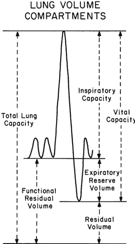
Restriction is a reduction of total lung capacity or one of its subvolumes (Fig I) as a result of collapse or volume displacement of the lung (fibrothorax), in-creased elastic recoil of the lungs (pulmonary fibro-sis), increased elastic recoil of the rib cage or abdo-men (obesity), or loss of forces needed to overcome normal elastic recoil (muscular dystrophy). It is
im-Correspondence and reprint requests to Dr. George W. Burke, III, Box 903, Medical College of Virginia, Richmond, VA 23298.
MCV QUARTERLY 14(4):163-168, 1978
portant to realize that a routine spirometric vital capacity measurement may not adequately describe the presence or degree of restriction. For example, some patients with advanced emphysema may have reduced vital capacity in the face of a large residual volume, with increased total lung capacity caused by obstructive air trapping; it is misleading to label such patients as "restricted" on the basis of reduced vital capacity when, in fact, they are overinflated. On the other hand, true early restriction may occur in the face of normal vital capacity. An example of this is obesity, a condition in which the abdominal mass renders the chest wall less compliant by hindering downward displacement of the diaphragm. The earliest change in the restrictive pattern of obesity is a marked reduction in expiratory reserve volume (ERV) (see Fig I) but only minor alterat.ions of the total lung and vital capacities. Therefore, accurate evaluation of patients with suspected restrictive dis-orders should include, in addition to routine spirome-try, a measurement of total lung capacity and its subdivisions by inert gas or plethysmographic tech-niques. The following case illustrates the use of pul-monary function studies in following the course of a restrictive lung disorder:
Case I. A 53-year-old white woman developed exertional dyspnea and cough in 1971. A chest roentgenogram re-vealed bilateral basilar "ground-glass" opacities. In April 1972, an open lung biopsy showed desquamative interstitial pneumonia. She was placed on 20 mg prednisone daily in August 1972, and sequential pulmonary function tests were performed (Table I). The initial vital and total lung capaci-ties were consistent with moderately severe restriction, and there was a correspondingly severe reduction in diffusing capacity. After two years of prednisone therapy, these ab-normalities improved substantially (October 1974). When steroids were stopped she experienced a severe relapse
164
(February 1976) which was reversed by their resumption (February 1977). In 1977 she was in remission while taking 20 mg prednisone on alternate days·. The pulmonary June· tion studies provided quantitative objective evidence that a prolonged course of steroid therapy was necessary.
Obstructive impairment, defined as a reduction in inspiratory or expiratory air-flow rates, is a result of intrinsic airway disease or loss of lung elastic re-coil. The latter causes airways to lose their structural
LUNG VOLUME
COMPARTMENTS
+
I
I I I
I I
I
Total Lung
Capacity
I I
I
Functional
Residual
Volume
I I I
t
*
I I I I I
I
I
I
lnspiratory
Capacity
+
I I
Vital
Capacity
I I
I
I I
t
:
Expiratory!
Reserve
IVolume I
•
I
Residual
Volume
I*
Fig 1-Spirogram with various subdivisions of total lung capacity. Initial deflections at left are tidal volumes recorded on slowly moving recording paper. Large deflection upward is inspiration to total lung capacity, followed by expiration to residual volume and resumption of tidal breathing. Residual volume is not measured directly, but has been previously determined by inert gas study. Time is on the horizontal axis, volume on the vertical.
BURKE: PULMONARY FUNCTION TESTS
support. Intrinsic airway disease may be caused by a variety of conditions ranging from totally reversible asthmatic bronchoconstriction to irreversible ad-vanced chronic bronchitis with bronchiolitis obliter-ans. Loss of elastic recoil almost always is a result of localized or generalized emphysema.
Because the conditions causing airways obstruc-tion tend to become more exaggerated during forced expiration (during which alveoli and airways are rap-idly diminishing in both length and diameter), the conventional method of assessing obstructive impair-ment is by measuring averaged flow rates on a vol-ume-time curve of an ordinary spirogram during a maximally forced expiration of the vital capacity. (Fig 2). Although one could arbitrarily measure flow at a multitude of points on the curve, three measure-ments have been standardized and used most fre-quently (see Fig 2): the timed vital capacity (FEY 1 );
the ratio of FEY, to total vital capacity; and the maximum mid-expiratory flow rate (MMEF) (the av-eraged flow over the mid-half of the vital capacity). Ordinarily all three measurements are made and ab-normalities of one tend to correlate with those of the others. Additional measurements such as peak flow, total expiratory time, and forced expiratory volume in three seconds, or "FEY3 ," add little new informa-tion but may be sensitive indicators of changes when evaluating drug effects in population samples.
Obstruction is conventionally quantitated dur-ing expiration; however, patients develop dyspnea from obstructive impairment because of the increased work of breathing during inspiration. In most cases, obstruction during inspiration is inferred by abnor-malities in the FEY, and MMEF; unfortunately, there are several important exceptions to this. Indi-viduals with obstruction isolated to the trachea or larynx (for example, tracheomalacia following endo-tracheal intubation) may not have evidence of
ex-TABLE I
Sequential Function Studies in a Patient with Desquamative Interstitial Pneumonia.
August October February February Pre· 1972 1974 1976 1977 dieted Vital capacity,
liters 2.06 3.45 2.30 3.12 3.97
Total lung capacity,
liters 3.55 5.24 3.79 4.95 6.32
Arterial Po,,
torr 66 73 67 76 90
Diffusing capacity,
BURKE: PULMONARY FUNCTION TESTS
4
(/)
3
' -a,-w
2
~
::::> ...J 0
>
0
FEV1
____ J__ __
0
Slope between
points A and 8 = MMEF ( liters/sec)
2
TIME (seconds)
Fig 2- Forced vital capacity shown on rapidly movlng recording paper. Total downward excursion of curve (vertical axis) is vital capacity. Volume exhaled during the first second is FEY,. MMEF is slope of line connecting points A and B. Time is on the h orizon-tal axis.
piratory obstruction if their defect is above the tho-racic outlet, or they may have spirometric
abnormalities which are misinterpreted as general-ized airways obstruction if their defect is below the thoracic outlet. In addition, individuals with s
ub-stantial narrowing of bronchioles but normal patency
of bronchi (for example, young smokers with early
chronic bronchitis) may have completely normal
spirometric flow rates but isolated abnormalities of
MM EF which are difficult to interpret because of the large standard deviation of this test in the normal
population.
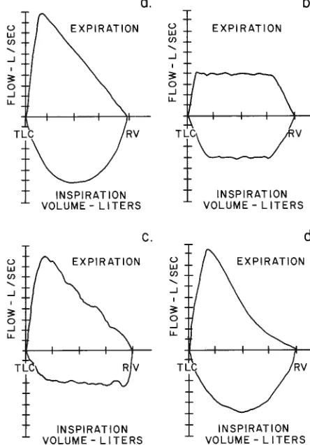
When either of these exceptions is suspected, additional information may be obtained by a record-ing of a flow-volume curve, or graphical representa
-tion of instantaneous mouth flow and expired volume during a forced inspiration and expiration of the vital
capacity (Fig 3 ). The normal configuration of this
curve is shown in Figure 3a. Fixed obstruction in the trachea produces a characteristic fixed limitation to
flow which is independent of volume (Fig 3b ). An
additional advantage of the flow-volume curve is its
depiction of inspiratory events, thus facilitating the
detection of variable extrathoracic upper-airway
ob-struction (Fig 3c ). Generalized bronchiolar
narrow-ing produces flow limitations which are detectable <.)
w (/)
'
_J
3: 0 _J
LL.
TLC
<.)
w
(/)
'
_J
3: 0 _J
LL.
t
EXPIRATION
INSPIRATION VOLUME - LITERS
EXPIRATION
a.
c
.
INSPIRATION
VOLUME - LITERS
<.)
w (/)
'
_J3: 0 _J
LL.
EXPIRATION
165
b
.
TLC\._______JV
I
INSPIRATIONVOLUME - LITERS
<.)
w (/)
'
_J 3: 0 _JLL.
TLC
I
d.
EXPIRATION
INSPIRATION
VOLUME - LITERS
Fig 3a- l nspiratory-expiratory flow-volume curve shows normal
configuration in a 28-year-old man.
Fig 3b-Fixed upper-airway obstruction seen in a 32-year-old man
with stenosis of intrathoracic trachea.
Fig Jc- Curve shows variable extrathoracic obstruction as a result
of loss of cartilagenous tracheal support in the subglottic region. The weakened area collapses on inspiration but is distended during
expiration by positive airway pressure.
Fig 3d- "I nwardly concave" distortion of the expired curve at lower lung volumes is seen in a 30-year-old chronic cigarette smoker with a normal spirograni. The distortion is due to
bronchiolar obstruction.
only on expiration at lower lung volumes and tend to
worsen as volume diminishes. This will result in an "inwardly concave" appearance to the flow-volume
curve in patients with obstruction limited to the
bron-chioles (Fig 3d). It should be pointed out that this "inwardly concave" configuration is not specific for
peripheral small airways obstruction; if the sp
-166
fut for differentiating generalized obstruction from that isolated to peripheral airways. The following case is an example of obstructive impairment and
also illustrates the value of lung volume
determina-tions by the helium dilutional method in differentia-ting obstructive from restrictive impairment:
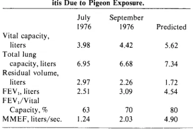
Case 2. A 19-year-old male gas station attendant abruptly developed exertional dyspnea in January 1976. Seven months later a carefully taken history revealed that he had been caring for his brother's homing pigeons since late December 1975. The ·chest roentgenogram was normal,
although symptoms persisted. Pulmonary function studies (Table 2, July 1976) revealed a reduced vital capacity; however, the total lung capacity and residual volume sug-gested probable obstructive air trapping rather than restric-tion. The FEV1 and MMEF confirmed obstruction. With-out specific treatment other than ceasing the pigeon exposure, his symptoms resolved and objective improvement was evident on sequential studies ( September 1976). Al-though both restriction and obstruction may occur in all er-gic alveolitis, restriction is usually the dominant impair-ment. However, in this patient's case, obstruction occurred without restriction.
Arterial hypoxemia is a sequela of impaired gas
exchange; however, measurement of resting arterial
oxygen tension (Po2) alone may not be sufficiently sensitive to detect such impairment. Impaired gas exchange is also present if there is an increase in the observed oxygen tension difference between alveolar air and arterial blood (DA-a 0 2) or if there is an abnormality in the diffusing capacity for carbon monoxide. For ambulatory patients breathing room
air, DA-a 0 2 can be estimated by obtaining a sample
of arterial blood under steady-state conditions. Al-veolar oxygen tension is calculated by the standard alveolar air equation:
PA02
=
F102(713)=
Paco2 (F 102+
1 - ;0
°
2)F102 = fraction of02 in inspired air
Paco2 arterial Pco2 (identical to mean alveolar
Pco2)
RQ = respiratory quotient
The achievement of steady-state conditions is an es-sential prerequisite for this test because the estima-tion of alveolar oxygen tension is based on the as-sumption that the ratio of C02 produced to 0 2 consumed (RQ) is 0.8. Transient hyperventilation caused by anxiety or pain will invalidate this assump-tion. Direct measurement of RQ may provide a more
BURKE: PULMONARY FUNCTION TESTS
TABLE 2
Sequential Function Studies in a Patient with Allergic Alveol-itis Due to Pigeon Exposure.
July September
1976 1976 Predicted
Vital capacity,
liters 3.98 4.42 5.62
Total lung
capacity, liters 6.95 6.68 7.34
Residual volume,
liters 2.97 2.26 1.72
FEY 1 , liters 2.51 3.09 4.54
FEV1/Vital
Capacity,% 63 70 80
MMEF, liters/sec. 1.24 2.03 4.90
refined estimation of PA 0 2; however, this also
re-quires sampling under steady-state conditions. For
either approach, hyperventilation may be avoided by
inserting an arterial cannula and allowing the patient
to rest prior to sample collection.
The measurement of carbon monoxide diffusing
capacity by the simplified single-breath technique
re-quires the patient to breath hold at total lung capacity
for ten seconds. Patients who are markedly dyspneic
at rest may not be able to perform this test. Despite these limitations, useful results are forthcoming for most cooperative subjects.
The precise pathophysiologic explanation for
impaired diffusion of carbon monoxide in lung
dis-eases is unknown. Most authorities agree that it is a
sign of alveolar or capillary destruction. Therefore, impaired CO uptake may occur in parenchymal de-structive processes such as idiopathic fibrosing al-veolitis, scleroderma, advanced sarcoidosis, and in vascular obliterative processes such as idiopathic pul-monary hypertension or multiple small pulpul-monary emboli. It is also impaired in processes causing re-versible loss of functioning alveoli, examples of which
are bacterial pneumonia and alveolar proteinosis. CO
uptake is impaired in generalized emphysema due to
emphysematous destruction of alveoli but remains.
nearly normal in chronic bronchitis because the
de-structive process is confined to airways and
centri-lobular elements of the parenchyma. Diffusing
capac-ity may be spuriously impaired by several conditions.
The first is the presence of severe anemia, which
limits the availability of hemoglobin CO receptors;
the second is that failure to achieve true total lung
capacity during the breathhold will reduce CO
up-take in proportion to the reduction in potential lung
BURKE: PULMONARY FUNCTION TESTS 167
TABLE 3
Pulmonary Function Studies in a Patient with Sarcoidosis
02 were also normal. However, a moderately severe reduc-tion in diffusing capacity suggested the presence of wide-spread interstitial disease despite the clear roentgenographic
lung fields. Hemoglobin was normal. A transbronchoscopic lung biopsy showed interstitial non-caseating granulomata consistent with sarcoidosis.
September 1978 Predicted
Vital capacity, liters 2.98 3.63
FEY" liters 2.68 2.88
FEY/vital
capacity,% 90 79
MMEF,
liters/second 4.03 3.37
Total lung capacity,
liters 3.85 5.12
Residual volume,
liters .76 1.49
Pao,, torr 92 90
DA-a 02 , torr 13 15
Diffusing capacity,
ml/min/torr 12.0 21.7
diffusing capacity in screening patients for the pres-ence of gas exchange impairment:
Although measured impairment in pulmonary function frequently provides an objective explanation for the presence of dyspnea, many patients may expe-rience this symptom because of abnormalities which are undetectable on screening evaluations. Negative screening studies should stimulate a careful search for vascular disease or nonpulmonary causes of dyspnea.
For example, "metabolically justified" hyperpnea and dyspnea may accompany hyperthyroidism; pa-tients with mitral stenosis or early congestive heart failure may have only mild impairment in lung me-chanics despite severe exertional dyspnea; and pa-tients with partial obliteration of the pulmonary vas-culature may have normal lung mechanics, arterial oxygen tensions, and diffusing capacities while at rest, only to show abnormalities during exercise.
Cas~ 3. A 32-year-old woman complained of mild ex-ertional dyspnea of two months duration. Chest roentgeno-gram q:vealed bilateral hilar adenopathy. Pulmonary func-tion studies (Table 3) showed a modest reducfunc-tion in total lung capacity (75% predicted) with normal vital capacity anq flow rates. Resting arterial oxygen tension and DA-a
For a variety of vascular and interstitial pul-monary disorders, it may be appropriate to evaluate
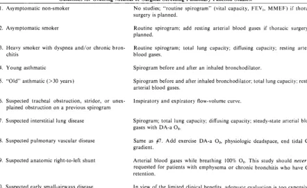
TABLE 4
Guidelines for Ordering Medical or Surgical Screening Pulmonary Function Studies. I. Asymptomatic non-smoker
2. Asymptomatic smoker
3. Heavy smoker with dyspnea and/or chronic bron-chitis
4. Young asthmatic
5. "Old" asthmatic (>30 years)
6. Suspected tracheal obstruction, stridor, or unex-plained obstruction on a previous spirogram 7. Suspected interstitial lung disease
8. Suspected pulmonary vascular disease
9. Suspected anatomic right-to-left shunt
I 0. Suspected early small-airways disease
No studies; "routine spirogram" (vital capacity, FEY" MMEF) if thoracic surgery is planned.
Routine spirogram; add resting arterial blood gases if thoracic surgery is
planned.
Routine spirogram; total lung capacity; diffusing capacity; resting arterial blood gases.
Spirogram before and after an inhaled bronchodilator.
Spirogram before and after inhaled bronchodilator; total lung capacity: resting arterial blood gases.
lnspiratory and expiratory flow-volume curve.
Spirogram; total lung capacity; diffusing capacity; steady-state arterial blood gases with DA-a 02 •
Same as #7. Add exercise DA-a 02, physiologic deadspace, end tidal CO,
gradient.
Arterial blood gases while breathing I 00% 02• This study should never be requested for patients with emphysema or chronic bronchitis who have CO,
retention.
168
physiology during exercise as resting abnormalities
frequently are exaggerated during exercise. However,
pulmonary exercise testing, unlike cardiac stress test-ing, should provide a graded low level of exercise
(doubling or tripling of the oxygen uptake) rather
than maximum stress. In fact, recent experimental
evidence' has suggested that maximum stress
mea-surements are potentially misleading because normal
subjects may demonstrate transient abnormalities in
Pao2 and DA-a 02 while performing temporary but
severe tasks such as rapid stair-climbing. Although a
complete discussion of exercise physiology testing is
beyond the scope of this paper, som'e recent reviews
of the relevance of pulmonary exercise testing in
clini-cal medicine are included in the bibliography.1-2 Table 4 is a suggested modus operandi for
order-ing screenorder-ing pulmonary function studies for specific
problems. Physicians ordering such tests must realize that measurements of lung mechanics require both
cooperation apd effort. Too much testing at a sing!~
sitting may fatigue the patient and cause spuriously
BURKE: PULMONARY FUNCTION TESTS
abnormal results. For example, it would be unwise to
subject a patient to both screening studies and
exer-cise physiology studies for suspected vascular disease
on the same day. Therefore, in the context of the
suggestions in Table 4, clinical wisdom and common
sense should dictate which tests are appropriate for
individual patients. .
BIBLIOGRAPHY
I. YOUNG I H, WOOLCOCK AJ: Changes in arterial blood gas
ten-sions during unsteady-state exercise. J Appl Physiol Respirat
Environ Exercise Physio/ 44(1):93-96, 1978.
2. WHIPP BJ, WASSERMAN K: Exercise physiology in health and
disease. A!Jl Rev Resp Dis 112:219-249, 1975.
3. GAENSLER EA, WRIGHT GW: Evaluation of respiratory
impair-ment. Arch Environ Hea/1h 12:146-186, 1966.