1Ahmed Omar Abdelnaeem, MSc
2Alaa Balbaa, PhD
3Nessreen Fawzy Mahmoud, MSc
CORRESPONDING AUTHOR
2Aliaa Rehan Youssef, PhD
Faculty of Physical Therapy,
Cairo University,
24 Mohammed Korium St. 6th district,
Nasr City, Cairo, Egypt, 11391
Int J Physiother. Vol 2(4), 581-586, August (2015) ISSN: 2348 - 8336
ABSTRACT
Background: Neck pain and dysfunction may be the consequence of adopting sustained non-neutral
spinal postures. Such postures are associated with increased activation of the neck-shoulder stabilizer muscles, which eventually increase the loading of cervical spine. Forward head posture is a common postural dysfunction that has been associated with many musculoskeletal disorders. The purpose of the study was to investigate the effects of deep cervical flexor muscles training on the severity of forward head posture in asymptomatic subjects.
Methods: Forty-one asymptomatic subjects volunteered in this study. Participants were randomly assigned into an intervention group (n= 20)that received a home-based training of deep cervical flexor muscles for 6-weeks, and a control group(n= 21) that received only the assessment procedure. Subjects were assessed at baseline and 6weeks later with regards to the severity of forward head as indicated by the cranio-vertebral angle. Also, the strength and endurance of the deep flexor muscles were assessed.
Results: After six weeks, participants in the intervention group showed significant improvement in all measured variables compared to the control group. Furthermore, participants in the intervention group showed significant difference in all measured variables after 6-weeks of training compared to baseline, whereas those in the control group remained the same.
Conclusion: Six-weeks of deep cervical training improves forward head posture and deep flexors strength and endurance in asymptomatic subjects. Thus, this exercise could be used as a preventive measure against the development of neck dysfunction in at risk population even before the onset of any symptoms.
Keywords: Cranio-vertebral angle, Forward head posture, Craniocervical flexion test, Electronic head posture instrument.
DOI: 10.15621/ijphy/2015/v2i4/67736
1Faculty of Physical Therapy, Cairo University
3Faculty of Physical Therapy, Cairo University
www.ijphy.org
INTRODUCTION
Correct posture permits efficient musculoskeletal function with minimal energy expenditure. An ideal head posture exists when the external auditory meatus is aligned with the vertical posture
line or the plump line.1
Forward head posture (FHP) is the most commonly seen faulty head posture in the sagittal plane. It is characterized by anterior protrusion of the head relative to the trunk. It is the most common musculoskeletal abnormality associated with neck
pain2which is a prevalent complaint3 that may
result in functional disability and substantial
socioeconomic burden.4 Neck pain is a common
occupation-related complaint such as seen in farm workers, dentists and operators at video display
terminals.5 Such occupations have been claimed to
increase cervical loading.6 Clinically, neck pain has
been associated with impaired activation of the deep cervical flexor (DCF) muscles; longus coli and longus capitis, in people with chronic neck pain, thus exercising these muscles as well as posture correction exercises is an integral part of managing
neck pain.7, 8
As pain induces neuromuscular adaptation in neck
muscle activation pattern.9 Thus, it could be
assumed that pain relief, associated with posture correction, would itself serve as a positive reinforcement, and therefore, facilitates the reversal of the adaptation initiated by the painful stimulation. This, in turn, may encourage patients to maintain a proper head posture throughout the day. However, it is not clear if a home-based craniocervical training would have the same effect on an asymptomatic subjects who do not actually experience any pain or complaint of neck
musculoskeletal dysfunction, and thereby,
whether prophylactic unsupervised training could be of a preventive value
Thus, the primary purpose of this study was to investigate the effects of unsupervised DCF muscles training on FHP severity in asymptomatic subjects. It was hypothesized that unsupervised home-based training of DCF would reduce the severity of FHP (or increase the cranio-vertebral angle (CVA)), and improve DCF muscles strength and endurance.
METHODS
Subjects: Forty two healthy physiotherapy students with FHP volunteered in this study. Subjects were included if they were asymptomatic and aged from 18 to 30 years old. Subjects were excluded if they had a positive past history of cervical or upper limb pain, neuromusculoskeletal disorders or surgery of
the upper quadrant, fixed or mobile spinal deformity, tempromandibular joint dysfunction, uncorrected impaired vision or audition, migraine, vertebro-basilar insufficiency, mouth breathers or if subject failed to comply with the proposed training or assessment. All subjects were required to sign up an informed consent prior to participation.
Assessment procedures:
The main outcome measures of this study were CVA (measured by the Electronic Head posture
instrument),10 and the strength and endurance of
the DCF (measured by a pressure biofeedback unit
(PBU; Chattanooga group, Inc, Hixson, TN).7
1. Craniovertebral angle (CVA):
The CVA was measured using the electronic head
posture instrument.11 Briefly, a digital level was
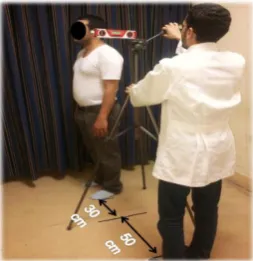
mounted on a camera tripod stand. The position of the tripod was adjusted until the bubble of the horizontal indicator and the central marking overlapped. The distance from the subject to the center of the camera stand was standardized at 0.3 m, while that between the operator and the camera stand was fixed at 0.5 m (figure 1).
Figure 1: Subject stands comfortably with body
weight evenly distributed on both feet while looking straight ahead as the tester measured the CVA. The floor was marked to standardize the instrument, subject and tester position.
Figure 2: Measuring CVA by the EHPI: the angle
Subject’s preparation: The seventh cervical (C7) spinous process was palpated and identified and an adhesive double sided tape with a pin marker was adhered to the skin. A second tape with a pin marker was fixed at the tragus of the ear. First, subject was instructed to stand comfortably with body weight evenly distributed on both feet. Then, the subject was asked to look straight ahead before he/she was asked to flex and extend the head for three times and finally to assume a comfortable
position to start the measurement.12
To determine the value of the CVA, the EHPI moved so that it was aligned parallel to an imaginary line passing between a mid-point of the tragus to C7 and reading of the digital display was
recorded(figure 2).11 The CVA was measured three
times and an average was calculated and used for statistical analysis.
2. Strength and endurance of DCF: the
cranio-cervical flexion test (CCFT) was used to assess the strength and endurance of DCF muscles (the longus capitis and colli). This is a valid and reliable
test.[13] Strength and endurance of the DCF muscles
were measured as follows:
Strength assessment: the subject was positioned in a crook lying position with PBU under the back of the head. The patient held the dial end of the unit to get instantaneous feedback during performance. The subject was then asked to slowly feel the back of his/her head slides up the bed in a head nod action, while elevating the target pressure at 2 mm Hg starting from 20 mm Hg. The amount of pressure that the subject was able to achieve and hold for 3-seconds with the correct craniocervical flexion action was recorded as the strength of the DCF. This test intra-rater reliability and inter rater reliability are 0.69 and 0.85, respectively. The assessor palpated the superficial flexors throughout the test to ensure that no substitution has occurred
throughout the test.7.14
Isometric endurance of the DCF: this test determines the pressure at which the subject is able to maintain the correct craniocervical flexion action. The subject performed the head nod action to first target pressure (the lowest level; 22 mm Hg) as described earlier, then the researcher instructed patients to hold that position for 10 seconds. If the subject could perform 3 repetitions of 10-second holds without substitution, the test was progressed at 2 mm Hg increments. This test intra-rater reliability and inter rater reliability are 0.68 and
0.70, respectively. 7.14
Home-based training: following baseline
assessment, subjects were randomly assigned into intervention and control groups using sealed
envelopes. The intervention group was instructed into a home based craniocervical training for 6 weeks; 2 times/day for 10 minutes. In addition, subjects in this group were seen once every week by the investigator to ensure proper application of exercise and to decide on exercise progression. Subjects in the control group were only assessed at baseline and 6-weeks later.
Craniocervical flexor muscles training: Subjects were instructed to gently nod their head as if they were saying 'yes'. The tester started with the target level on the CCFT that the subject could hold steadily for 10 seconds without substituting with the superficial neck flexor muscles or quick jerking craniocervical flexion movement. The tester monitored the activation of the superficial muscles by palpation. The training commenced at the pre-determined pressure level. The subject was then taught how to perform a slow and controlled craniocervical flexion action before being trained to progressively increase the amount of pressure using the feedback from the pressure sensor that was placed under the neck.
For each pressure target level, the contraction duration progressively increased to reach 10 seconds. Also, the number of repetitions performed increased to 10 times. When the duration of holding and number of repetitions were achieved, training progressed to the next pressure level in 2mmHg increments starting from baseline of 20 mmHg to the final level of 30 mmHg until the
5 pressure target levels were completed.9
Statistical analysis was done using SPSS for windows, version 21.0 (SPSS, Inc., Chicago, IL). Unpaired t-test was used to compare the demographic characteristics (age, sex, weight and height) at baseline. MANOVA test was done to compare the CVA as well DCF muscles strength and endurance as a function of patient’s grouping and time. All data are presented as means± standard deviation (SD). Bonferroni correction was done to adjust for repeated comparisons.
RESULTS
A total of 41 subjects completed the study. One subject dropped out from the training group because he failed to continue the exercise program. The primary outcome measures were CVA as well as DCF strength and endurance.
kg and height of 172.2(±8.46) cm. There were no significant differences between the two groups with regards to participants’ age, weight and height (p= 0.91, 0.27, 0.38, respectively).
1. Craniovertebral angle:
Regarding the intervention group, the mean CVA
at baseline was 49.63±5.89ᵒ. After six weeks of
training, this angle significantly increased to reach
a mean of 52.48±6.83ᵒ (p-value= 0.001).For the
control group, the mean CVA at baseline was
48.56±4.76ᵒ. After six-weeks follow up, the CVA
was not statistically different (48.50±4.55ᵒ;
p-value=0.95).At baseline, the CVA of intervention and control groups was not significantly different (p-value=0.63). However, after six weeks of
training, the intervention group showed
significantly greater CVA angle compared to that of the control group (p-value=0.03) (figure 3).
Figure 3: Baseline and six-weeks mean CVA for
intervention and control groups. * Indicates that the intervention group showed significant difference than the control group at week 6 (P<0.05).** Indicates that the Intervention group showed significant improvement in the CVA after 6 weeks compared to baseline values (p<0.05); denoting decreased forward head severity.
2. DCF strength
At baseline, the intervention group mean DCF strength was 24.05±1.67 mmHg. Strength significantly increased with training to reach 31.15±4.29 mmHg (p-value<0.001).For the control group, the strength of DCF was 23.52±1.66 mmHg at baseline. After six weeks, DCF strength did not significantly change (24.14±1.49 mmHg; p-value=0.40). Comparing the DCF strength at baseline between the two groups showed no significant difference (p-value=0.32). Six weeks later, the strength of DCF significantly increased in the intervention compared to that of the control group (p-value <0.001) (figure 4).
Figure 4: Baseline and six-weeks mean DCF
strength for intervention and control groups. * Indicates that the intervention group showed significant difference than the control group at week 6 (P<0.05). ** Indicates that the Intervention group showed significant improvement in the strength after 6 weeks compared to baseline values (p<0.05).
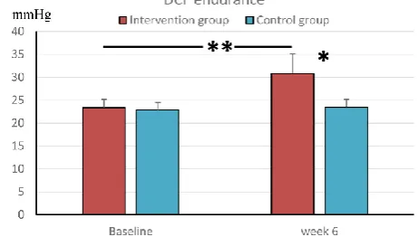
3. DCF endurance
For the intervention group, the mean endurance was 23.35 mm Hg±1.84 at baseline. After six weeks of training, the endurance significantly increased to reach 30.85mm Hg±4.24 (p-value<0.001).For the control group, the mean endurance measured at baseline was 22.86 mm Hg±1.62. Six weeks later, the endurance was not significantly different from baseline values (23.48 mm Hg±1.72) (p=0.79).Although the DCF endurance at baseline were not significantly different between the two groups (p=0.68), six weeks of training significantly improved the endurance in the intervention compared to the control group (p<0.001) (figure 5).
Figure 5: Baseline and six-week mean DCF
endurance for intervention and control groups. * Indicates that the intervention group showed significant difference than the control group at week 6 (P<0.05). ** Indicates that the Intervention group showed significant improvement in the endurance after 6 weeks compared to baseline values (p<0.05).
In this study, the primary outcome was the CVA. This angle was measured to assess the severity of FHP. There are many instruments that are used to assess FHP such as the Cervical Range of Motion
(CROM) instrument,[15] the plumb line and
photographic imaging.[16] However, these methods
have the disadvantages of being complicated procedures, expensive and inconvenient to use
clinically.[17] The EHPI was used to assess CVA.
This instrument was reported to have a high intra-rater reliability (ICC ranged from 0.86 to 0.94) and inter-rater reliability (ICC ranged from 0.85 to
0.91).[11]
It was hypothesized that the CVA would increase as a result of training. Based on the results, this hypothesis was accepted as evident by the significant increase of CVA in the intervention compared to the control group. Increased CVA is
an indicator of improved head posture.[15,18] The
improvement of CVA as a function of training shown in the current study is consistent with those findings reported by Falla et al, 2007 who studied head and thoracic postures during 10 minutes static computer posture after six weeks of DCF muscles
strength and endurance training.[9] Results showed
that strength training was associated with significant increase in CVA compared to endurance training. This indicates that despite the simplicity of this minimally supervised home-based training program, yet increasing subjects awareness of proper head posture and DCF muscle action may have had improved their proprioception and hence to the improvement seen in participants of the intervention group.
In this study, the control group did not show significant changes in CVA as a function of time (6 weeks). This is in an agreement with the finding that the CVA remained stable within a session, a
day, and over a 7-days period.[19]
Secondary outcome measures included the strength and endurance of DCF muscles. Strength and endurance were assessed by the CCFT. Several methods are used to evaluate the DCF function. These include craniocervical flexion test (CCFT), and electromyography (EMG). However, the CCFT is an easy, noninvasive, low-cost clinical test to
specifically assess and retrain DCF.[7,20]
For DCF strength and endurance, we hypothesized that DCF training would significantly improve DCF strength and endurance. Based on our results, these hypotheses were accepted as evident by the significant increase in DCF muscles strength and endurance in the intervention group compared to the control values. This could be explained by improvement of DCF muscle as a function of
training. Craniocervical flexion targets mainly the longus capitis and longus colli muscles rather the
superficial flexor muscles such as
sternocleidomastoid muscles and anterior scalene muscles. It is believed that such muscles play an important role in maintaining cervical lordosis and
improving cervical posture.[7–9,21,22]
It must be noted that it is not known whether the improvements in CVA and DCF muscles strength and endurance that were observed following 6-weeks of exercise intervention would be maintained in the long term. Additional research is warranted to address the long-term effect of this training. Also, different age population may have different responses to exercise, and thus the effect of age on response should be investigated.
CONCLUSION
Based on the current results, 6-weeks of home-based unsupervised deep cervical flexor training reduces forward head, as measured by the craniovertebral angle, and improves muscle strength and endurance in asymptomatic young adults. Thus, this exercise could be used as a preventive measure against the development of neck dysfunction in at risk population even before the onset of any symptoms.
REFERENCE
1. Haughie, L. J., Fiebert, I. M., & Roach KE.
Relationship of forward head posture and cervical backward bending to neck pain. J Man Manip Ther. 1995;3(3):91–7.
2. Bryden L. The influence of posture and
alteration of function upon the craniocervical
and craniofacial region. Craniofacial
Dysfunction and Pain. In: Craniofacial dysfunction and pain: manual therapy,
assessment, and management. London:
Butterworth-Heinemann; 2001. p.164-71.
3. Fejer R, Kyvik KO, Hartvigsen J. The
prevalence of neck pain in the world population: a systematic critical review of the literature. Eur Spine J. 2006;15(6):834–48.
4. Walker-Bone K, Reading I, Coggon D, Cooper C,
Palmer KT. The anatomical pattern and determinants of pain in the neck and upper
limbs: an epidemiologic study. Pain
2004;109:45–51.
5. Sommerich CM, Joines SM, Hermans V, Moon
SD. Use of surface electromyography to estimate neck muscle activity. J Electromyogr Kinesiol. 2000;10(6):377–98.
6. Szeto GPY, Straker LM, O’Sullivan PB. EMG
workers when challenged by different physical
stressors. J Electromyogr Kinesiol.
2005;15(6):544–55.
7. Jull, G. A., O’Leary, S. P., & Falla DL. Clinical
assessment of the deep cervical flexor muscles: the craniocervical flexion test. J Manip Physiol. 2008;31(7):525–33.
8. Jun I, Kim K. A Comparison of the Deep
Cervical Flexor Muscle Thicknesses in Subjects
with and without Neck Pain during
Craniocervical Flexion Exercises. J Phys Ther Sci. 2013;25(11):1373–5.
9. Falla D, Farina D, Dahl MK, Graven-Nielsen T.
Muscle pain induces task-dependent changes in cervical agonist/antagonist activity. J Appl Physiol. 2007;102(2):601–9.
10.Mullaney MJ, McHugh MP, Johnson CP, Tyler
TF. Reliability of shoulder range of motion comparing a goniometer to a digital level. Physiother Theory Pract. 2010;26(5):327–33.
11.Lau HMC, Chiu TTW, Lam T-H. Measurement
of craniovertebral angle with Electronic Head Posture Instrument: Criterion validity. J Rehabil Res Dev. 2010;47(9):911–8.
12.Wilmarth, M. A., & Hilliard TS. Measuring head
posture via the craniovertebral angle. Orthop Phys Ther Pr. 2002;14:13–5.
13.Arumugam, A., Mani, R., & Raja K. Interrater
reliability of the craniocervical flexion test in asymptomatic individuals—a cross-sectional
study.J Manipulative Physiol Ther.
2011;34(4):247–53.
14.Juul T, Langberg H, Enoch F, Sogaard K. The
intra- and inter-rater reliability of five clinical muscle performance tests in patients with and without neck pain. BMC Musculoskelet Disord. 2013;14:339.
15.Agustin CTS, Wilmarth MA, Raymond J,
Hilliard TS. The Amount and Variation of Craniovertebral Angle Changes in College-aged
Students Using One-strapped and Two-strapped
Backpacks and Bags. Orthop Pract.
2003;15(3):30–33.
16.Silva AG, Punt TD, Johnson MI. Reliability and
validity of head posture assessment by observation and a four-category scale. Man Ther. 2010;15(5):490–5.
17.Yip CHT, Chiu TTW, Poon ATK. The
relationship between head posture and severity and disability of patients with neck pain. Man Ther. 2008;13(2):148–54.
18.Watson DH, Trott PH. Cervical headache: an
investigation of natural head posture and upper
cervical flexor muscle performance.
Cephalalgia 1993;13:272–84; discussion 232.
19.Silva, A. G., Punt, T. D., & Johnson MI.
Variability of angular measurements of head posture within a session, within a day, and over a 7-day period in healthy participants. Physiother Theory Pract. 2011;27(7):503–11.
20.Falla D. Relationship between cranio-cervical
flexion range of motion and pressure change during the cranio-cervical flexion test. Man Ther. 2003;8(2):92–6.
21.Gupta BD, Aggarwal S, Gupta B, Gupta M,
Gupta N. Effect of Deep Cervical Flexor Training vs. Conventional Isometric Training on Forward Head Posture, Pain, Neck Disability Index In Dentists Suffering from Chronic Neck Pain. J Clin Diagn Res. 2013;7(10):2261–4.
22.Iqbal ZA, Rajan R, Khan SA, Alghadir AH. Effect
of deep cervical flexor muscles training using pressure biofeedback on pain and disability of school teachers with neck pain. J Phys Ther Sci. 2013;25(6):657–61.
Acknowledgments
The authors would like to thank Radwan, H, Balbaa for their scientific input and critical reading
Citation
Abdelnaeem, AO, Alaa Balbaa, A & Mahmoud, NF. (2015). THE SHORT-TERM EFFECT OF A HOME-BASED PROGRAM TO CORRECT FORWARD HEAD POSTURE IN ASYMPTOMATIC SUBJECTS.