IBBJ
Winter 2019, Vol 5, No 1
A General Overview of Chitosan and its Use in Dentistry
Begüm Erpaçal
1*, Özkan Adigüzel
2, Suzan Cangül
1, Musa Acartürk
11.Department of Restorative Dentistry, Faculty of Dentistry, Dicle University, Diyarbakir, Turkey.
2.Department of Endodontics, Faculty of Dentistry, Dicle University, Diyarbakir, Turkey.
Submitted 24 Mar 2019; Accepted 6 May 2019; Published 23 May 2019
Chitosan is a biocompatible material that has been researched in many areas in recent years. As it can be obtained
from renewable sources, has antimicrobial properties, is biocompatible, and has no toxic effect, chitosan is
currently a widely-used material. The use of chitosan was investigated in different areas related to dentistry.
Chitosan is used in the prevention of caries and wear, to increase the regeneration capability of the dentin pulp
complex, in pulpotomy to accelerate osteogenesis in guided tissue regeneration due to its hemostatic property,
and primarily to benefit from its antimicrobial activity by adding it to materials such as glass ionomer cement,
calcium hydroxide, and adhesive systems. The aim of this review was to examine the areas of use of chitosan in
dentistry, and to explain its antimicrobial activity in particular, and also its capability for regeneration of dental
tissues.
Keywords: Chitosan, dentistry, biopolymer, regeneration
hitin is the second most common biopolymer
found in the natural world after cellulose.
Chitosan, which is almost the only polysaccharide in
nature, is produced upon deacetylation of chitin in
the basic environment. Chitosan is the most
important product of chitin, which is a skeletal
material of arthropods, and is also found in the cell
walls of fungi. The general term is used to refer to
chitin which has reached approximately 50%
de-acetylation, although some sources only name it
chitosan when it has undergone 70% de-acetylation
(Figure 1).
Beta-1, 4-O-glycosyl associated with the
remnants of glycosamine is found in the basic
structure of chitosan, which has a linear structure.
The active regions of chitosan include 3 different
functional groups of amino and amido groups found
in C-2 location, and hydroxyl groups found in C-3
and C-6. Therefore, it can be easily derived.
Chitosan is actually a general term used for
compounds of different molecular weight obtained
in this way (2-6). Enzymatic hydrolysis following
deacetylation transforms chitosan into an
oligosaccharide, which is water-soluble and has low
molecular weight (7).
While chitosan is not itself soluble in water and
basic solvents, it dissolves well in water-based
solvents of organic acids and shows limited
solubility in inorganic acids. It is a material that is
resistant to digestive enzymes, and cannot be
digested by humans. It can be fragmented by some
bacteria, and can be bound in mammalian cells and
micro-organisms. At the same time, chitosan has
high viscosity, and may appear in gel form, and has
high water binding capacity. The biocompatibility of
this material renders it suitable for use in many
areas, and it has the additional advantage of being
obtained from renewable sources (4, 8, 9).
C
Review Article
Chitosan has anti-microbial, anti-oxidant, and
anti-tumor effects. Bone formation is accelerated by
chitosan through increasing osteoblasts formation in
bone tissue, and it has also the capability of
connective tissue regeneration. It has a suppressing
role on the central nervous system, and may act as a
stimulant of the immune system. It also has effects
on intestinal motility, gastrointestinal system and
liver functions regulation, and blood pressure
reduction. Chitosan has also been reported to have
an assistive role in the control of cholesterol and
hemostasis (4, 8).
Areas of use
Chitosan is widely used in several areas such
as healthcare, pharmaceuticals, agriculture, food
production, tissue engineering and cosmetics. The
most important areas requiring the specificity of
chitosan are cosmetic products and pharmaceutical
and biomedical fields. It is used as a hemostatic and
anticoagulant agent in acne treatment and in hair
care products to preserve moisture in the hair and
skin. The use of chitosan is increasing especially in
wound plasters, as absorbable suture material, in
contact lenses, cosmetic products, food packaging,
medical textiles, drug distribution systems and even
as an artificial skin scaffold in a water filtration
environment. In the food industry, it is used in many
areas because of the antimicrobial effect, and when
used as an additive material or with the aim of
increasing stabilisation of the nutrients, the shelf-life
of the product is increased.
Chitosan is also useful for colour stabilisation
or to increase the effect of sweeteners in foodstuffs
(4, 6, 10). It is used in water purification, as a
flocculator in clarification, and in the removal of
metal ions from water (11, 12). In agriculture it is
used for the stimulation of fruit and vegetable
defence systems in fertilizers, and to accelerate
Figure 1. The chemical structure of chitosan (1).
Figure 2. The properties of chitosan and areas of application.
growth (13). Benefit is gained from chitosan in the
pharmaceutical industry in creating capsules for
drugs to increase absorption by binding to
negative-charged cell membranes, in enzyme immobilisation,
surface modification, bone regeneration, nerve
regeneration, wound healing, and wound care.
Recently, chitosan has also become a popular
material for weight loss by reducing absorption as it
binds to fatty acids (6, 14) (2).
Antimicrobial activity
The most important property of chitosan
derivatives is the antimicrobial activity exerted
against bacteria, viruses, fungi, and even algae.
Yeasts and moulds are most sensitive to chitosan,
followed by gram positive and gram negative
bacteria. Chitosan inhibits bacteria mostly through
bacteriostatic routes. It has been shown to have an in
vitro antibacterial effect on Streptococcus mutans,
Aggregatibacter actinomycetemcomitans, and
Porphyromonas gingivalis (8, 15,16). A study by
Fujiwara et al. was the first to report that
water-soluble chitosan showed an inhibitory effect on
Streptococcus mutans related to caries.
It has been said that the antibacterial activity
of chitosan could originate from a combination
of bacteria cell binding and DNA binding
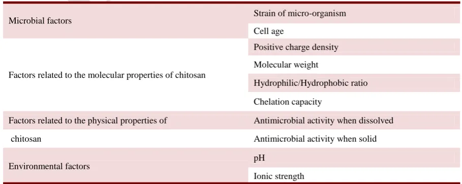
mechanisms (17). Factors affecting the antibacterial
activity of chitosan can be classified into 4 groups
(8) (Table 1).
Chitosan shows antioxidant activity by
transforming different components into stable
molecules through neutralisation of free radicals and
binding to metal ions. Hydroxyl OH) and amino
(-NH) groups are the basic groups responsible for this
antioxidant activity (8, 18).
The antibacterial effect of restorative materials
renders bacteria ineffective and prevents recurrent
caries. Therefore, the addition of chitosan which is
biocompatible with a proven antibacterial effect, to
materials is extremely important in dentistry (16).
The use of chitosan in dentistry
Studies conducted in the field of dentistry have
used chitosan for the prevention of tooth decay as it
eliminates bacteria and/or provides bacteriostatic
properties (Table 2). Low molecular weight chitosan
has been reported to prevent the adsorption of
Streptococcus mutans to hydroxyapatite. It has also
been suggested that it is a bio-adhesive polymer that
can be retained for a long time on oral mucosa (16,
19). The use of chitosan in dentistry is increasing as
it both triggers regeneration and has antibacterial
properties (Figure 3).
Guided tissue regeneration
The application of chitosan has been studied in
the formation of new bone in orthopaedics and
periodontology. It has superior properties compared
Factors affecting the antibacterial activity of Chitosan
Microbial factors Strain of micro-organism
Cell age
Factors related to the molecular properties of chitosan
Positive charge density
Molecular weight
Hydrophilic/Hydrophobic ratio
Chelation capacity
Factors related to the physical properties of Antimicrobial activity when dissolved
chitosan Antimicrobial activity when solid
Environmental factors pH
Ionic strength
Table 1. Clinical characteristics of recurring patients and two months after relapse patients
to synthetic membranes. These advantages render it
more flexible in a wet environment, and its structure
is similar to that of glycosaminoglycan, which are
found in the bone extracellular matrix. Also,
chitosan has a positive surface charge and the
hydrophilic property affects the differentiation,
production and adhesion of the cell to the surface
(10, 20).
Bone cements are used in orthopaedic surgery
and dentistry and they are widely prepared from
polymethylacrylate. Chitosan is added to the
prepared bone cement to increase biocompatibility.
A positive effect on mechanical properties has been
observed with the addition of chitosan (21).
Chitosan is an ideal membrane material for guided
tissue regeneration and guided bone regeneration,
but the mechanical resistance is insufficient and
bioactivity must be improved. Therefore, chitosan is
widely used in combination with various osteogenic
materials. When itis used in combination with
inorganic materials such as hydroxyapatite,
tricalcium phosphate, silica and bioactive glass, it
has been shown to have better mechanical resistance
and osteogenic properties (9, 10).
Implant coating
Coating dental implants with chitosan may
affect the surface-bone interface by changing the
surface properties of the implant. For example,
chitosan coating changes the elasticity module,
thereby reducing the compatibility between the
implant surface and the alveolar bone and the stress
concentration areas. Chitosan coatings may also be
potentially used for the localised application of
various drugs such as antibiotics around the implant
area (7, 22). Several studies have reported promising
results for chitosan coating of dental implants (23).
Hemostasis and pulpotomy
Chitosan is one of the commonly used
substances to bring bleeding under control. Celox
(SAM Medical Products, Newport, OR, USA) is an
effective hemostatic agent that contains chitosan.
The positive charged Celox binds to negative
charged erythrocytes and drives a reaction in direct
contact with blood. Many studies have shown the
efficacy of chitosan as a hemostatic agent for mass
bleeding. While chitosan granules are successful in
controlling hemorrhage, they continue to have the
majority of assistive properties of an ideal
hemostatic wound cover. This agent is a fine granule
product, which works by entering into a direct
interaction with red blood cells and thrombocytes to
form a cross-linked clot barrier independently of
natural factors (5, 24-26).
Chitosan has started to be used as a hemostatic
agent in the treatment of milk tooth pulpotomy. In
pulpotomy, chitosan diluted with sterile saline is
applied to the pulp chamber following removal of
the crown pulp, left for 15-20 seconds and
Figure 3.Uses of Chitosan in dentistry. GIC: glass ionomer cement; GTR: guided tissue regeneration.
hemostasis is obtained. Chitosan increases the
formation of reparative dentin and the formation of
hard tissue, and has therefore been reported to be an
appropriate material for pulpotomy (6, 27).
Modification of glass ionomer cement
Chitosan can be added to traditional glass
ionomer cement (GIC) to increase the effect of
protein and growth factor release, primarily used in
the treatment of vital pulp (7). Various filling
materials have been added to improve the
mechanical properties of traditional GICs. In a
previous study, the effect of the addition of chitosan
nanoparticles to GIC (NCH-GIC) content was
evaluated on fluoride release and resistance to
pressure, bending and wear, and these results were
compared with traditional GIC (TGIC). The results
of the study showed that the resistance to bending of
NCH-GIC was much higher than that of TGIC, and
it was concluded that the addition of chitosan had
increased the resistance to wear. Fluoride release
throughout 7 days was found to be significantly
higher in NCH-GIC than in TGIC. From the results
of that study, it can be said that the addition of
nano-chitosan to GIC could develop the anti-cariogenic
and mechanical properties for high-resistance
applications (28). The results of another study,
which examined the bovine serum albumin (BSA)
expression profile from chitosan-modifed GIC,
showed that there was no cytotoxic effect on pulp
cells when compared with TGIC.
These results, which show a protective effect
against toxicity originating from remnant traces of
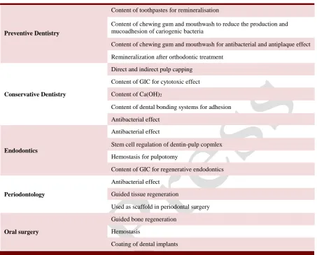
Chitosan uses in dentistry specialities
Preventive Dentistry
Content of toothpastes for remineralisation
Content of chewing gum and mouthwash to reduce the production and mucoadhesion of cariogenic bacteria
Content of chewing gum and mouthwash for antibacterial and antiplaque effect
Remineralization after orthodontic treatment
Conservative Dentistry
Direct and indirect pulp capping
Content of GIC for cytotoxic effect
Content of Ca(OH)2
Content of dental bonding systems for adhesion
Antibacterial effect
Endodontics
Antibacterial effect
Stem cell regulation of dentin-pulp copmlex
Hemostasis for pulpotomy
Content of GIC for regenerative endodontics
Periodontology
Antibacterial effect
Guided tissue regeneration
Used as scaffold in periodontal surgery
Oral surgery
Guided bone regeneration
Hemostasis
Coating of dental implants
Table 2. The uses of chitosan in different areas of dentistry
GIC: glass ionomer cement.
GIC and from hydroxyethyl metacrylate elements,
may represent a significant advance in the
development of pulp-friendly restorative materials.
Thus, chitosan-modified GIC may be able to be
presented for routine use in bioactive dental
restorations as an effective pulp treatment material
and in the field of regenerative endodontics (7).
In a study that was conducted to evaluate the
efficacy of chitosan nanoparticles (CNP) added to a
calcium hydroxide material to eliminate bacterial
biofilms, it was seen that when this combination was
used it could be potentially useful in respect of
eliminating bacteria in long and short-term
exposures (29).
Prevention of caries
The addition of chitosan to chewing gum and
mouth washes reduces cariogenic bacteria, and
stimulates the flow and amount of saliva.
Consequently, bacterial plaque is inhibited at the
rate of 80% (8, 9, 30). The antibacterial properties
and biocompatibility of chitosan are highly desired
properties in dental materials, and as chitosan is not
water-soluble, the material can be retained in the
oral cavity (16, 17).
The chewing of gum containing chitosan
inhibits the proliferation of Streptococcus mutans
which is a cariogenic bacteria. In a study related to
gum containing chitosan, a reduction in the total
number of bacteria was observed in the subjects who
chewed gum in comparison with those who did not,
and the expression of saliva was also found to have
significantly increased (9). In another study
conducted to evaluate whether or not gum
containing chitosan effectively suppressed the
development of oral bacteria in the saliva, a
significant reduction in the amount of oral bacteria
was found in the chitosan group. These results
showed that chitosan was an effective material for
controlling the number of cariogenic bacteria in
situations where brushing the teeth is difficult (31).
Water-soluble chitosan is known to directly inhibit
the growth of the typical cariogenic bacteria,
Streptococcus mutans, at pH 6.5 without causing
demineralisation of the tooth surface. When chitosan
is used as a mouthwash, it shows an antibacterial and
plaque-reducing effect (32). However, the
application of chitosan as a chemical agent in
mouthwashes or toothpaste is limted as it is not
water-soluble (16, 33).
With the use of a chitosan-based, non-fluoride
toothpaste (Chitodent, B&F Elektro GmbH, Filsum,
Germany), studies have shown a significant loss of
dental hard tissue. By developing the anti-wear
property of tin-based toothpastes in acid oral
environments, the addition of chitosan has increased
the effect of reducing tissue loss (7). In a study
which evaluated toothpastes containing chitosan and
propolis in respect of remineralisation of
demineralised enamel, the lowest wear value was
observed in the group using Chitodent containing
chitosan (34). Another study investigated the wear
prevention potential of materials, and it was reported
that the AmF / NaF / SnCl2 /chitosan formula reduced tissue loss by 20-25% in the absence of an
organic matrix (35).
When the effects on enamel wear of
toothpastes containing fluoride, tin and chitosan
(F/Sn/chitosan) were compared, the substance loss
of the chitosan group was found to be significantly
lower than that of the other groups (36). It has been
shown that toothpaste containing F/Sn/chitosan
could provide good protection for patients who
frequently consume acidic food (37). All the studies
support the view that chitosan is an effective
material that could be used to decrease dental hard
tissue losses.
Mucoadhesive nanofibre polymers have been
developed using chitosan and thiolated chitosan
(CS-SH). While CS-SH causes higher
mucoadhesion, the synergic antibacterial activity of
the agents has shown rapid release of the active
substances. It reduced bacteria in the oral cavity and
provided good mucoadhesion without cytotoxicity.
The results of a previous study showed that
polymers containing chitosan had the potential to
protect oral hygiene by reducing the production of
bacteria that cause dental caries (38).
Adhesion and dentin bonding
Previous studes have recommended chitosan as an element of an “etch and rinse” adhesive system to effectively increase the resistance of dental
restorations (39). The application of
chitosan-antioxidant gels on dentin has a positive effect on
impermeability because it increases the bonding of
composite resins. In a study conducted to evaluate
the capability of increasing bonding strength after
the adhesives of chitosan-antioxidant hydrogels, it
was reported that both the application of phosphoric
acid of the material and the form of application
significantly improved adhesion to the tooth (40).
Chitosan-based alternative materials are also
noticeable in adhesive systems. Relative values have
been accepted as significantly higher than those of
traditional adhesive systems with or without the
application of phosphoric acid (7). In one study,
chitosan added to adhesives reduced collagen
destruction starting with endogenous matrix
metalloproteinase, and prevented water permeation
in hybrid layers suggesting a role for chitosan in
eliminatingbacteria from dentin surfaces (41).
However, in another study, an experimental
adhesive system containing chitosan showed that the
effects against S. mutans and L. casei were similar
to those of the traditional 2-stage adhesive system,
and it was stated that the addition of chitosan did not
affect the antimicrobial effect against these bacteria
(42). According to previous studies, the amount of
chitosan affects the bonding strength. In comparison
of adhesives containing 0.12% and 0.25% chitosan
with a control group, no signficant difference was
observed in shear bonding strength, while the
addition of 0.5% and 1% chitosan significantly
reduced bonding (43).
Enamel and dentin repair with remineralization
Chitosan is used as a scaffold in various tissue
engineering applications. Degraded products are not
toxic or allergenic, but their interactions with cells
are disrupted by less hydrophilicity and poor
cytocompatibility. There are studies showing that
chitosan monomers have the capacity to support the
regeneration of dental pulp injuries, and chitosan is
accepted as a potential scaffold for dental pulp cells.
To improve the biological properties of chitosan,
collagen is added to support cell adhesion and
migration (44-46).
Research related to chitosan
The aim of regenerative dentistry is the
development of biomaterials that can accelerate the
regeneration of the dentin-pulp complex (47). The
combination of the good biomaterial properties
found in chitosan structure scaffolds with the
cellular properties desired in human dental pulp
stem cells (hDPSC) provides an interesting
approach for tissue engineering applications.
However, when there is no adhesion property to
support direct cell fixation, chitosan requires
scaffold surface modification to support stem cell
adhesion (48).
In a study that evaluated the behaviour of
human dental pulp cells (DPC) which had been
cultured on chitosan membranes, it was reported that
chitosan membranes supported the collection of
DPCs for the formation of multi-cell spheroids. The
growing of DPCs on chitosan membranes has been
recommended as a promising method for the
production of 3D multi-cell spheroids in DPC-based
tissue regeneration and therapeutic applications
(49).
In an experimental canine study, no
statistically significant difference was observed
radiologically in apical thickening and root length
between structure scaffolds containing and not
containing chitosan. Histologically, while
pulp-dentin tissue regeneration was observed in teeth with
the scaffold containing chitosan, no tissue
regeneration was seen in the absence of chitosan. It
was concluded that hDPSC and growth factors
added to chitosan hydrogel could contribute to
regeneration by forming pulp-dentin-like tissue
(50). It was reported in another study that chitosan
supported cell vitality and proliferation throughout
14 days in scaffold culture (51).
In a study that investigated the differentiation
of pulp cells through the combination of hDPSC
with chitosan gel scaffolds, rapid binding to
collagen and gelatine and proliferation were
observed, but chitosan did not support appropriate
cell growth. Chitosan could trigger only 61% of the
cell mass of cells coated with type I collagen, which
demonstrates that chitosan is less appropriate than
collagen in respect of cell growth. However, this
does not show that chitosan is an unsuccessful
material, as it was 39% less successful than collagen
(45). In studies that have compared chitosan,
gelatine and collagen, cell retention and alkaline
phosphatase activation were mostly observed in
collagen and gelatine, whereas no regular cell
increase was seen in chitosan. Therefore, hybrid
tissue scaffolds have been developed. It has been
reported that hybrid tissue scaffolds containing bone
morphoprotein-7-weighted chitosan/collagen have
increased odontogenic differentiation (52).
Amelogenin-chitosan (CS-AMEL) hydrogel
has been reported as a promising material for in situ
enamel growth in the future. During the enamel
remineralisation of CS-AMEL hydrogel, the
amelogenin mechanisms promote a micro-structure
formation with a similar organisation to enamel,
which is very important for a successful
reconstruction. CS-AMEL hydrogel has been
reported to be a promising material for surface
enamel repair (53). In another study, chitosan-based
hydrogel was used as the distribution medium for
the application of amelogenin to the enamel. The use
of chitosan did not affect the enamel crystal
orientation and provided a protective effect against
the development of secondary caries, corresponding
to antibacterial properties. Chitosan-based
restor-ative formulations have been shown to be able to
provide enamel regeneration by delivering organic
amelogenin to the area of the enamel defects (7).
In a study that evaluated the cytotoxic effects
of an experimental plaster-based biomaterial
prepared with chitosan, no cytotoxic effects of
chitosan on the cultured stem cells of an extracted
milk tooth was reported. The fact that there was no
cytotoxicity of such a promising material has
widened the area of use for the material in
restorative dentistry (54). Another study performed
in vitro evaluations of the cytoxicity of chitosan
derivatives on cells, and all the chitosan derivatives
tested showed low cytotoxicity. These results
demonstrated that chitosan derivatives carrying
quaternary ammonium salts could be used as good
biomaterials (55).
The findings available in literature show that
the application of natural materials such as chitosan
is useful both for oral health and quality of life (9).
There is a need for many further studies including
disciplines such as tissue engineering, biomolecular
science and material science, to investigate the
potential of chitosan in regeneration applications on
dental tissues (7).
Conclusion
Chitosan, which has come to the forefront in
the development of dental materials with
antibacterial activity and biocompatibility, is a
promising agent in this field. There is an increase in
studies related to materials which have no toxic
effects while accelerating the regeneration of the
dentin-pulp complex, especially in regenerative
dentistry. The regeneration capability is valid not
only for the dentin-pulp complex, but is also
effective on other dental tissues with mineral
remineralisation. While previous studies have
supported odontogenic differentiation, it has also
been clearly shown that there is no cytotoxic effect.
In the light of current findings, it can be said that
increased use of chitosan in these areas will be
beneficial in respect of treatment success.
Nevertheless, there is a need for further studies for
the effective use of chitosan.
Conflict of interest
The authors declared no conflict of interest.
References
1. Oğuzhan Yıldız P and Yangılar F. The Use of Chitosan in the
Food Industry. Erciyes University Journal of the Institute of Science and Technology. 2014;30:198-206 [Article In Turkish]. 2. Ekici S, Işıkver Y, Saraydın D. Poly(Acrylamide-Sepiolite) Composite Hydrogels: Preparation, Swelling and Dye Adsorption Properties. Polymer Bulletin. 2006;57:231-41.
3. Li D H, Liu L M, Tian K L. Synthesis, biodegradability and cytotoxicity of water-soluble isobutylchitosan. Carbohydr Polym. 2007;67:40-5.
4. Bostan K, Aldemir T, Aydın A. Chitosan and its antimicrobial activity. Turkish Microbiology Society Journal. 2007;37:118-27 [Article In Turkish].
5. Pozza M and Millner R W. Celox (chitosan) for haemostasis in massive traumatic bleeding: experience in Afghanistan. Eur J Emerg Med. 2011;18:31-3.
6. Rinaudo M. Chitin and chitosan: properties and applications. Prog Polym. 2006;31:603-32.
7. Husain S, Al-Samadani K H, Najeeb S, et al. Chitosan Biomaterials for Current and Potential Dental Applications. Materials (Basel). 2017;10:602.
8. Yildirim Z, Oncul N, Yıldırım M. Chitosan and its antimicrobial properties. Omer Halisdemir University Journal of Engineering Sciences. 2016;5:19-36 [Article In Turkish]. 9. Hayashi Y, Ohara N, Ganno T, et al. Chitosan-containing gum chewing accelerates antibacterial effect with an increase in salivary secretion. J Dent. 2007;35:871-4.
10. Hurt A P, Getti G, Coleman N J. Bioactivity and biocompatibility of a chitosan-tobermorite composite membrane for guided tissue regeneration. Int J Biol Macromol. 2014;64:11-6.
11. Kuzgun N and Inanlı G. Chitosan production and properties of chitosan. Turkish Journal of Scientific Compilations. 2013;6:16-21 [Article In Turkish].
12. Demir A and Seventekin N. Chitin, chitosan and general application areas. Electron J Textile Techn. 2009;3:92-103. 13. Sharif R, Mujtaba M, Ur Rahman M, et al. The Multifunctional Role of Chitosan in Horticultural Crops; A Review. Molecules. 2018;23:872.
14. Younes I and Rinaudo M. Chitin and chitosan preparation from marine sources. Structure, properties and applications. Mar Drugs. 2015;13:1133-74.
15. Kong M, Chen X G, Xing K, et al. Antimicrobial properties of chitosan and mode of action: a state of the art review. Int J Food Microbiol. 2010;144:51-63.
16. Kim J S and Shin D H. Inhibitory effect on Streptococcus mutans and mechanical properties of the chitosan containing composite resin. Restor Dent Endod. 2013;38:36-42.
17. Chen C Y and Chung Y C. Antibacterial effect of water-soluble chitosan on representative dental pathogens Streptococcus mutans and Lactobacilli brevis. J Appl Oral Sci. 2012;20:620-7.
18. Dutta P K, Dutta J, Tripathi V S. Chitin and chitosan: Chemistry, properties and applications. J Sci Ind Res. 2004;63:20-31.
19. Rabea E I, Badawy M E, Stevens C V, et al. Chitosan as antimicrobial agent: applications and mode of action. Biomacromolecules. 2003;4:1457-65.
20. Cheung R C, Ng T B, Wong J H, et al. Chitosan: An Update on Potential Biomedical and Pharmaceutical Applications. Mar Drugs. 2015;13:5156-86.
21. Zou Q, Li Y, Zhang L, et al. Characterization and cytocompatibility of nano-hydroxyapatite/chitosan bone cement with the addition of calcium salts. J Biomed Mater Res B Appl Biomater. 2009;90:156-64.
22. Leong K F, Chua C K, Sudarmadji N, et al. Engineering functionally graded tissue engineering scaffolds. J Mech Behav Biomed Mater. 2008;1:140-52.
23. Bumgardner J D, Chesnutt B M, Yuan Y, et al. The integration of chitosan-coated titanium in bone: an in vivo study in rabbits. Implant Dent. 2007;16:66-79.
24. Kozen B G, Kircher S J, Henao J, et al. An alternative hemostatic dressing: comparison of CELOX, HemCon, and QuikClot. Acad Emerg Med. 2008;15:74-81.
25. Fathi P, Sikorski M, Christodoulides K, et al. Zeolite-loaded alginate-chitosan hydrogel beads as a topical hemostat. J Biomed Mater Res B Appl Biomater. 2018;106:1662-71.
26. Koksal O, Ozdemir F, Cam Etoz B, et al. Hemostatic effect of a chitosan linear polymer (Celox(R)) in a severe femoral artery bleeding rat model under hypothermia or warfarin therapy. Turkish Journal of Trauma and Emergency Surgery. 2011;17:199-204.
27. Delikan E. Hemostatic agents for pulpotomy treatment. Yediyepe Klinik. 2018;14:109-16 [Article In Turkish]. 28. Kumar S R, Ravikumar N, Kavitha S, et al. Nanochitosan modified glass ionomer cement with enhanced mechanical properties and fluoride release. Int J Biol Macromol. 2017;104:1860-5.
29. Del Carpio-Perochena A, Kishen A, Felitti R, et al. Antibacterial Properties of Chitosan Nanoparticles and Propolis Associated with Calcium Hydroxide against Single- and Multispecies Biofilms: An In Vitro and In Situ Study. J Endod. 2017;43:1332-6.
30. Ş. T M and Ş. A. Evaluation of the effects of various gums on oral and dental health. Acta Odontologica Turcica. 2015;32:42-6. 31. Hayashi Y, Ohara N, Ganno T, et al. Chewing chitosan-containing gum effectively inhibits the growth of cariogenic bacteria. Arch Oral Biol. 2007;52:290-4.
32. Fujiwara M, Hayashi Y, Ohara N. Inhibitory effect of water-soluble chitosan on growth of Streptococcus mutans. New Microbiol. 2004;27:83-6.
33. Bae K, Jun E J, Lee S M, et al. Effect of water-soluble reduced chitosan on Streptococcus mutans, plaque regrowth and biofilm vitality. Clin Oral Investig. 2006;10:102-7.
34. Ozalp S and Tulunoglu O. SEM-EDX analysis of brushing abrasion of chitosan and propolis based toothpastes on sound and artificial carious primary enamel surfaces. Int J Paediatr Dent. 2014;24:349-57.
35. Ganss C, Klimek J, Schlueter N. Erosion/abrasion-preventing potential of NaF and F/Sn/chitosan toothpastes in dentine and impact of the organic matrix. Caries Res. 2014;48:163-9. 36. Carvalho T S and Lussi A. Combined effect of a fluoride-, and chitosan-containing toothpaste and stannous-containing rinse on the prevention of initial enamel erosion-abrasion. J Dent. 2014;42:450-9.
37. Schlueter N, Klimek J, Ganss C. Randomised in situ study on the efficacy of a tin/chitosan toothpaste on erosive-abrasive enamel loss. Caries Res. 2013;47:574-81.
38. Samprasit W, Kaomongkolgit R, Sukma M, et al. Mucoadhesive electrospun chitosan-based nanofibre mats for dental caries prevention. Carbohydr Polym. 2015;117:933-40. 39. Diolosà M, Donati I, Turco G. Use of methacrylate-modified chitosan to increase the durability of dentine bonding systems. Biomacromolecules. 2014;15:4606-13.
40. Perchyonok V T, Zhang S, Grobler S R, et al. Insights into and relative effect of H, H-propolis, chitosan-H-propolis-nystatin and chitosan-H-nystatin on dentine bond strength. Eur J Dent. 2013;7:412-8.
41. Gu L S, Cai X, Guo J M, et al. Chitosan-Based Extrafibrillar Demineralization for Dentin Bonding. J Dent Res. 2019;98: 186-93.
42. Lobato M F, Turssi C P, Amaral F L, et al. Chitosan incorporated in a total-etch adhesive system: antimicrobial activity against Streptococcus mutans and Lactobacillus casei. Gen Dent. 2017;65:62-6.
43. Elsaka S E. Antibacterial activity and adhesive properties of a chitosan-containing dental adhesive. Quintessence Int. 2012;43:603-13.
44. Yang X, Han G, Pang X, et al. Chitosan/collagen scaffold containing bone morphogenetic protein-7 DNA supports dental pulp stem cell differentiation in vitro and in vivo. J Biomed Mater Res A. 2012.
45. Kim N R, Lee D H, Chung P H, et al. Distinct differentiation properties of human dental pulp cells on collagen, gelatin, and chitosan scaffolds. Oral Surg Oral Med Oral Pathol Oral Radiol Endod. 2009;108:e94-100.
46. Matsunaga T, Yanagiguchi K, Yamada S, et al. Chitosan monomer promotes tissue regeneration on dental pulp wounds. J Biomed Mater Res A. 2006;76:711-20.
47. Soares D G, Anovazzi G, Bordini E a F, et al. Biological Analysis of Simvastatin-releasing Chitosan Scaffold as a Cell-free System for Pulp-dentin Regeneration. J Endod. 2018;44:971-6.e1.
48. Sana A F, Capkin Yurtsever M, Kaynak Bayrak G, et al. Spreading, proliferation and differentiation of human dental pulp stem cells on chitosan scaffolds immobilized with RGD or fibronectin. Cytotechnology. 2017;69:617-30.
49. Hsieh H Y, Young T H, Yao C C, et al. Aggregation of human dental pulp cells into 3D spheroids enhances their migration ability after reseeding. J Cell Physiol. 2018;234:976-86. 50. El Ashiry E A, Alamoudi N M, El Ashiry M K, et al. Tissue Engineering of Necrotic Dental Pulp of Immature Teeth with Apical Periodontitis in Dogs: Radiographic and Histological Evaluation. J Clin Pediatr Dent. 2018;42:373-82.
51. Bakopoulou A, Georgopoulou A, Grivas I, et al. Dental pulp stem cells in chitosan/gelatin scaffolds for enhanced orofacial bone regeneration. Dent Mater. 2019;35:310-27.
52. Erişken C, Aksel H. Tissue Scaffolds Used in Pulp Regeneration (Pulp Regeneration). Turkey-Special Topics Clinical J Endod. 2017;3:187-96 [Article In Turkish].
53. Ruan Q, Siddiqah N, Li X, et al. Amelogenin-chitosan matrix for human enamel regrowth: effects of viscosity and supersaturation degree. Connect Tissue Res. 2014;55 Suppl 1:150-4.
54. Subhi H, Reza F, Husein A, et al. Cytotoxicity of gypsum-based biomaterial for direct pulp capping using stem cells from human exfoliated deciduous teeth. J Conserv Dent. 2018;21 :21-5.
55. Wei L, Li Q, Chen Y, et al. Enhanced antioxidant and antifungal activity of chitosan derivatives bearing 6-O-imidazole-based quaternary ammonium salts. Carbohydr Polym. 2019;206:493-503.