1S. Tulasi Ratna
2Praveen Dowle
3Dr.V.B.N.Prasad
4Dr. Rakhee K. Paruchuri
CORRESPONDING AUTHOR
1 S. Tulasi Ratna
MPT (Musculoskeletal Sciences),
College of Physiotherapy, Nizam’s Institute of Medical Sciences
Int J Physiother. Vol 2(4), 587-593, August (2015) ISSN: 2348 - 8336
ABSTRACT
Background: Plantar fasciitis is one of the most common musculoskeletal disorders of foot. The pain and discomfort associated with this condition has a dramatic impact on physical mobility and function. Treatment of this condition is usually conservative; however, review of literature revealed no clinical studies demonstrating the efficacy of any targeted treatment for this condition.
Methods: This was a prospective study which included 60 subjects with plantar fasciitis, who were randomly divided into two groups. Subjects in group I received ultrasound and exercise therapy while subjects in group II received kinesio tape in addition to ultrasound and exercise therapy. Patients were evaluated at the beginning of the study and at the end of three weeks using VAS (visual analogue scale) for pain intensity, PFPS (plantar fasciitis pain / disability scale) for functional ability and ultra sonography for any change in the caliber of plantar fascia.
Results: A statistically significant difference in improvement was noted within the groups and between the groups in terms of visual analogue scale and plantar fasciitis pain /disability scale score (p<0.05). Although a statistically significant difference was seen in the plantar fascia thickness by ultrasound in both the groups pre and post treatment, it was insignificant when both groups were compared to each other (p>0.05).
Conclusion: Kinesio taping can be used as an adjunct to conventional therapy in clinical application for greater improvement in pain levels and functional ability in patients with plantar fasciitis.
Key words:plantar fasciitis, kinesio tape, ultrasonography, stretching.
DOI: 10.15621/ijphy/2015/v2i4/67737
2MPT (Musculoskeletal Sciences), Faculty of Physiotherapy, College of Physiotherapy, Nizam’s Institute of Medical Sciences. 3Professor and HOD, Department of Orthopeadics, Nizam’s Institute of Medical Sciences.
4Assistant Professor, Department of Radiology and Imagelogy, Nizam’s Institute of Medical Sciences.
www.ijphy.org
INTRODUCTION
The plantar fascia is a thick, fibrous, relatively inelastic sheet of connective tissue which originates from the medial heel, passes over the superficial musculature of the foot and inserts onto the base of each toe and acts like a bowstring to maintain and provide support for the longitudinal arch of the foot and to assist with dynamic shock absorption.1
Plantar fasciitis is the most common cause of inferior heel pain.2 The word ‘fasciitis’ assumes inflammation of the plantar fascia, however recent research suggests that plantar fasciitis may manifest as non-inflammatory, degenerative process and should more aptly be termed as “plantar fasciosis”.3
Plantar fasciitis affects about 10% of the population at least once in a lifetime and non athletic population are affected more than the athletic population.4 Under normal conditions, the plantar fascia performs its function appropriately without incurring injury but in the presence of risk factors, such as, excessive foot pronation (pes planus), obesity (body mass index greater than 30 kg per m2), prolonged standing/walking occupations (e.g., teachers, construction workers etc), excessive running etc., may cause micro-tears in the plantar fascia. This repetitive trauma exceeds the fascia’s ability to recover and may lead to degenerative changes with an increased risk of injury ultimately leading to calcification of the fascia and enthesopathy.5
Clinical presentation is pain in the medial plantar region of the heel with the first few steps taken on rising in the morning or after an extended refrain from weight-bearing activity. Diagnosis of plantar fascitiis is usually made on the basis of history and physical examination. Diagnostic imaging is helpful to rule out other causes of heel pain. Ultrasonography is an inexpensive tool which can objectify the plantar fasciitis and rule out any other soft tissue pathology of the heel. Findings that support the diagnosis of plantar fasciitis include proximal plantar fascia thickness greater than 4 mm and hypoechoic areas within the plantar fascia.6
Treatment of this condition is usually conservative. Several research studies using different conservative treatments both individually and in combination have concluded that conservative treatment yielded better response in terms of relief from worst pain and also reduction from the first step of morning pain.7 Invasive management such as fenestration and steroid injections are also available, however, so far, there are no clinical
studies to demonstrate the efficacy of any particular intervention i.e., there are no studies to determine which particular treatment approach is the most effective for treating plantar fasciitis. Recently published clinical practice guidelines reported that there is level 1 evidence for kinesio taping in treating heel pain.8 The purpose of this study was to evaluate the efficacy of kinesio tape in adjunct to conventional therapy which includes therapeutic ultrasound and exercise therapy. Kinesio taping was originally developed by Japanese chiropractor Dr. Kenso Kase in the 1970s.9 Kinesio tape is 100% cotton, hypoallergenic, latex free, non-restrictive elastic adhesive tape designed to have the same amount of stretch as human skin and provides a multitude of uses as a protective and rehabilitative taping technique. Kinesio tape, has unique elastic properties that allow it to provide dynamic support, protecting the muscles and/or joints, while still allowing a safe and functional range of motion compared to other athletic or medical tapes which are fairly thick, non-elastic and provide rigid support to an injured area. It is the only tape that actually has therapeutic benefits that include enhancing muscle function and accelerating the healing process.10
Thus, application of kinesio tape during the reparative process of fascial injury, creates convolutions in the skin, which are believed to increase the interstitial spaces between sheets of fascia, thereby reducing stiffness, improving joint range of motion, and decreasing pain by reducing the mechanical load on free nerve endings within the fascia (fascia unloading).11
METHODOLOGY
syndrome, corticosteroids injection in past three months and skin diseases were excluded.
OUTCOME MEASURES
VISUAL ANALOGUE SCALE (VAS): Pain was quantitatively measured by visual analogue scale. Here the subjects were shown a 10 centimeter line where one end is marked ‘0’ and other end is marked ‘10’. They were explained that ‘0’ represents no pain and ‘10’ represents the maximum pain and they were instructed to mark their level of pain over that 10 centimeter scale.12
PLANTAR FASCIITIS PAIN/DISABILITY SCALE (PFPS):
This includes a series of key questions that relate to symptoms and control questions for plantar fasciitis. It also includes visual analogue scale and questions to measure the effect the pain has on activities of daily living. The PFPS includes unique symptomatic questions in differentiating PF and control questions as well, which make scores of 0 or 100 points to be invalid.13
ULTRASONOGRAPHY:
Ultrasounds were performed by a single radiologist with 12 years of experience with training in musculoskeletal sonography. Sonograms were performed on MylabTM 50, Esaote, USA with a 4-13 MHz linear probe. Both heels were imaged irrespective of laterality of pain. The patients were in prone position with their feet hanging over the end of the examination couch and ankles were dorsiflexed to 900 degrees. The ultrasound probe was placed on the plantar surface to measure plantar fascia thickness at 1cm distal to the anterior calcaneal margin where maximum inflammation is usually found. Plantar fascia thickness greater than 4mm was considered abnormal. Heterogeneity within the plantar fascia was also looked for.14, 15
Figure 1: Sonogram showing normal caliber
plantar fascia
ALLOCATION
Following the baseline examination, patients were randomly allocated into two groups by simple random sampling using lottery method. Allocation was concealed.
PROCEDURE
CONTROL GROUP: Received conventional
therapy which includes,
1. Therapeutic ultrasound: Ultrasound with the output of 1W/cm2 for 7 minutes using a pulsed mode 1: 1 ratio with frequency of 1MHz was given every alternate day for three weeks.16 2. Stretching exercises: Plantar fascia stretching-
10 seconds hold for 10 repetitions along with Tendo Achilles stretching (10 seconds hold for 10 repetitions) thrice a day for 3 weeks.17 3. Strengthening exercises: Exercises for intrinsic
muscles strengthening.
a. Towel curls up for 10 repetitions 3 sets in a day for 3 weeks.
b. Lifting pebbles with the toes 4. MCR foot wear.
EXPERIMENTAL GROUP: Received kinesio taping along with conventional therapy as mentioned in control group.
Kinesio tape application procedure: The length of the kinesio strip was measured from the metatarsal heads to little below the origin of gastrocnemius. On one end, a 4-strip fan technique was cut from the metatarsal heads to the calcaneal tubercle cutting a little shorter than measured to remove all tension from the tape. The kinesio strip paper backing was torn just superior to the fan. With the foot in dorsiflexion, kinesio strip was begun on the heel. Keeping the foot in dorsiflexion, one of the four strips was applied with severe to full tension (75-100% of available) from the calcaneus to the space between the first and second ray (toe) on the metatarsal head. Glue was activated before the additional strips were placed. The same was continued by placing a strip of kinesio tape between the second and third, then third and fourth, and lastly fourth and fifth toes. On the other end, a Y cut was made on the kinesio tape. Medial strip of this Y tape was applied around the medial head and lateral strip around the lateral head of the grastrocnemius. This was an insertion- to- origin technique using very light to light tension (15-25% of available) or paper off tension.
using light to moderate tension (25-50% of available), tape was applied from the base of the 5th metatarsal to the tarsal navicular joint region.9 Kinesio tape was applied every alternate day for 3 weeks. Assessment of all parameters was done on first day and at the end of three weeks of treatment
using VAS, PFPS questionnaire and
ultrasonography.
Figure 2: Kinesio taping application
Figure 3: Kinesio taping application
STATISTICAL ANALYSIS:
To determine within group difference between pre and post treatment, Wilcoxon signed rank test for VAS score and paired sample test for PFPS score and changes in plantar fascia thickness were used. Mann Whitney test for VAS score and independent sample test for PFPF score and changes in plantar fascia thickness were used to determine between group differences.
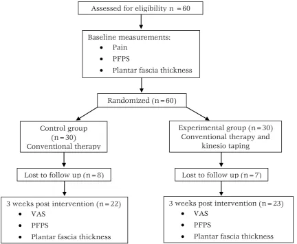
Figure 4: Flow diagram of patients throughout the course of the study
Assessed for eligibility n =60
Baseline measurements:
Pain
PFPS
Plantar fascia thickness
Randomized (n=60)
Control group (n=30)
Conventional therapy
Experimental group (n=30) Conventional therapy and
kinesio taping
Lost to follow up (n=8) Lost to follow up (n=7)
3 weeks post intervention (n=22)
VAS
PFPS
Plantar fascia thickness
3 weeks post intervention (n=23)
VAS
PFPS
RESULTS
Within group analysis revealed a significant difference between pre and post treatment in both the groups in terms of:
a) VAS: group I mean pre (6.3) and post (3.8) and calculated Z-value is -4.053 and P-value 0.000(<0.05); group II mean pre (6.4) and post (1.6) and calculated Z-value is -4.274, P-value 0.000(<0.05).
b) PFPS score: group I mean pre (59.06) and post (45.67) and calculated t-value is 10.373 and P-value 0.000(<0.05) ; group II mean pre(61.25) and post(36.46) and calculated t-value is 58.863, P-value 0.000(<0.05)
c) Ultrasonography: group I mean pre (4.477) and post (4.268) and calculated t-value is 7.967 and P-value 0.000 (<0.05) ; group II mean pre(4.595) and post(4.108) and calculated t-value is 23.037,P-t-value 0.000(<0.05).
At baseline, there was no significant difference between the two groups in terms of VAS, PFPS score and plantar fascia thickness. Between group analysis revealed a significant difference between pre and post treatment in terms of VAS score: Z-value -4.456, P-Z-value 0.000(<0.05), PFPS score: t-value 5.542, P-t-value 0.000(<0.05) but not under ultrasonography: t-value is 1.748, p-value 0.088(>0.05).
VAS
GRAPH 1: VAS score
GRAPH 2: PFPS score
GRAPH 3: Changes in plantar fascia thickness
Figure 5: Sonogram showing thickened plantar
fascia (pre-treatment)
Figure 6: Sonogram showing reduction in the
plantar fascia thickness (post-treatment)
DISCUSSION
The chief objective of this study was to determine the effect of kinesio taping as an adjunct to conventional therapy in reducing pain and improving functional ability in individuals with Plantar fasciitis. Both the groups were assessed for pain, functional ability and thickness of plantar fascia using visual analogue scale, plantar fasciitis
6.32
3.82 6.39
1.70
0.00 1.00 2.00 3.00 4.00 5.00 6.00 7.00
Pre Post
Control Experimental
59.06
45.67 61.26
36.47
0.00 10.00 20.00 30.00 40.00 50.00 60.00 70.00
Pre Post
PFPS SCORE
Control Experimental
4.48
4.27 4.60
4.11
3.80 3.90 4.00 4.10 4.20 4.30 4.40 4.50 4.60 4.70
Pre Post
CHANGES IN THE PLANTAR FASCIA THICKNESS(mm) UNDER ULTRASONOGRAPHY
pain and disability scale and ultrasonography respectively. All the parameters were measured at baseline (day 1) and at the end of three week following the completion of the intervention. Over all 60 subjects who met with the inclusion and exclusion criteria were randomly allocated into two groups. The subjects who fell into the age group of 20-55 yrs, of both sexes and who were suffering from plantar fasciitis were selected but the study was analyzed only on 45 subjects due to 15 drop outs (due to personal and transport reasons). 22 subjects from group I were treated with conventional therapy while 23 subjects from group II were treated with kinesio taping along with conventional therapy. Taping resulted in no adverse reactions for any of the participants. On comparing the pretreatment and post treatment values of the present study, it revealed that there was a statistically significant difference (p<0.05) in both the groups in terms of pain intensity and functional ability but there was more improvement in group II than group I and there was no statistically significant difference (p>0.05) in terms of changes in plantar fascia thickness between the two groups on ultrasound.
Therapeutic ultrasound, by its non-thermal effects of pulsed mode, has the potential to accelerate normal resolution of inflammation, ultimately relieving pain.18 This is in agreement with the study done by Hana Hroncova et al,2000 in which, the group which received ultrasound showed significant reduction in terms of pain.
Stretching reduces the tension in the fascia which becomes tight in plantar fasciitis. A study performed by DiGiovanni et al to determine the effectiveness of plantar fascia stretching versus calf stretching concluded that the plantar fascia stretching group had greater improvement in functional outcome compared to calf stretching group. Stretching plantar fascia recreates windlass mechanism, limits repetitive micro trauma and associated inflammation by performing exercise prior to the first step in the morning or after any prolonged sitting or inactivity.19
Strengthening the intrinsic foot muscles helps in supporting the arches of the foot as plantar fasciitis is often attributable to poor intrinsic muscle strength.20
The significant improvement in control group in terms of pain intensity and functional ability were thus, due to the effects produced by the ultrasound, stretching and strengthening exercises.
When a fascial injury occurs, proliferation and activation of fibroblasts will result in deposition of
collagen at the location of injury which later assembles into fibers and aligns with mechanical tension in the tissue. If such tissue is immobilized, dense connective forms. Thus, application of kinesio tape, which is a very good alternative to non-elastic tape because of the elasticity that it provides, over the plantar fascia during reparative process of fascial injury, creates convolutions in the skin. When skin is lifted by this technique, there is increase in the interstitial spaces between sheets of fascia, increase in the subcutaneous blood flow and allow the lymphatic system to channel more freely. The result is that pressure and irritation are taken off the neural and sensory receptors and thus alleviating pain, reducing stiffness, improving range of motion and finally reducing the injury recovery time. Application of kinesio tape over gastrocsoleus will reduce pain and tightness associated with plantar fasciitis. Reduction in pain intensity in group II is in agreement with the study done by Daniel O Sullivan et al, stating kinesio tape may alleviate pain through a reduction in mechanical stress on the tissue (i.e., fascia unloading).11 As the pain reduces disability also reduces.
Thus, the more significant results in experimental group compared to control group would be due to addition of kinesio tape to the conventional therapy which provided additional benefits.
There was no statistically significant difference (p>0.05) in terms of changes in thickness of plantar fascia between the two groups on ultrasound. The reason for this is probably due to small sample size and short duration of treatment in this study. A long follow up period may be necessary to see the greater changes.
The limitations of the present study include small sample size, short duration (3 weeks) of treatment and lack of no long term follow up. Further research can be done by including parameters like BMI in the study. The authors can use a control group for whom intervention will not be given so that there will be a chance to know the effects of kinesio tape alone in a more significant manner. In this study, subjects were tested for pain and foot function, similar studies can also be done to detect the strength of the intrinsic foot muscles. Further studies can be done with a long term follow up to assess the long term benefits.
CONCLUSION
was found in subjects who received kinesio taping along with conventional therapy. Therefore, kinesio taping can be used as an adjunct to conventional therapy in clinical application for greater improvement in pain levels and functional ability in patients with plantar fasciitis.
Acknowledgements: We would like to sincerely
thank Dr. S. Ram Murti, MD, FICR, MAMS (HOD of Radiology and Imageology, NIMS) for his kind support.
REFERENCES
1. Roxas M. Plantar fasciitis: diagnosis and therapeutic considerations. Alternative medicine review. 2005; 10(2):83-93.
2. McPoil TG, Martin RL, Cornwall MW, Wukich DK, Irrgang JJ, Godges JJ. Heel Pain -Plantar Fasciitis: Clinical Practice Guidelines Linked to the International Classification of Functioning, Disability, and Health from the Orthopaedic Section of the American Physical Therapy Association. J Orthop Sports Phys Ther. 2008; 38(4):2-18.
3. Lemont H, Ammirati K, Usen N. Plantar Fasciitis: A Degenerative Process (Fascoisis) Without Inflammation. J Am Podiatr Med
Assoc.2003; 93(3):234-37.
4. Simon J. Bartold. Plantar heel pain syndrome: overview and management. Journal of
Bodywork and Movement Therapies.2004;
214-226.
5. Charles Cole, Craig Seto, Gazewood J. Plantar Fasciitis: Evidence-Based Review of Diagnosis and Therapy. Am Fam Physician. 2005; 72(11):2237-2242.
6. Goff JD, Crawford R. Diagnosis and treatment of plantar fasciitis. Am Fam Physician. 2011; 84(6):676-682.
7. Stuber K, Kristmanson K. Conservative therapy for plantar fasciitis: a narrative review of randomized controlled trials. J Can Chiropr
Assoc. 2006; 50(2):118-133.
8. Martin RB, Davenport TE, Reischl SF, Mcpoil TG, Matheson JW, Wukich DK etal. Heel Pain -Plantar Fasciitis: Revision 2014 Clinical Practice Guidelines Linked to the International Classification of Functioning, Disability, and Health from the Orthopaedic Section of the
American Physical Therapy Association. J
Orthop Sports Phys Ther. 2014; 44(11).
9. Kase K, Wallis J, Kase T. Clinical therapeutic
applications of the kinesio taping method. Tokyo,
Japan: Ken Ikai Co Ltd; 2003.
10.Thelen MR, Dauber JA, Stoneman PD. The clinical efficacy of kinesio tape for shoulder pain. J Orthop Sports Phy Ther. 2008; 38(7):389-395.
11.Daniel O’Sullivan. Utilization of Kinesio Taping for Fascia Unloading. Internatinal journal of
Atheltic therapy and training. 2011; 16(4):21-27.
12.Bijur PE, Silver W, Gallagher J. Reliability of visual analogue scale for measurement of acute pain. Academic emergency medicine. 2001; 8(12):1153-1157.
13.Buck Willis, Angel Lopez, Andres Perez, Larry Sheridan, Stanley R Kalish. Pain Scale for Plantar Fasciitis. The Foot and Ankle Online
Journal. 2009; 2(5):3.
14.Sabir N, Demir lenk S, Yagci B, Karabulut N, Cubukcu S. Clinical utility of sonography in diagnosing plantar fasciitis. J Ultrasound Med. 2005; 24(8):1041-8.
15.Mahowald S, Legge BD, Grady JF. Correlation between plantar fascia thickness and symptoms of plantar fasciitis. J Am Podiatr Med
Assoc. 2011; 101(5):385-389.
16.Apeksha O, Lakshmiprabha R. Comparison of the effects of therapeutic ultrasound v/s myofascial release technique in treatment of plantar fasciitis. Indian Journal of Physiotherapy
&Occupational Therapy. 2012; 6(2):13-16.
17.Benedict FD, Deborah AN, Daniel PM, Petra AG, Taryn TW, Gregory EW etal. Plantar Fascia-Specific Stretching Exercise Improves Outcomes in Patients with Chronic Plantar Fasciitis. J Bone Joint Surg.2006; 88(8):1775-81 18.Robertson V, Ward A, Low J, Reed A.
Electrotherapy explained: principles and practise. 4th edition; 2005.
19.DiGiovanni BF, Nawoczenski DA, Lintal ME etal. Tissue specific plantar fascia stretching exercise enhances outcomes in patients with chronic heel pain. A prospective, randomized study. J Bone Joint Surg Am. 2003; 85-A: 1270-77. 20.Furey J. Plantar fasciitis: The painful heel syndrome. J Bone Joint Surg Am.1975; 57:762-73.
Citation
Tulasi Ratna, S., Dowle, P., Prasad, V., & Paruchuri, R. (2015). EFFECT OF KINESIO TAPING IN ADJUNCT TO CONVENTIONAL THERAPY IN REDUCING PAIN AND IMPROVING FUNCTIONAL ABILITY IN INDIVIDUALS WITH PLANTAR FASCIITIS – A RANDOMIZED CONTROLLED TRIAL.