Case Report
Management Therapy in Chronic Ectopic Pregnancy
Manajemen pada Kehamilan Ektopik Lama
Aryando Pradana, Sandhy Prayudhana Department of Obstetrics and Gynecology Medical Faculty of Indonesia University/
Dr. Cipto Mangunkusumo Hospital Jakarta
INTRODUCTION
Ectopic pregnancy generally occurs in 2 out of every 100 pregnancies.1 In Industrial countries, the
inciden-ce showed a six-fold increase in the last twenty years, though there were also some decrease in these last few years. In United States, the rate during the last three decades was increased from 4.5 per 1000 preg-nancies in 1970 to a rate of 19.7 per 1000 pregpreg-nancies in 1992.2 However, there was a decline in maternal
mortality rate due to ectopic pregnancy, from 13% between 1979 and 1986 to 9% in 1992.2
The real incidence of chronic ectopic pregnancy is unknown, with only a few done studies reported that the frequency was about 6 - 20%.2 The rate could
be higher if the study was done by observing it from the clinical point of view, the low titre of β-hCG, and the non-diagnostic early ultrasound examination. None-theless, the diagnosis was usually not made until after the surgery was done. Clinical appearance with pain and bleeding is a sign of a subacute or even chronic process, which may be overlap with many other cau-ses of pelvic pain in women.
The treatment and management of ectopic preg-nancy depends on the patient’s clinical conditions. In an acute setting with unstable hemodynamic condi-tion, the treatment of choice should be radical sur-gery, to immediately control the bleeding. While for a stable hemodynamic condition, there are various conservative treatments that can be choose to main-tain the tubal pregnancy.1,3
In this case, we will discuss the tubal conservative treatment for a chronic ectopic pregnancy. There will
be discussions about diagnosis and treatment methods available for chronic ectopic pregnancy. The ectopic pregnancy in the case illustration was found concur-rently with an acute ectopic pregnancy, which is a rare thing to happen. Literatures available are usually in the format of case reports.
CASE ILLUSTRATION
Female, 35 years old, came to RSCM (Dr. Cipto Ma-ngunkusumo Hospital) complaining of lower abdomi-nal accompanied by vagiabdomi-nal bleeding since 17 hours before admission. The patient was referred from Ke-mayoran PKM (Community Health Center), diagno-sed with abortion. Patient admitted that she was two months pregnant and her LMP (Last Menstrual Pe-riod) was on November 8th, 2009 ~ pregnancy week
eleven. She had never gone for any antenatal care vi-sits nor ultrasound examinations. The lower abdomi-nal pain accompanied by vagiabdomi-nal bleeding occurred since 17 hours before admission. The abdominal pain was described as a hot/burning feeling, radiating to the waist, occurring alternatingly on and off. Patient said that there was no fever.
Both past and family medical history of diabetes mellitus, hypertension, asthma, and heart disease were denied. The patient was currently in her second mar-riage. The first marriage was in 1993, no children, and ended up with a divorce. The second one was in 2008. Patient was gravida 2, with a history of 1 abor-tion during her previous pregnancy in 2009. The pre-vious abortion happened at pregnancy month three Abstract
Objective: Improving skill in making a diagnosis and manage-ment therapy in chronic ectopic pregnancy.
Method: Case Report.
Conclusion: Diagnosis of chronic EP is difficult to establish be-fore surgery. Conservative treatment using medication (methotre-xate) can not be applied to chronic EP because the β-hCG level is difficult to be detected.
[Indones J Obstet Gynecol 2010; 34-4: 199-203]
Keywords: ectopic pregnancy, chronic ectopic pregnancy
Abstrak
Tujuan: Meningkatkan pengetahuan dalam membuat diagnosis
dan melakukan manajemen terhadap kehamilan ektopik lama.
Metode: Laporan kasus.
Kesimpulan: Diagnosis untuk kehamilan ektopik lama tidak
mu-dah ditegakkan sebelum operasi. Terapi konservatif menggunakan methotrexate sulit digunakan karena kadar β-hCG sulit terdeteksi.
[Maj Obstet Ginekol Indones 2010; 34-4: 199-203]
Kata kunci: kehamilan ektopik, kehamilan ektopik lama
Correspondence: Aryando Pradana, Department of Obsetrics and Gynecology Dr. Cipto Mangunkusumo Hospital, Central Jakarta. Telephone: 0811877598 Email: [email protected]
and was not treated by curettage because patient only went to a witchdoctor. Patient had never used any contraception before.
Physical examination on arrival at RSCM revealed that the blood pressure was 120/90 mmHg, heart rate 92 beats per minute, respiratory rate 20 breaths per minute, temperature 36.7°C, body weight 55 kg, body height 154 cm, and BMI (body mass index) 23.19. General examination showed that both conjunctivas were pale and there was lower abdominal tenderness during palpation. Others were within normal limit. From the gynecologic examination, vulva and urethra were normal under inspection, no active bleeding was found. Speculum examination showed that the portio was smooth and livide, external uterine ostium was closed, fluxus was positive but no fluor was found. Bimanual examination could not assess uterine corpus due to the pain felt when pressure was applied. Ex-ternal uterine ostium was closed, cervical motion ter-derness was positive, and protrusion of pouch of Dou-glas was felt. From rectal touche, we found that the rectal ampulla was not collapse, tone of the spincter ani was good, and the anal mucose was smooth.
Voluson ultrasound examination revealed that the uterus size was 81 x 44 x 54 mm. Endometrial thick-ness was about 20 mm ~ Aria Stella reaction. Left ovary was of size 1.8 x 1.2 cm and the right one was of 1.56 x 1.1 cm. At the right adnexa, a mass of size 7 x 2 cm, haematocele, and free fluid were also found. Conclusion: suggestive of right tubal disrupted ec-topic pregnancy.
Patient was diagnosed with acute abdomen e.c. haemoperitoneum e.c. disrupted ectopic pregnancy at G2A1 pregnancy week eleven. Diagnostic plan inclu-des vital signs and acute abdomen signs observation per hour, CBC (complete blood count), complete uri-nalysis, blood glucose levels, and bleeding time/clot-ting time test. The initial plan was to do an evacuation by laparotomy with blood preparation of 500 cc pack red cells and antibiotics injection of 1 x 2 g ceftriax-one.
The laboratory results were as follow: haemoglobin 8.7, haematocrit 26, leucocyte 16100, thrombocyte 262000, BT/CT 3’/13’, complete urinalysis was with-in normal limit, and the blood glucose levels was 116. Two hours later, a procedure of laparotomy, bilat-eral salpingectomy, and cystectomy were being per-formed. During the operation, we found a ruptured right pars ampullaris tube, chronic infected mass at the left pars ampullaris tube, and left paraovary cyst. The amount of blood lost during operation was about 450 cc and urine production was 150 cc. Postopera-tive laboratory studies showed the following results: haemoglobin 6.0, haematocrit 18, leucocyte 10200, thrombocyte 185000, MCV 86, MCH 29, and MCHC 34. The results were suggestive of normocytic nor-mochrome anemia e.c. bleeding (haemoperitoneum). The next plan was to do a blood transfusion until the haemoglobin level ≥ 8g/dl.
After the transfusion of 1 bag of PRC (270 cc), we reran the blood test and got the following results: hae-moglobin 8.0, haematocrit 23, leucocyte 13700, thrombocyte 238000, MCV 86, MCH 30, and MCHC 34. Patient was then sent home in a goood condition after being admitted for 3 days.
DISCUSSION
Acute ectopic pregnancy (EP) is a common clinical problem, generally diagnosed using a combination of clinical findings, sonographic imaging, and laboratory results.1,2 Chronic ectopic pregnancy is an uncommon
condition, can be a result from a minor rupture which developed into a haematocele. Such patient may pre-sent with symptoms of subacute or chronic pain and a low or negative β-hCG level.2 Chronic EP has
vari-ous sonographic images, ranging from amorphvari-ous, avascular mass, and high vascularity complex. For some clinical conditions, sonography plays an impor-tant role in confirming the preoperative diagnosis of the disease.1 In this case illustration, patient came
with acute abdominal pain which is a symptom of acute EP. From the initial examination, there was no findings nor reasoning suggestive that there was chronic EP coexisted with acute EP. The diagnosis of chronic EP was confirmed during the operation which also accompanied by a ruptured right tube in which a mass resembling a residual conception was found.
The weakness in this case illustration was that the patient did not send the tissue taken from the opera-tion to the anatomy pathology for further evaluaopera-tion due to economic problem. The diagnosis of chronic EP can only be done by doing a macroscopic exami-nation which shows a cystic mass containing a pla-centa-like mass with no active bleeding.
Ectopic pregnancy itself is a common diagnosis based on clinical findings and sonographic imagings. In this case, we got a typical (classic) acute findings including amenorrhea, pelvic pain, and bleeding. To help confirming the diagnosis of chronic EP, some literatures stated that the clinical appearances are: some patients may have a nonspecific history, low or slowly raising β-hCG level, intermittent or chronic pelvic pain, and light bleeding. Sometimes the β-hCG level can be negative or about normal.2 The
sono-graphic imaging can overlap with acute pelvic inflam-matory disease, pelvic abscess, vascular tumor, and endometriosis.2
Ultrasonographic images in chronic EP are various and nonspecific. The descriptions consist of nondiag-nostic sonographic images such as heterogenous adnexal mass and high vascularity adnexal com-plex.2,4 Doppler’s flow is usually found at the margin
or the external area of the mass, often with abnormal blood vessels and arteriovenous shunting. In some rare cases, there may be some free fluid in the pouch of Douglas or at other places in the pelvic.2,4 In this
case illustration, a mass was found at the right adnexa with the size of 7 x 2 cm, accompanied by a haema-tocele and free fluid at the pouch of Douglas. Due to the presence of classic triad of EP with acute abdo-men, the Doppler examination was not done in this case, indicating that initially there was no suspect to-wards chronic EP.
Case Distribution
pullas of Falloppian tubes. These are the sites EP ge-nerally occur.1
One literature explained that the distributions of tubal ectopic pregnancies based on the locations are: 70% at the ampulla, 12% at isthmus, 11% at fimbriae, and 2 - 3% at interstitial. Pregnancies outside of the tubes are very rare, sometimes found in the abdomen. Of all the ectopic pregnancies, 3% was found at the ovary and < 1% at cervix.1,3
Risk Factors
Previous history of disrupted tubes, be it from tubal ectopic pregnancy or tubal surgery related to sterili-zation or infertility treatment, plays as the highest risk factor for having an ectopic pregnancy (Table 1). Af-ter having an EP, the chance of having another one is about 10%.1 Infertility and the application of
as-sisted reproductive technology as its treatment signifi-cantly increase the risk for ectopic pregnancy.1,3 A
typical implantation - corneal, abdominal, cervical, ovarian, and heterotrophic pregnancies - are more common after undergoing the assisted reproductive technology.
Table 1. Risk factors for ectopic pregnancy.1
Risk Factors Relative Risk Previous history of ectopic pregnancy 3 - 13
Corrective tubal surgery 4
Tubal sterilization 9
Intrauterine contraceptive device 1 - 4.2 Documented tubal pathology 3.8 - 21
Infertility 2.5 - 3
Assisted reproductive technology 2 - 8 Previous history of genital infection 2 - 4
Chlamydia 2
Salpingitis 1.5 - 6.2
Smoking 1.7 - 4
Previous history of abortion 0.6 - 3 Multiple sexual partners 1.6 - 3.5 Previous history of sectio caesarea 1 - 2.1
In this case, the patient had a previous history of infertility, which increased her risk 2.5 to 3 times higher. Previous history of infection had not been able to be excluded although the patient did not complaint of any previous pelvic pain because pelvic pain itself can be asymptomatic.4 Another risk factor found in
this patient was the previous history of abortion which could increase her risk by 0.6 to 3 times.
Pathogenesis
Chronic ectopic pregnancy is considered to be derived from small recurrent ruptures of tubal ectopic preg-nancy which develop into haematocele and tropho-blastic tissue that may be active or inactive.2 The
hae-matocele is usually surrounded by adhesion and in-duces inflammatory response. In this case, a peritubal adhesion was found, accompanied by a cystic mass at the ampulla of the left tube. During the incision of the cystic mass, a placenta-like tissue was revealed. In a chronic EP, β-hCG level is generally low, though in some rare cases it can be normal. In one previously published case report, the negative β-hCG level was stated to be about 50%, which may be due to a less sensitive β-hCG level examination. An acute rupture can happen but is extremely rare.2,5
In this case, the chronic EP occurred along with the acute EP, resulting in a positive β-hCG level and making the diagnosis of concurrent chronic EP even more difficult. Some different theories have been spe-culated to explain the mechanism of the negative pregnancy test:6
N Reduced level of β-hCG hormone produced by the living and active trophoblasts.
N Small persistent mass of active trophoblasts pro-duces β-hCG in a very little amount, resulting in an undetectable low serum level (even with the mo-dern test pack).
N Increased clearance of hormonal serum (unknown process).
N Involution of active trophoblasts tissue brings about a low β-hCG level, but the tubal haematoma can still exist, grow, and rupture.
Tubal 95-96%
Isthmic 12% Caesarean scar < 1%
Cervical < 1%
Abdominal 1%
Fimbrial 11% Ampullary 70%
Ovarian 3% Interstitial and cornual 2-3%
Figure 1. Distribution of ectopic pregnancies based on the lo-cation.1
0 500 1000 1500 2000 2500 3000 3500 4000 4500 5000 5500
C
on
cent
ra
tio
n of
hum
an
c
hor
io
ni
c go
na
do
trop
hi
n (
IU
/l)
Weeks after last menstrual period
0 4 8 12 16 20 24 28 32 36 40
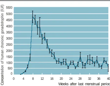
Figure 2.β-hCG level during pregnancy. β-hCG level peak at 8 - 12 weeks and slowly reduced after that.1
N Implantation may happen late in relation to the woman’s expected menstrual date, showing the li-mitation of β-hCG pregnancy test pack on the first day of the missed period.
Options for Treatments
Conservative Surgery
The conservative management of unruptured EP con-sist of two choices; linear salpingotomy or segmental resection. Conservative surgery approach is possible if the diagnosis of EP is confirmed in such an early stage that the tubal rupture has not yet happened.3,5
Segmental resection is done for the tubal pregnan-cy occurred at the isthmus, because the muscularis layer there is thicker compared to the relatively nar-row lumen. After the resection was done, a reanasto-mosis may also be carried out. These procedures can only be done by an experienced surgeon.3
For those women who want to keep their repro-ductive ability, the conservative surgery by doing a linear salpingotomy is considered the gold standard management of distal tubal pregnancy. Most recent studies have reported that the uninvolved tube can be abnormal, grossly or subclinically, in at least 50% EP cases. Even though there are no randomized stu-dies comparing the fertility outcome after conserva-tive and radical surgery for EP, the already available information shows that chance of having intrauterine pregnancy for the next pregnancy is higher after the conservative surgery (linear salpingotomy).3,5,7
There are many studies about salpingotomy that has been done but unfortunately they do not describe the condition of the uninvolved tubes. Langer and friends described the uninvolved tubes in their studies of 30 patients undergoing salpingotomy. Of all the patients having a normal contralateral tube, 80% will have a normal pregnancy. When the contralateral tube is disrupted or has peritubal adhesion, only 11 (55%) patients able to have a viable pregnancy.3,7
Another technique besides salpingotomy is salpi-ngostomy. The salpingostomy procedure is usually done to evacuate EP less than 2 cm in length and located at the distal lower third of the tube. A linear incision of 10 - 15 mm is done with the unipolar cau-ter needle at the antimesencau-teric area above the EP site.1 Techniquely, salpingostomy is the same with
salpingotomy except that in salpingostomy, there is no suturing. According to Tulandi and Saleh, there was no difference in prognosis with or without sutu-ring.1,8
Radical Surgery
Total salpingectomy is needed when the tubal preg-nancy has been ruptured and haemoperitoneum hap-pened. In such case, the intraabdominal bleeding must be controlled quickly and there is no place to try to do a conservative surgery. Extensive haemoperitone-um can puts the patient in a critical cardiopulmonal state. Salpingectomy can also be indicated for another condition, including recurrent EP at the same tube, EP at a poorly disrupted tube, and EP in a women who has already had enough children.3,9
Medical Conservative
There is an alternative medication treatment, using ac-tinomycin D, intratubal methotrexate, intratubal pro-staglandineba, and hyperosmolar glucoser. However, those treatment have not been standardized.9
Methotrexate is an antagonist of folic acid which inactivates the dihidrofolate reductase that reduces te-trahidrofolate, an essential cofactor for the synthesis of DNA dan RNA, causing a disruption in cell divi-sion. Actively dividing cells are very sensitive to me-thotrexate, which is the why drug is used as a treat-ment for malignant trophoblastic disease.1,9
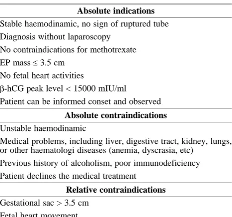
Table 2. Considerations for using methotrexate.7
Absolute indications Stable haemodinamic, no sign of ruptured tube Diagnosis without laparoscopy
No contraindications for methotrexate EP mass ≤ 3.5 cm
No fetal heart activities
β-hCG peak level < 15000 mIU/ml
Patient can be informed conset and observed
Absolute contraindications Unstable haemodinamic
Medical problems, including liver, digestive tract, kidney, lungs, or other haematologi diseases (anemia, dyscrasia, etc)
Previous history of alcoholism, poor immunodeficiency Patient declines the medical treatment
Relative contraindications Gestational sac > 3.5 cm
Fetal heart movement
Administration of methotrexate uses the β-hCG le-vel as indicator of success. In a chronic EP case with a very low or even undetectable β-hCG, it is difficult to use this drug as a choice of treatment.6
Outcomes
Chronic EP has various outcomes. Some patients show immediate or gradual improvement after the medication treatment. In some cases, the acute rupture may happen. Some others may have a persistent pel-vic pain, which needs salpingooophorectomy surgery. Ultrasonography plays an important role, not only to exclude intrauterine pregnancy, but also to recognize any adnexal abnormalities. Ultrasonography is able to exclude other causes of pelvic pain in such populati-on, such as ovarian torsipopulati-on, hemorrhagic cyst, endo-metrioma, and dermoid. The ultrasonography images of acute pelvic inflammatory disease with piosalping or tuboovarian abscess can overlap with the ones of chronic EP, and the negative β-hCG level here may be a problem. If patient has a persistent adnexal ab-normalities, a surgery is needed to exclude tumors.2
Tubal pregnancy is related to a poor prognosis of the next reproductive function. In most cases, extrau-terine pregnancies showed a failure of the fertilized ovum to migrate throught rugae within the tube as a result from the altered tubal function.3
In 1975, a study by Shoen and Nowak concluded that about 70% patients having EP at their first preg-nancy could not give another living birth. About 30% patients having EP would again have another EP, compared to 10 - 15% total prevalence of recurrent EP in the general population of reproductive age.3,4
More than a half of recurrent EP cases occurred with-in 2 hour time and 80% occurred 4 years after the first episode. Potential reproductive capacity of pa-tients having had an EP depends on their reproductive history. If the EP were their first pregnancy attempt, the prognosis of the next pregnancy would be poorer than if the EP were a complication occurred 1 year or more after a successful pregnancy.3,4
According to the study done by Yao and Tulandi in 1999, of 1514 patients trying to get pregnant after having a linear salpingostomy, 61.2% successfully achieved intrauterine pregnancy and 15.5% had a re-current EP. On the other hand, only 38.1% of 3584 patients trying to get pregnant after salpingectomy who successfully had another intrauterine pregnancy though the incidence of EP is relatively lower (9.8%).3
Among the reports of salpingotomy done on the only one tube left, pregnancy rate of about 50% was recorded by some researchers. However, the reports showed various results and some reports had only a few numbers of patients. Recurrent EP in salpingo-tomy patients with only one tube is about 20%, higher than in those patients who had both tubes.3
In one study, it is reported that a previous history of infertility has the same significant risk for a failure of the next pregnancy attempt after an EP conserva-tive treatment. Even if there was a little differences for the successful pregnancy between after conserva-tive and radical treatment, it was better to reduce the recurrent risk by applying an assisted reproductive technology, which could increase the successful rate of having an intrauterine pregnancy.10
CONCLUSION
The diagnosis of chronic ectopic pregnancy was done based on macroscopic appearance of the tubal cystic mass during the operation of the acute EP. Patient did not sent the tissue taken from the operation to the anatomy pathology laboratory for further examina-tion. Diagnosis of chronic EP is difficult to establish before surgery. This patient may be subjected to a conservative surgery (due to economic problem faced by the patient) to maintain her reproductive capacity even though the chance of having an intrauterine pregnancy was low, since the patient had a previous history of infertility. Conservative treatment using medication (methotrexate) can not be applied to chro-nic EP because the β-hCG level is difficult to be de-tected.
REFERENCES
1. Cunningham FG, Leveno KJ. Ectopic pregnancy. In: Wil-liam’s Textbook of Obstetrics. 23rd ed. Electronic book.
USA: McGraw-Hill. 2010
2. Talavera MD, Horrow MM. Chronic ectopic pregnancy. J Diag Med Son. 2008; 24: 101-3
3. Rock JA, Jones HW. Ectopic pregnancy. In: Te Linde’s Operative Gynecology. 10th ed. Electronic book. USA:
Lip-pincott Williams and Wilkins. 2008
4. Condous G, Okaro E. The accuracy of transvaginal ultra-sonography for the diagnosis of ectopic pregnancy prior to surgery. Hum Rep. 2005; 20(5): 1404-9
5. Cust MP, Filshie GM. Modern management of ectopic pregnancy. Current Obstetrics and Gynaecology. 1991; 1: 210-16
6. Alfhaily Fadi, Whitlow Barry. Laparoscopic removal of a large 8-cm ectopic pregnancy with a negative pregnancy test. Gynecol Surg. 2009; 6: 173-5
7. RCOG. The management of tubal pregnancy (guideline). RCOG Practice Bulletin. 2004; 21: 1-6
8. Tulandi T, Saleh A. Surgical management of ectopic preg-nancy. Clin Obstet Gynecol. 1999; 42(1): 31-8
9. Vivek N, Isaac M. Tubal ectopic pregnancy: diagnosis and management. Arch Gynecol Obstet. 2009; 279: 443-53 10. Dela Cruz A, Cumming DC. Factors determining fertility
after conservative or radical treatment for ectopic preg-nancy. Fertility and sterility. 1997; 66(5): 871-4