1
Folding of a 10 Ala Peptide
2
The Anfinsen Experiments
Fig. 5.24
•Urea disrupts H-bonds &
hydrophobic interactions.
•-ME reduces disulfides.
(urea) H2N–C–NH2
II O
+
HS–CH2CH2–OH (-mercaptoethanol) Native (active)
Ribonuclease
Denaturation
The disruption (or unfolding) of tertiary &
secondary protein structure that leads to
loss of protein function.
Notes:
Denaturation can be caused by a variety of
treatments and/or reagents.
“Denaturation” also refers to the disruption
of nucleic acid structure & function.
Denaturing Agents &
Treatments
Heat / Temperature
Increases the rate of molecular motion/vibration, causing disruption of H-bonds, leads to protein unfolding.
Strong Acids / Bases
Cause protonation/deprotonation of sidegroups, alters H-bonding & salt bridges.
Proteins become insoluble & precipitate as they reach their isoelectric points.
Water-soluble Organic Solvents (alcohols)
Interfere with hydrophobic interactions; substitute or interfere in H-bonding.
Detergents
Disrupt hydrophobic interactions, leading to protein unfolding.
Denaturing Agents &
Treatments
High Salt Concentration
Interferes with salt bridges / charge interactions in proteins.
Can “extract” water molecules from protein surface, leading to protein aggregation & precipitation.
Reducing Agents
(e.g. -ME, DTT) reduce disulfide bridges; unfolding.
Mechanical Stress
(e.g. stirring, grinding) disrupts weak forces that stabilize tertiary protein structure.
6
Proteins Start Folding As They
Form During Translation
DNA
RNA
Protein
Replication
Transcription Translation
“Information Processing”
Traits
Polyribosomes and
folding nascent peptides. Translation:
The ribosome-mediated biosynthesis of a polypeptide based on the genetic information encoded in mRNA.
7
Energy Landscape Model
for Protein Folding
8
Protein Folding is Assisted by
“
Molecular Chaperones
”
Background on Chaperones
Originally discovered in 1980’s in relation to heat shock stress in higher plants and Drosophila.
Given the descriptive name “Heat Shock Proteins” or “hsp’s”.
Hsp’s conferred thermo-tolerance to organisms subjected to non-lethal high temperature stress.
Molecular chaperones assist protein folding in two ways:
Preventing inappropriate protein-protein interactions
Helping folding occur rapidly and precisely
Two major classes: Hsp70s and Hsp60s
(chaperonins) Release of polypeptides from hsp70 & hsp60 requires ATP.
Hsp’s also promote re-folding of proteins
resulting from stress conditions.
If re-folding is not possible, hsp’s promote
protein degradation
Figure 5.31 Space-Filling Model of the E. Coli
Chaperonin, called the GroES-GroEL Complex
Section 5.3:
Proteins
Section 5.3:
Proteins
Some Diseases Associated with
Mis-folded Proteins
Alzheimer’s Disease
Associated with insoluble fibrous aggregates of
-amyloid protein (plaques) in brain neurons involved in memory & cognition.
-Amyloid protein is derived from the proteolytic
cleavage of “amyloid precursor protein” (a
transmembrane protein with unknown function).
Proteolytic Processing of Amyloid
Precursor Protein in Alzheimer’s
13
Alzheimer’s:
Aggregation of -amyloid protein to form plaques.
Normal:
Some Diseases Associated with
Mis-folded Proteins
Mad Cow Disease (Bovine Spongiform
Encephalitis)
14
Mad Cow Prions- Proteinaceous
Infectious Particles
17
Normal
Folding Pattern of Pre-prion
Abnormal
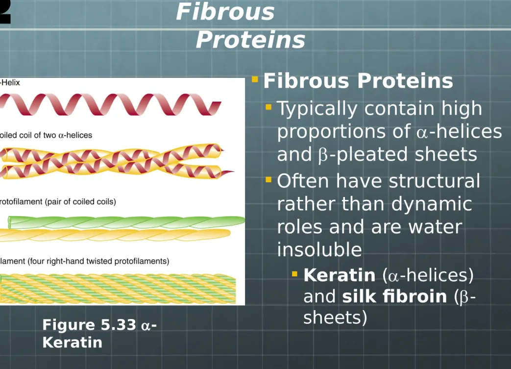
Fibrous Proteins
Typically contain high
proportions of
a
-helices
and
-pleated sheets
Often have structural
rather than dynamic
roles and are water
insoluble
Keratin
(
a
-helices)
and
silk fibroin
(
-sheets)
Figure 5.33 a -Keratin
Fibrous
Proteins
Figure 5.34 Molecular Model of Silk Fibroin
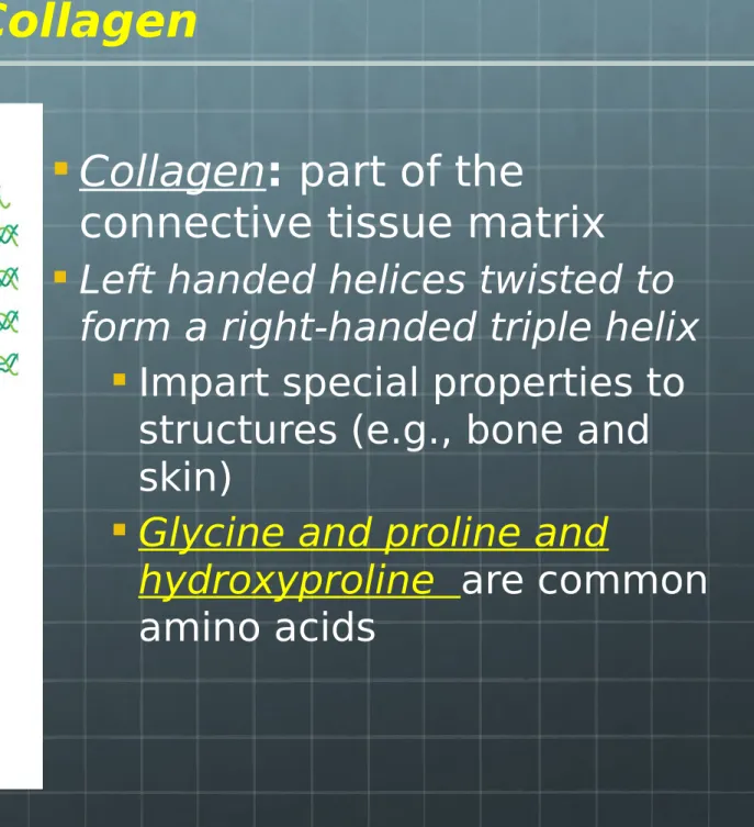
Fibrous Proteins
Collagen
:
part of the
connective tissue matrix
Left handed helices twisted to
form a right-handed triple helix
Impart special properties to
structures (e.g., bone and
skin)
Glycine and proline and
hydroxyproline
are common
amino acids
Figure 5.35 Collagen Fibrils
Collagen
Collagen and aging
22
Globular proteins- Structure &
Function
of Myoglobin vs. Hemoglobin
Very similar polypeptide tertiary
structures.
Same heme “prosthetic group” to bind
oxygen.
Very similar functions (oxygen
transport).
Myoglobin is a monomer; Hemoglobin
is a tetramer.
Different physiological locations,
affinities for O
2,
kinetics of binding &
23
Functional Relations Between
Myoglobin & Hemoglobin
Hb-O2
Hb-CO2 Mb-O2
Mb
Cells or Storage
MYOGLOBIN
HEMOGLOBIN
• 8 α helices
• Found in skeletal and cardiac muscles • Stores oxygen; has higher affinity for oxygen compared to hemoglobin
• 2 α and 2β helices • Found in RBCs
25
Heme Groups of Globins
101
26
The Hemoglobin
Allsosteric Transition
Oxygen binding causes a 15 rotation of the
a11 dimer relative to the a22 dimer.
Change in conformation increases the affinity / binding of subsequent O2 molecules.
27
The Hemoglobin
Allsosteric Transition
Oxygen binding causes a 15 rotation of the
a11 dimer relative to the a22 dimer.
28
Factors Affecting
the Amount of 0
2Bound by Hemoglobin
1. Availability of oxygen
(i.e. concentration or partial
pressure of O
2).
2. Binding of first O
2molecule
(i.e. cooperativity)
3. pH
29
Oxygen-Binding Equilibrium Curves
for Myoglobin vs. Hemoglobin
% S a tu ra ti o n
Change in pp O2 causes
changes in O2 affinity & binding.
working muscle
30
Effects of pH on O
2Binding
H+ Ions decrease hemoglobin’s affinity for O 2.
Sources of H+ ions include bicarbonate buffering system
and lactic acid production.
Both CO2 and lactic acid accumulate in tissues that are in need of O2.
Low pH promotes release of O2; high pH stabilizes HbO2.
-CO2 + H2O HCO3 + H+
Glucose Pyruvate CH3 – C – COOH O
I
H H
-31
Effects of pH on O
2Binding
The Bohr Effect
Low pH (high CO2, high lactic acid) decreases Hb affinity for O2.
H+ ions displace O 2
bound to Hb.
32
Effects of Carbon Monoxide
on O
2Binding
CO (carbon monoxide) binds very
tightly to Fe2+ in the heme of
hemoglobin (250 x’s tighter than O2).
CO-Hb can not carry O2.
CO binding is cumulative & virtually “permanent”.
CO poisoning leads to asphyxiation. “Mainstream” cigarette smoke has
500 ppm CO.
Cigarette smokers have 5 – 15% CO-Hb.
C III O
33
Effects of CO
34
Other Types of Hemoglobin
Adult (HBA) –
a
2
2configuration
(
-
His143,
-
Glu6)
Fetal (HBF) –
a
2
2(
-143 Ser)
configuration
Sickle Cell (HBS) –
a
2
2(
-6 Val)
35
Sickle Cell Hemoglobin
HBS has Val substituted for Glu at position 6 in subunits. Position 6 is located at surface of subunits.
Hydrophobic Val binds to hydrophobic pockets in other
subunits.
Cross binding polymerizes HBS into sickle shaped
36
Features of Sickle Cell Anemia
HBS fibers distort cells into sickle shapes.
Sickle-shaped cells are more rigid, pass through capillaries with greater
difficulty, impair circulation, cause tissue damage & pain.
Sickle cells are more fragile, targeted by immune system, & turn over faster.
Afflicted people are anemic (i.e. they produce only about half the normal number of blood cells).
HBS is less efficient in carrying O2.
Sickling is promoted in the absence of O2.
37
Study Questions / Objectives- Lectures 6&7
1. Understand primary structure of proteins
2. Define/describe different secondary structures and
super-secondary structures. Understand how to classify them in a Ramachandran plot. What are turns and loops in the context of secondary structures?
3. Define “tertiary protein structure” and describe how hydrophobic &
electrostatic interactions and hydrogen & covalent bonds can all be involved in stabilizing tertiary protein structure.
4. Describe the nature of the “Anfinsen experiments”, and how or why
they were important in protein folding.
5. What are heat-shock proteins and/or molecular chaperones, and
what is their role in protein folding and turnover.
6. Define “protein denaturation” and describe the main reagents or
treatments that denature proteins.
7. Define “quaternary protein structure” and describe how or why it
38
Study Questions / Objectives
8. How is protein degradation facilitated?
9. Describe prions and how they influence misfolding of proteins causing neurological disorders?
10. Know structures of collagen and its function
11. Describe the structural and functional similarities &
differences between hemoglobin and myoglobin, including the mechanisms of action of any physiological factors that can affect O2 binding.