Original Article
Generation of a CRISPR/Cas9-Based Vector Specific for Gene
Manipulation in
Leishmania
major
Ghodratollah SALEHI SANGANI 1,2, *Vahid JAJARMI 3,4, Ali KHAMESIPOUR 5, Mahmoud MAHMOUDI 6, Abdolmajid FATA 2,7, *Mehdi MOHEBALI 1,8
1. Department of Medical Parasitology and Mycology, School of Public Health, Tehran University of Medical Sciences, Tehran, Iran 2. Department of Medical Parasitology and Mycology, Faculty of Medicine, Mashhad University of Medical Sciences, Mashhad, Iran 3. Department of Medical Biotechnology, School of Advanced Technologies in Medicine, Shahid Beheshti University of Medical
Sci-ences, Tehran, Iran
4. Cellular and Molecular Biology Research Center, Shahid Beheshti University of Medical Sciences, Tehran, Iran 5. Centre for Research and Training in Skin Diseases and Leprosy, Tehran University of Medical Sciences, Tehran, Iran 6. Department of Epidemiology and Biostatistics, School of Public Health, Tehran University of Medical Sciences, Tehran, Iran 7. Cutaneous Leishmaniasis Research Center, Emam Reza Hospital, Mashhad University of Medical Sciences, Mashhad, Iran
8. Research Center for Endemic Parasites of Iran, Tehran University of Medical Sciences, Tehran, Iran
Received 15 Jan 2018 Accepted 23 Apr 2018
Abstract
Background: Gene manipulation strategies including gene knockout and editing are becoming more sophisticated in terms of mechanism of action, efficacy and ease of use. In classical molecular meth-ods of gene knockout, homologous arms are designed for induction of crossing over event in double strand DNA. Recently, CRISPR/Cas9 system has been emerged as a precise and powerful tool for gene targeting. In this effort, we aimed to generate a CRISPR/Cas9-based vector specific for targeting genes in Leishmania parasites.
Methods: U6 and DHFR promoters and neomycin-resistance gene were amplified from genome of
L. major (MHRO/IR/75/ER) and pEGFP-N1, respectively. U6 promoter was cloned in pX330 vec-tor which is named as pX330-U6. DHFR promoter and neo resistance gene sequence fragments were fused using a combination of SOE (Splicing by overlap extension)-PCR and T/A cloning techniques. To generate pX-leish, fused fragments su-bcloned into the pX330-U6. Two sgRNAs were designed to target the gp63 gene and cloned in pX-leish.
Results: The pX-leish vector was designed for simultaneous expression of cas9 and G418 resistance
proteins along with a self-cleaving 2A peptide for efficient separation of the two proteins. In this study pX-leish was designed with 3 features: 1) Compatible promoters with Leishmania parasites. 2) Insertion of antibiotic selection marker 3) Designing an all-in-one vector containing all components required for CRISPR/Cas9 system.
Conclusion: This modified system would be valuable in genome manipulation studies in Leishmania
for vaccine research in future.
Keywords:
Leishmania major;
CRISPR/Cas9; Gene manipulation
*Correspondence
Emails:
[email protected] [email protected]
Iranian Society of Parasitology http://isp.tums.ac.ir
Iran J Parasitol
Open access Journal at http:// ijpa.tums.ac.ir Tehran University of Medical
Introduction
eishmaniasis is a disease caused by intracellular obligate parasites with clinical manifestation from self-healing skin sores as found in cutaneous Leishmaniasis to life threatening form related to visceral Leishmaniasis (1). Chemotherapy currently is the main control strategy for Leishmaniasis, but toxic side effects and in-creasing drug resistance lead to failure of con-trol measures (2). Hence development of a vaccine against Leishmaniasis has considered by researchers in recent years. Different types of vaccines have been used to protect against
Leishmania in the past. Inoculation with live
promastigotes so called leishmanization (3), killed promastigotes (4), non-pathogen
Leish-mania (5), recombinant immunogenic antigens
of Leishmania (6), DNA vaccines (7) and vari-ous attenuated parasites (8) have been tested for their immunogenicity and protective effi-cacy (9). Genetically attenuated parasites and targeted elimination of virulent genes have proven to confer good protection in animal models and could be promising to find effec-tive vaccines (10, 11).
Gene targeting is a process in which differ-ent strategies may be applied to alter or elimi-nate the function of a gene. These modifica-tions can be obtained through deletion, inser-tion or replacement of endogenous sequence with alternative sequences (12). Homologous recombination is one of the main pathways for DNA repairing which is utilized for target-ed gene replacement to generate gene knock-out parasites (13).
During recent years, generation of targeted mutations has been facilitated by new gene editing systems. These systems specifically are able to induce DNA double-strand breaks (DSBs) at a target nucleotide sequence by en-gineered nucleases and subsequently cell repair system is activated such as CRISPR (Clustered regularly interspaced short palindromic re-peats)-Cas9 system (14). The CRISPR/Cas9
system is an adaptive immune system in pro-karyotes that confers resistance to viruses and other foreign genetic elements. Targeted gene disruption is created by cas9 protein directed by guide RNA (gRNA). The guide RNA se-quence is complementary to the target gene sequence which is located upstream of a triple nucleotide NGG known as the proto spacer adjacent motif (PAM). Cas9 nuclease creates a double strand break three nucleotides up-stream of the PAM site. In mammalian cells, DSBs are generally repaired through the Non-homologous end joining (NHEJ) leading to small deletions and insertions (Indels) (12). But leishmania parasites exploit different strate-gy called micro-homolostrate-gy mediated end join-ing (MMEJ) to repair DSBs (15).
Recently the CRISPR/Cas9 system has been introduced as a therapeutic technology for treating genetic disorders and cancers, fur-thermore it is considered as a promising ap-proach for developing effective vaccines against infectious diseases such as leishmania-sis (15). The CRISPR/Cas9 system is an ap-propriate and powerful tool which speed our studies up in this way. But it requires to im-prove the CRISPR/Cas9 system for
Leishma-nia studies. Generally, the transcription of the
sgRNA is under the control of U6 promoter which is recognized by RNA polymerase III, however studies have indicated that such RNA polymerase has not been characterized in trypanosomes (15, 16). Since the original vector does not possesses a Leishmania-driven promoter, we intended to replace the promot-er with a Leishmania-originated promotpromot-er. On the other hand, we intend to improve pX330 vector by insertion of drug selection marker under Leishmania-specific promoter region.
Several Leishmanial antigens and proteins such as gp63, LmsTI1, LeIF, TSA, P27, A2,
HSPs, Centrin and etc. play an important role
developed system in this study and can be studied as potential vaccine candidates. Also the identification of the novel therapeutic tar-gets will be more feasible and efficient com-pared to conventional methods.
Material and Methods
Preparation of parasites and DNA extrac-tion
L. major Promastigotes MHRO/IR/75/ER
strain were grown at 22±1 °C in RPMI 1640 medium (Gibco®, BRL) supplemented with 10% heat inactivated fetal calf serum, and 100U/ml of penicillin and 100 μg/ml of strep-tomycin. DNA was extracted from pro-mastigotes by phenol chloroform method as previously described (17).
Plasmid construction
a) Digestion-free preparation of PCR products and cloning
The 262-bp fragment of L. major U6 pro-moter was amplified from L. major genomic DNA by two parallel PCRs with two sets of primers U6LmF1/U6LmR1 (reaction 1) and U6LmF2/U6LmR2 (reaction 2). Primers were designed so that BpiI restriction site sequences were bearing by primers U6LmR1 and U6LmR2, also sticky ends compatible with BpiI restriction enzyme embedded in the 5-ends of primers U6LmR1 and U6LmF2. Equimolar amounts of two PCR products (re-action 1 and 2) were mixed and heated to 95 °C for 5 min, after that was allowed to cool down to room temperature for few minutes. Reannealed DNA fragments were cloned into the BpiI site of pX330 plasmid and the result-ing plasmid was called pX330-U6. All primers used are listed in Table 1.
b) Fusion of the DHFR promoter with
Neomycine resistance gene
ɪ. The 974-bp fragment of upstream of the DHFR gene of L. major was amplified by PCR with primers DHFR-F and DHFR-R.
ɪɪ. The neomycin resistance gene sequence was obtained from pEGFP-N1 by PCR reac-tion with primers Neo-F/Neo-R1 and subse-quently the 2A peptide sequence of Leishmania was added to the construct by latter PCR with primers F/R2. (R1 and Neo-R2 are long primers contain 2A peptide se-quence of Leishmania).
ɪɪɪ. The 974-bp fragment of regulatory region of DHFR gene from step "i" and 883-bp frag-ment of neomycin resistance gene sequence followed by the 2A sequence of Leishmania from step "ii" were fused together by SOE-PCR with primer pair DHFR-F and Neo-R2 containing AgeI enzyme restriction site.
c) Cloning of the construct
SOE-PCR product (DHFR -Neo-2A frag-ment) was first ligated into the linearized pTG19 vector (T/A cloning). We named this vector as pTG19-DHFR-Neo.
Then pTG19- DHFR-Neo was digested by Age I restriction enzyme. The 1825-bp frag-ment was extracted from agarose gel by Nu-cleoSpin® Gel and PCR Clean-up kit (MA-CHEREY-NAGEL GmbH, Germany) and cloned into Age I restriction site of pX330-U6. The resulting plasmid was named pX-leish (Fig. 1).
Preparation of the vector containing sgR-NAs
Designing of sgRNAs
Two guide RNAs were designed for gp63 gene of L. major on plus and minus strand. Complementary oligonucleotides of each guide RNA sequence were first mixed at equimolar concentration and annealed by heating at 95 °C for 5 min and then shaked gently and immediately cooled down and in-cubated on ice for few minutes. The annealed oligonucleotides were then cloned into BpiI restriction sites of pX-leish.
mamma-lian cells. U6 snRNA regulatory elements have been already utilized for transcription of the sgRNA in P. falciparum and L. major (18, 19).
Leishmania transfection and antibiotic selection
Leishmania transfection was performed by
electroporation method by an electroporation system (Eppendorf® Multiporator® 36205-10) at 2500V and 5 ms. An electroporation reaction was also subjected as control in the same conditions without addition of DNA. G418 treatment was started with concentra-tion of 15 µg/ml and increased up to 50µg/ml in the next days.
Evaluation of the pX-Leish vector effi-ciency
Genomic DNA was extracted from electro-porated parasites and wild parasites and uti-lized as the template for amplification of an 800bp fragment containing the target se-quences of sgRNA and PAM for targeting by Cas9 protein. Disruption of gp63 gene was evaluated through the comparison of ampli-fied fragment in transfected and wild type par-asites. Also purified PCR products of trans-fected and WT parasites were mixed well to-gether and denatured at 95 °C for 10 min.
Then cooled down to room temperature. Di-gestion of annealed PCR products was carried out by T7 endonuclease I for 1 hour at 37 ºC. T7 Endonuclease I was inactivated using pro-teinase K and incubation at 37 °C for 10 min. Analysis of experiment was performed by gel electrophoresis of digestion products.
Results
Modification of U6 promoter
For sgRNA transcription, we also used the upstream sequences of U6 snRNA on chro-mosome 24 of L. major (KEGG T01014: LMJF24_snRNA_01). Amplification of 262-bp sequences of upstream region of U6 snR-NA by two parallel PCR reactions and mixing of their products (as described above) to gen-erate sticky ends required for ligation in 25% of re-annealed fragments. As shown in Fig. 2, resulting sticky ends are complementary to Bpi I restriction site on the pX330. Cloning was confirmed by amplification of the insert (by primers hU6F and U6LmR2) and enzyme digestion of the plasmid. Digestion by NdeI restriction enzyme produced two fragments corresponding in size to the 8079bp and 678 bp bands (Fig. 3).
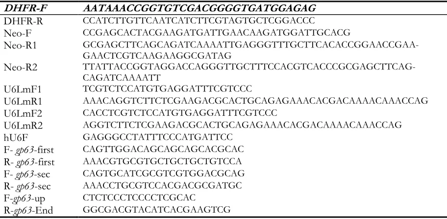
Table 1: The sequence of primers used in the study
DHFR-F AATAAACCGGTGTCGACGGGGTGATGGAGAG
DHFR-R CCATCTTGTTCAATCATCTTCGTAGTGCTCGGACCC
Neo-F CCGAGCACTACGAAGATGATTGAACAAGATGGATTGCACG
Neo-R1 GCGAGCTTCAGCAGATCAAAATTGAGGGTTTGCTTCACACCGGAACCGAA-GAACTCGTCAAGAAGGCGATAG
Neo-R2 TTATTACCGGTAGGACCAGGGTTGCTTTCCACGTCACCCGCGAGCTTCAG-CAGATCAAAATT
U6LmF1 TCGTCTCCATGTGAGGATTTCGTCCC
U6LmR1 AAACAGGTCTTCTCGAAGACGCACTGCAGAGAAACACGACAAAACAAACCAG
U6LmF2 CACCTCGTCTCCATGTGAGGATTTCGTCCC
U6LmR2 AGGTCTTCTCGAAGACGCACTGCAGAGAAACACGACAAAACAAACCAG
hU6F GAGGGCCTATTTCCCATGATTCC
F- gp63-first CAGTTGGACAGCAGCAGCACGCAC
R- gp63-first AAACGTGCGTGCTGCTGCTGTCCA
F- gp63-sec CAGTGCATCGCGTCGTGGACGCAG
R- gp63-sec AAACCTGCGTCCACGACGCGATGC
F-gp63-up CTCTCCCTCCCCTCGCAC
R-gp63-End GGCGACGTACATCACGAAGTCG
Fig. 2: Preparation of U6 snRNA promoter, A) PCR products of reaction 1 and 2. B) Re-annealing fragments indicates that 25% of them bearing sticky ends of interest. (BpiI enzyme restriction sites are shown colored
Fig. 3: Confirmation of U6 cloning. A)Lane 1; DNA marker, Lane 2;digestion of pX330-u6 by Ndei B) Lane 1; DNA marker, Lane 2; PCR product of U6 promoter using primers hU6 and U6LmR2 in the reaction
Preparation of spliced fragments
Fusion of the two fragments regulatory re-gion of the DHFR gene and the neomycin resistance of SOE-PCR product displayed an expected band in size, but did not show a clear and single band and adequate intensity as well. Subsequently, a PCR reaction was performed using the SOE-PCR product but no bands observed following gel electrophoresis (Fig. 4).
Fig. 4: SOEing PCR. Lane1; DNA marker, Lane2; DHFR promoter fragment, Lane3; Neo gene fragment, Lane4; Unsuccessful SOE-PCR. Lane5; Re-amplification from SOEing PCR product
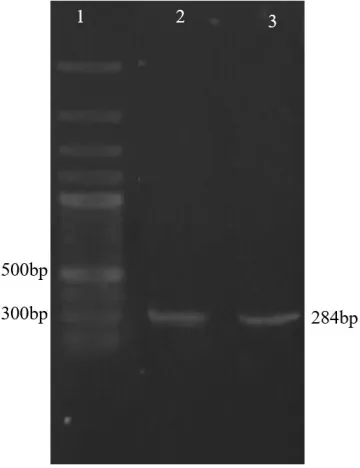
In an attempt to alleviate the problem follow-ing a SOE-PCR reaction, the construct (SOE-PCR product) was cloned into a T-vector (lin-earized pTG19). T/A cloning was confirmed by PCR experiments using M13 and specific primers as forward and reverse primers, re-spectively (Fig. 5). Amplification of SOE con-struct was successfully performed from pTG19-DHFR-Neo. SOE construct was ob-tained in high yield in this way and successful-ly sub-cloned into pX330-U6.
Cloning of the SOE-PCR product
The insert (construct) for ligation was pre-pared in high yield with enzymatic digestion of pTG19-DHFR-Neo by Age I that showed the 1825-bp DNA fragment (Fig. 5). Colony PCR results showed successful cloning of SOE-construct into pX330-U6. Resulting plasmid was described as pX-leish. Cloning was con-firmed by amplification of SOE-construct from extracted pX-leish and enzymatic diges-tion. Moreover correct orientation of con-struct was confirmed by amplification of a 2200-bp fragment using primers U6LmF1/DHFR-R. pX-leish has a XhoI re-striction site on the SOE construct sequence but not on the pX330-U6. pX-leish was linear-ized by XhoI restriction enzyme (Fig. 6).
Cloning of specific guide RNAs for gp63 Following the preparation of the vector con-taining a neomycin resistance gene fused to Cas9 under the regulatory elements of the DHFR gene, the two sgRNAs for targeting the gp63 Gene were cloned into the site of BpiI restriction enzyme downstream of the U6 promoter in the vector (Fig. 7). Amplification of U6 fragment using a strand of sgRNA se-quence as reverse primer demonstrated the accuracy of cloning of sgRNAs (Fig. 8). Assessment of double-strand DNA breaks (DSBs)
To evaluate the capability of the vector to target the gene of interest at the regions spe-cific for guide RNAs, the L. major parasites were transfected with pX-Leish and incubated at 22 ºC for 24 hours. Then the culture medi-um was replaced with RPMI 1640 containing
15 µg/ml of G418. Unlike what we expected, after electroporation and allowing parasites to grow, the transfected cell growth was arrested. Different protocols were applied for transfec-tion with no success in survival. Comparison of the amplified fragments from extracted DNA of transfected and WT parasites verified the pX-Leish efficiency. The expected size of amplified fragment was 800bp as observed in PCR from DNA of WT parasites, whereas amplified fragment from DNA of transfected parasites was 500bp (Fig. 9). Furthermore, de-letion or insertion was evaluated by digestion of the re-annealed heterogenic PCR products with T7 Endonuclease I. Gel analysis indicat-ed multiple bands corresponding to detectindicat-ed fragments about 800 bp, 500 bp, 400 bp and 100 bp sizes (Fig. 9).
Fig. 6: Confirmation of sub-cloning. A) Lane1; DNA marker, Lane 2; PCR reaction using primers U6LmF1/DHFR-R B) Lane1; DNA marker, Lane 2; leish digestion by XhoI Lane 3; circular pX-leish
Fig. 8: Confirmation of sgRNA cloning. Lane1; DNA marker, Lane 2; PCR reaction using U6LmF1 primer and sgRNA (R-gp63-first) as reverse primer, Lane 3; PCR reaction using U6LmF1 primer and sgRNA (R-gp63-second) as reverse primer
Fig. 9: Confirmation of pX-Leish efficiency. A) Agarose Gel detection of PCR amplicon resulted from MMEJ. Lanes 1 and 2; PCR products of transfected and WT parasites by primers Fgp63-up/Rgp63-end respectively; Lane 3, DNA Marker; B) Digestion of PCR products by T7 Endonuclease: Lane 1; Digestion of PCR product of DNA extracted from WT parasites; Lane 2,
Digestion of mixed PCR products of WT and transfected parsites; Lane 3, DNA marker
The 800 bp length is original size related to WT parasites. The 400 bp- and 100 bp- frag-ments resulted from digestion of 500 bp fragment in size that confirm the deletion oc-curred in target sites.
In a different experiment we allowed to growth electroporated parasites in an antibi-otic free RPMI medium, passaged in a fresh RPMI in a ratio of 1:10, gradually a population of promastigotes grew up during 2 weeks. Amplification of 800 bp-fragment was per-formed to evaluate mutation of gp63 gene in this population. As expected, we observed the same electrophoresis pattern in PCR amplicon of WT parasites. This indicates transfected promastigotes could not growth in the antibi-otic free medium and therefore new popula-tions were untransfected promastigotes.
Discussion
Until recently, homologous recombination has been one of the main techniques in genet-ic manipulation of Leishmania. Generation of knockout parasites has been common method via targeted gene replacement with antibiotic selection marker.
Although tolerance of homologous recom-bination by Leishmania is considered as an ad-vantageous trait for generation of knockout parasites, but this method is labor, time con-suming with low rate of recombination (15).
Over the last few years, a more efficient ge-nome editing technology known as CRISPR/Cas9 system has been used in mammalian cells and other organisms such as protozoan parasites. This method has im-proved our ability in genome editing of
Leish-mania. The current available vectors are
marker. For this end, we modified Px330 vec-tor and constructed an all-in-one vecvec-tor con-taining single guide RNA expression cassette and Neo-Cas9 expression cassette.
For transcription of the sgRNA, U6 small nuclear RNA (snRNA) regulatory elements have been already used in P. falciparum and L.
major(18, 19). RNA polymerase I promoter
was also implemented in CRISPR/Cas9 sys-tem for expression of gRNA in L. donovani (15). Valerian Nakaar and his team demon-strated that two intragenic regulatory elements of tRNA genes namely A and B boxes act as extragenic regulatory elements for U6 snRNA gene in Trypanosomes. They discovered A and B boxes at upstream of U6 snRNA gene are essential for expression of U6 snRNA gene and deletion of these boxes resulted in undetectable levels of the U6 snRNA gene expression (20). We found U6 snRNA se-quences of L. major on the minus strand of chromosome 24 using BLAST alignment of U6snRNA sequences of T. brucei. Therefore we implemented a 262-bp sequence of 5´-upstream region of the transcriptional start site of U6 snRNA as the sgRNA promoter. On the other hand, we re-established the BpiI enzyme restriction site by addition of the 12-bp fragment at the 5´-end of reverse primers (Fig. 1). The designed sgRNAs in this study were cloned successfully in to the pX-leish.
Current vectors are used for targeting the mammalian genomes. Different promoters have been applied for expression of cas9 gene in parasites such as: the alpha-tubulin promoter in Toxoplasma gondi(21), plasmodial regulatory elements in Plasmodium falciparum(18), Leishmania tubulin intergeneic region in L. donovani (15), DHFR-TS promoter in L. major (19) and ribo-somal promoter in Trypanosoma cruzi (22).
Dihydrofolate reductase (DHFR) is an es-sential enzyme was express in the both pro-mastigote and apro-mastigote forms of Leishmania (23). Hence we employed 5´-flanking region of DHFR gene of L. major for expression of antibiotic resistance marker and cas9 protein. In order to simultaneous expression of Cas9
and antibiotic resistance proteins a self-cleaving 2A peptide was used to mediate effi-ciently co-translational “cleavage” between the upstream of cas9 and downstream of Neo gene. The 2A self-cleaving peptide allows them to be encoded as a polyprotein and dis-sociate from each other during translation. Function of 2A peptide has already proved in various parasites such as Eimeria tenella,
Plas-modium yoelii, Trypanosoma cruzi (24-27).
In mammalian cells, DSBs are repaired through either introducing homologous DNA sequences (homologous recombination) or NHEJ. High rates of DSBs increases the fre-quency of homologous recombination in presence of the homologous template. Also repair of DNA occurs by NHEJ when ho-mologous template is absent. Small deletions and insertions (Indels) occurred during NHEJ may lead to mutation at the target site with subsequent alteration or disruption of protein function (12). Observations has been indicated that NHEJ is absent in Leishmania, but it has been demonstrated that L. donovani utilizes MMEJ to repair the DSB-induced by Cas9-nuclease(15). Distance of microhomology se-quences to the DSB site determines the length of deletion (15, 28).
In the current study selected PAMs were located near the 5´end of the gp63 coding se-quence close to the start codon. The guide RNAs were designed against sequences up-stream of the PAM regions. T7 endonuclease digestion proved a deletion in the region of the PAM sequence. This assay was performed as a primary confirmation of the CRISPR/cas9 activity. However western blot-ting would confirm the absence of the protein.
efficacy of pX-Leish, to understand the reason which we not able to obtain mutant live para-sites, we speculate that designed sgRNAs probably have been non-specific for gp63 gene and generated the various off targets on the Leishmania genome and induced apoptosis and death of parasites. For this purpose we evaluated sgRNAs again with online special softwares and they were not ranked with high score. Although we cannot certainly assert that off targets have been the arrest cause of para-sites, but it is a strong possibility and only rea-son we speculate that may cause multiple DNA fragmentation and death.
Conclusion
The pX-leish can be utilized for gene target-ing of any virulent antigens in Leishmania or even it may be possible to design sgRNAs or targeting different genes simultaneously. The CRISPR/cas9 technique benefits from this fact that it is capable of targeting gene of in-terest simply by designing specific sgRNAs followed by an easy method for cloning the fragments into the vector. Ease and high effi-ciency potential of this method may help for example generation of attenuated parasites as a vaccine, identification of therapeutic targets and function of genes. The current study was conducted to improve this system which may able to open promising ways for control measures of leishmaniasis.
Acknowledgements
This research was a part of student thesis, funded by Tehran University of Medical Sci-ences and Shahid Beheshti University of Med-ical Sciences under grants No.29599 and No.1395-400 respectively. We appreciate the Vice-Chancellor for Research of both univer-sities, for financial support of this study.
Conflict of interest
The authors declare that there is no conflict of interests.
References
1. Mohebali M. Visceral leishmaniasis in Iran: review of the epidemiological and clinical features. Iran J Parasitol. 2013; 8(3):348-58. 2. Ahmadi M, Fata A, Khamesipour A et al. The
efficacy of hydro alcoholic extract of Seidlitzia rosmarinus on experimental zoonotic cutaneous leishmaniasis lesions in murine model. Avicenna J Phytomed. 2014;4(6): 385-91.
3. Nadim A, Javadian E, Mohebali M. The experience of leishmanization in the Islamic Republic of Iran. 1997. http://www.who.int/iris/handle/10665/117339 4. Mohebali M, Khamesipour A, Mobedi I, Zarei Z,
Hashemi-Fesharki R. Double-blind randomized efficacy field trial of alum precipitated autoclaved
Leishmania major vaccine mixed with BCG against
canine visceral leishmaniasis in Meshkin-Shahr district, IR Iran. Vaccine. 2004; 22(29-30):4097-100. 5. Pirdel L, Farajnia S. A non‐pathogenic
recombinant Leishmania expressing Lipophosphoglycan 3 against experimental infection with Leishmania infantum. Scand J Immunol. 2017: 86(1):15-22.
6. Shaddel M, Ormazdi H, Akhlaghi L, Kazemi B, Bandepour M. Evaluating the Cloning of
Leishmania major p4 Gene in Production of
Vaccine. Razi J Med Sci. 2009;15(60):115-20. 7. Rezvan H, Rees R, Ali S. Leishmania mexicana
Gp63 cDNA using gene gun induced higher immunity to L. mexicana infection compared to soluble Leishmania antigen in BALB/C. Iran J Parasitol. 2011; 6(4):60-75.
8. Avishek K, Kaushal H, Gannavaram S, Dey R, Selvapandiyan A, Ramesh V, et al. Gene deleted live attenuated Leishmania vaccine candidates against visceral leishmaniasis elicit pro-inflammatory cytokines response in human PBMCs. Sci Rep. 2016;6.
9. Kumar R, Engwerda C. Vaccines to prevent leishmaniasis. Clin Trans Immunol. 2014;3(3):e13.
11. Fiuza JA, Santiago Hda C, Selvapandiyan A, Gannavaram S et al. Induction of immunogenicity by live attenuated Leishmania
donovani centrin deleted parasites in dogs.
Vaccine. 2013; 31(14):1785-92.
12. Maeder ML, Gersbach CA. Genome-editing technologies for gene and cell therapy. Mol Ther. 2016; 24(3):430-46.
13. Almani PG, Sharifi I, Kazemi B et al. The role of GlcNAc-PI-de-N-acetylase gene by gene knockout through homologous recombination and its consequences on survival, growth and infectivity of Leishmania major in in vitro and in vivo conditions. Act Trop. 2016; 154:63-72. 14. Kurnaz IA. Techniques in Genetic
Engineering.CRC press;2015. 194 p.
15. Zhang WW1, Matlashewski G. CRISPR-Cas9-mediated genome editing in Leishmania donovani. MBio. 2015; 6(4):e00861.
16. Das A, Banday M, Bellofatto V. RNA polymerase transcription machinery in trypanosomes. Eukaryot Cell. 2008; 7(3):429-34. 17. Mohebali M, Motazedian M, Parsa F, Hajjaran
H, Yaghoobi-Ershadi M. Identification of
Leishmania species from different parts of Iran
using a random amplified polymorphic DNA in humans, animal reservoirs and vectors. Med J IRIN. 2002;15(4):243-6.
18. Ghorbal M, Gorman M, Macpherson CR et al. Genome editing in the human malaria parasite
Plasmodium falciparum using the CRISPR-Cas9
system. Nat Biotechnol. 2014; 32(8):819-21. 19. Sollelis L, Ghorbal M, MacPherson CR et al.
First efficient CRISPR-Cas9-mediated genome editing in Leishmania parasites. Cell Microbiol. 2015;17(10):1405-12.
20. Nakaar V, Dare AO, Hong D, Ullu E, Tschudi C. Upstream tRNA genes are essential for expression of small nuclear and cytoplasmic RNA genes in trypanosomes. Mol Cell Biol. 1994; 14(10):6736-42.
21. Sidik SM, Hackett CG, Tran F, Westwood NJ, Lourido S. Efficient genome engineering of
Toxoplasma gondii using CRISPR/Cas9. PLOS One. 2014; 9(6):e100450.
22. Lander N, Li Z-H, Niyogi S, Docampo R. CRISPR/Cas9-induced disruption of paraflagellar rod protein 1 and 2 genes in
Trypanosoma cruzi reveals their role in flagellar
attachment. MBio. 2015;6(4):e01012-15. 23. Gilbert IH. Inhibitors of dihydrofolate
reductase in Leishmania and trypanosomes. Biochim Biophys Acta. 2002; 1587(2-3):249-57. 24. Tang X, Liu X, Tao G, Qin M, Yin G, Suo J, et al. “Self-cleaving” 2A peptide from porcine teschovirus-1 mediates cleavage of dual fluorescent proteins in transgenic Eimeria tenella. Vet Res. 2016;47(1):68.
25. Zhang C, Xiao B, Jiang Y, Zhao Y, Li Z, Gao H, et al. Efficient editing of malaria parasite genome using the CRISPR/Cas9 system. MBio. 2014;5(4):e01414-14.
26. Heras SR, Thomas MC, García-Canadas M et al. L1Tc non-LTR retrotransposons from
Trypanosoma cruzi contain a functional viral-like
self-cleaving 2A sequence in frame with the active proteins they encode. Cell Mol Life Sci. 2006;63(12):1449-60.
27. Tang X, Liu X, Tao G et al. "Self-cleaving "2A peptide from porcine teschovirus-1 mediates cleavage of dual fluorescent proteins in transgenic Eimeria tenella. Vet Res. 2016;47(1):68
28. Peng D, Kurup SP, Yao PY, Minning TA, Tarleton RL. CRISPR-Cas9-mediated single-gene and single-gene family disruption in Trypanosoma
cruzi. MBio. 2015; 6(1):e02097-14.