MECHANISMS MEDIATING CYCLIN E/CDK2-REGULATED REPLICATION-DEPENDENT HISTONE mRNA BIOSYNTHESIS
Anne Eileen White
A dissertation submitted in the faculty of the University of North Carolina at Chapel Hill in partial fulfillment of the requirements for the degree of Doctor of Philosophy in the
Department of Biology.
Chapel Hill 2010
Approved by:
Dr. Robert J. Duronio Dr. William F. Marzluff Dr. Mark Peifer
ABSTRACT
Anne E. White: Mechanisms mediating Cyclin E/Cdk2-regulated replication-dependent histone mRNA biosynthesis
(Under the direction of Dr. Robert J. Duronio)
Nuclear organization is a dynamic modulator of gene expression. Subcellular compartments called nuclear bodies concentrate factors regulating gene expression and change in size, composition, and activities in response to cellular inputs. Thus,
understanding the mechanisms that assemble and organize nuclear bodies is important for appreciating how they contribute to genome function. We explored the relationship between histone locus bodies (HLBs) and replication-dependent histone mRNA biosynthesis as a model for understanding nuclear body function. HLBs are localized near the histone genes and are enriched with factors required for histone mRNA biosynthesis. Cyclin E/Cdk2 is necessary for cell cycle-dependent histone mRNA
biosynthesis. However, the molecular mechanisms of this regulation are not fully known. Using the MPM-2 monoclonal antibody as a tool for exploring cell cycle-mediated
ACKNOWLEDGEMENTS
TABLE OF CONTENTS
LIST OF TABLES ... ix
LIST OF FIGURES ...x
LIST OF ABBREVIATIONS ... xii
Chapter I. INTRODUCTION ...1
Histone locus bodies as paradigms of nuclear bodies ...2
Cell cycle regulation of replication-dependent histone mRNA biosynthesis ...3
Studying histone mRNA biosynthesis in Drosophila melanogaster ...6
Dissertation goals ...8
References ...10
II. DEVELOPMENTAL AND CELL CYCLE REGULATION OF THE DROSOPHILA HISTONE LOCUS BODY...16
Preface...16
Abstract ...16
Introduction ...17
Materials and Methods ...20
Drosophila stocks ...20
Cultured cell immunostaining and RNAi ...22
Microscopy ...23
Measurement of MPM-2 focus size ...23
Results ...24
MPM-2 labels the histone locus body ...24
Embryonic MPM-2 foci depend on Cyclin E/Cdk2 activity ...29
MPM-2 foci co-localize with nascent histone transcripts ...32
The HLB disassembles during mitosis ...34
MPM-2 foci assemble in the absence of the histone pre-mRNA processing factors SLBP and U7 snRNP ...37
Embryonic MPM-2 foci do not require stringcdc25 or histone transcription ...39
MPM-2 foci occur coincidentally with activation of zygotic histone gene expression ...41
MPM-2 foci form independently of the histone locus ...43
Discussion ...46
HLB behavior during early Drosophila development ...47
Cell cycle regulation of the HLB ...49
Summary ...50
Acknowledgements ...50
References ...51
III. IDENTIFICATION OF NOVEL CELL CYCLE-REGULATED COMPONENTS OF THE DROSOPHILA HISTONE LOCUS BODY ...58
Preface...58
Introduction ...59
Material and Methods ...62
Cell culture, RNA interference, and cell staining ...62
Screen imaging, analysis, and candidate selection for secondary screening ...63
Histone GFP reporter assay ...64
Constructs and transfections ...64
Immunoprecipitations and Western blotting ...65
Protein identification by mass spectroscopy ...65
Drosophila strains ...66
Immunofluorescence of embryos and larval brains ...66
Confocal microscopy ...67
Results ...67
A genome-wide screen for MPM-2 reactive proteins co-localizing with HLBs ...67
Several proteins identified in the RNAi screen are novel components of HLBs ...75
FLASH, Mxc, and Mute localization in the embryo ...76
A proteomic approach to identify MPM-2 reactive HLB proteins ...78
Spt6 co-localization with HLBs correlates with active histone gene transcription ...81
A core HLB remains assembled during mitosis ...83
HLB assembly is ordered in the early embryo ...85
Mxc is a novel regulator of HLB assembly and
histone gene expression ...93
Secondary screen exploring inter-dependencies of HLB components ...97
Discussion ...99
HLB assembly during early Drosophila embryogenesis ...99
HLB dynamics during mitosis ...100
Identification of a Drosophila S phase MPM-2 antigen ...102
Mxc is a novel regulator of histone mRNA biosynthesis ...103
Summary ...104
Acknowledgements ...104
References ...105
IV. DISCUSSION AND FUTURE DIRECTIONS ...111
Cell cycle regulation of HLB dynamics ...112
HLB assembly during early Drosophila development ...114
Mxc is a novel regulator of histone mRNA biosynthesis ...116
HLBs coordinate histone gene transcription and pre-mRNA processing ...118
LIST OF TABLES Table
2.1 Characterization of the HLB in histone deletion embryos ...46 3.1 Top hits from primary screen ...71 3.2 Gene products required for HLB localization in order of
necessity ...98 3.3 Classification of histone pre-mRNA processing GFP reporter
LIST OF FIGURES
Figure
1.1 Histone locus bodies in Drosophila ...3
2.1 MPM-2 labels the histone locus body ...28
2.2 Embryonic MPM-2 foci depend on Cyclin E activity ...31
2.3 MPM-2 foci co-localize with the histone locus body in replicating cells ...33
2.4 MPM-2 and Lsm11 foci are cell cycle dependent ...35
2.5 MPM-2 foci depend on the cell cycle ...36
2.6 MPM-2 foci form independently of the U7 snRNP ...38
2.7 MPM-2 foci are not dependent on Lsm11 ...39
2.8 MPM-2 foci do not depend on string or Slbp ...40
2.9 The histone locus body forms for the first time during nuclear cycle 11 ...42
2.10 The histone locus body first forms and co-localizes with MPM-2 foci during nuclear cycle 11 ...43
2.11 Aberrant histone locus bodies form in the absence of the histone locus ...45
3.1 Genome-wide RNAi screen for MPM-2 co-localization with HLBs ...70
3.2 Spt6 is a novel HLB component identified by mass spectroscopy ...80
3.3 Spt6 co-localization with HLBs is cell cycle-dependent ...82
3.4 A core HLB remains assembled during mitosis ...84
3.5 A core histone locus body begins assembling during nuclear cycle 10 ...87
LIST OF ABBREVIATIONS Act5C actin 5C
ATAC Ada2A containing
CB Cajal body
CDK Cyclin-dependent kinase
Cy cyanine
DAPI 4',6-diamidino-2-phenylindole DNA deoxyribonucleic acid
dsRNA double stranded ribonucleic acid ECL enhanced chemiluminescence EMS ethyl methanesulfonate
E2F E2-Factor
FISH fluorescent in situ hybridization FLASH FLICE-associated huge protein GFP green fluorescent protein
HA hemagglutinin
HAT histone acetyltransferase HLB histone locus body HRP horseradish peroxidase
H3 histone H3
IP immunoprecipitate
ISWI Imitation SWI
LSM laser scanning microscope MPM-2 Mitotic protein monoclonal 2 MS mass spectroscopy
Mute Muscle wasted Mxc Multi sex combs NE nuclear extract
NPAT Nuclear Protein Ataxia-Telangiectasia locus
IP immunoprecipitate
PBS phosphate Buffer Saline PCR Polymerase Chain Reaction PH3 phospho-histone H3
PMSF phenylmethanesulphonylfluoride pRb Retinoblastoma protein
PTB Polypyrimidine Tract Binding protein RNA ribonucleic acid
RNAi RNA interference
RSC Chromatin structure remodeling complex
SDS-PAGE sodium dodecyl sulfate polyacrylamide gel electrophoresis
S2 Schneider 2
SLBP stem loop binding protein SnRNP small nuclear ribonucleoprotein
stg string
TOF time of flight
ubi ubiquitin
WT wild-type
CHAPTER I
INTRODUCTION
the cell cycle. For example, mammalian HLBs and nucleoli disassemble during mitosis and reform during the subsequent interphase (Hernandez-Verdun, 2006; Ma et al., 2000; Zhao et al., 2000). Thus, understanding how nuclear bodies control gene expression by responding to various cellular inputs, such as cell cycle inputs, will increase our
understanding of how genome function is modulated. This dissertation uses histone locus bodies (HLBs) as a paradigm of nuclear body function to explore how nuclear bodies are regulated by the cell cycle, assemble, and contribute to gene expression.
Histone locus bodies as paradigms of nuclear bodies
Histone locus bodies (HLBs) were first discovered in Drosophila as nuclear bodies enriched with factors necessary for efficient histone gene expression, such as U7 snRNP and FLASH (Liu et al., 2006; Yang et al., 2009). HLBs associate with the replication-dependent histone genes, which are encoded in a 5-kb repeat present in approximately 100 tandem copies in Drosophila (Liu et al., 2006). They are distinct from and
frequently situated next to Cajal bodies (CBs) in Drosophila (Liu et al., 2006; Liu et al., 2009). CBs are involved in many aspects of RNA metabolism, including small nuclear ribonucleoprotein (snRNP) maturation (Cioce and Lamond, 2005; Kiss, 2004; Matera and Shpargel, 2006; Stanek and Neugebauer, 2006). In mammalian cells and Xenopus
oocytes, CBs contain the U7 snRNP and can transiently associate with the snRNA, small nucleolar RNA (snoRNA), and histone genes (Matera et al., 2009).
performed in primary human cells revealed that U7 snRNP co-localizes with histone gene expression-specific factors FLASH and NPAT (Ghule et al., 2008). The ubiquitous presence of U7 snRNP in CBs seen in human cancer cell lines, therefore, may be a consequence of transformation and an exception to the norm.
HLBs serve as a good model for understanding nuclear body function, because they assemble at a discrete nuclear location, the histone genes, and are likely a conserved feature of the metazoan nucleus. In addition, the cell cycle is a critical cellular input regulating HLB function, which limits replication-dependent histone gene expression to S phase when DNA is being replicated. For these reasons, studying HLBs will both
increase our understanding of nuclear body function and how the cell cycle modulates gene expression.
Figure 1.1. Histone locus bodies in Drosophila. Syncytial blastoderm (cycle 13) embryos were hybridized
with an H3/H4 gene probe (red in merge) and stained with anti-Lsm11 (green in merge) antibodies and the
DNA labeling dye DAPI (blue in merge). Arrows indicate a single histone locus body. Scale bar: 20 µm.
Cell cycle regulation of replication-dependent histone biosynthesis
cell divides during mitosis. The coupling of histone biosynthesis to S phase is crucial for cell viability. DNA replication without concomitant histone protein synthesis is lethal in budding yeast (Han et al., 1987; Kim et al., 1988). Further, in vitro reconstitution
experiments have shown that production of histones in excess to DNA can cause insoluble histone-nucleic acid aggregates (Carruthers et al., 1999; Detke et al., 1979; Dyer et al., 2004), which could lead to incorrect DNA segregation should these
conditions arise in the cell. Thus, the timing and amount of histone protein biosynthesis must be carefully controlled to ensure that cell division is successful.
Cyclin-dependent kinase (Cdk) complexes coordinate the major events of cell division. In animal cells Cyclin E/Cdk2 phosphorylates proteins, such as pRb, that mediate changes in gene expression associated with the onset of S phase, including proteins that regulate histone gene expression (Du and Pogoriler, 2006). Two Cyclin E/Cdk2 substrates, NPAT and HIRA, activate and repress, respectively, histone gene expression in human cell culture (Hall et al., 2001; Ma et al., 2000; Miele et al., 2005; Nelson et al., 2002; Zhao et al., 2000). How the activity of such factors is modulated by Cyclin E/Cdk2 and integrated into cell cycle-regulated histone gene expression is incompletely understood. Even less is known about how histone gene expression is regulated during animal development, because studies have mainly been done in cell culture.
For most organisms the amount of replication-dependent histone protein
1979; Harris et al., 1991; Heintz et al., 1983; Parker and Fitschen, 1980). This sharp rise in mRNA levels is achieved by increases in the rate of transcription initiation, pre-mRNA processing, and half-life of the mature mRNA (Marzluff and Duronio, 2002). Whether all of these mechanisms are under cell cycle control is unknown.
Transcription of the histone genes in mammalian cells is stimulated by the Cyclin E/Cdk2 substrate NPAT (Ma et al., 2000; Wei et al., 2003; Ye et al., 2003; Zhao et al., 2000). NPAT is found in HLBs associated with the histone genes in both primary human cells and cancer cells (Ma et al., 2000; Zhao et al., 2000). Phosphorylated NPAT acts with histone subtype-specific transcription factors, such as HiNF-P and OCA-S, which bind subtype-specific regulator elements in the promoters of the histone H4 and H2B genes, respectively (Miele et al., 2005; Mitra et al., 2003; Ye et al., 2003; Zheng et al., 2003). While several phosphorylation sites on NPAT have been mapped, how this phosphorylation regulates NPAT activity is incompletely understood (Ma et al., 2000; Zhao et al., 2000). It was recently proposed that NPAT plays a role in promoting histone H4 acetylation by recruiting the TRRAP-Tip60 complex to the histone genes (DeRan et al., 2008). A connection between NPAT and histone pre-mRNA processing has also been made, whereby NPAT interacts with CDK9 at the histone genes. CDK9 is a component of the P-TEFb transcription elongation complex that also monoubiquitinates histone H2B, which in turn recruits the histone pre-mRNA processing machinery (Pirngruber and Johnsen, 2010). Cyclin E/Cdk2 could also promote histone mRNA biosynthesis by regulating HLB dynamics as well as the activity of other HLB proteins.
polyadenylated (Marzluff and Duronio, 2002; Marzluff et al., 2008). Instead they end in a conserved stem loop structure that regulates all aspects of replication-dependent histone mRNA metabolism, including biosynthesis, translation, and stability (Marzluff, 2005). Binding of Stem Loop Binding Protein (SLBP) to the stem loop in the 3’ UTR and U7snRNP to the purine-rich downstream element recruits a cleavage endonuclease complex. This complex cleaves the histone pre-mRNA 4-5 nucleotides after the stem loop producing the unique 3’ end of the mature histone mRNA (Dominski and Marzluff, 1999; Marzluff, 2005). In mammalian cells SLBP protein levels are cell cycle regulated, yet this mode of regulation is not universal as SLBP protein is expressed uniformly through the cell cycle in Drosophila cells (Lanzotti et al., 2004b; Marzluff et al., 2008). Moreover, it is uncertain whether other components of the histone pre-mRNA processing machinery are cell cycle regulated. Cyclin E/Cdk2 does, however, regulate pre-mRNA splicing in cultured mammalian cells. Three subunits of the essential splicing factor SF3, SAP114, SAP145, and SAP155, as well as the U2 snRNA and the snRNP core protein B/B’ have been shown to immunoprecipitate with cyclin E (Seghezzi et al., 1998). Of these proteins, Cyclin E/Cdk2 has been shown to phosphorylate SAP155 in vitro
(Seghezzi et al., 1998). Similarly, Cyclin E/Cdk2 may interact with and/or phosphorylate proteins specifically involved in histone pre-mRNA processing.
Studying histone mRNA biosynthesis regulation in Drosophila melanogaster
Drosophila has provided fundamental insights into the regulation of the cell cycle,
model system to explore how histone mRNA biosynthesis is regulated. As part of normal fly development, the first 13 embryonic cycles after fertilization consist only of rapid, synchronous S-M nuclear division cycles that occur in a common cytoplasm without cytokinesis. These early cycles depend on maternally contributed factors. During
interphase 14 nuclei pause in G2 for the first time and cellularization takes place. Groups of cells then enter mitosis 14 asynchronously in a manner dependent on zygotic
transcription of an activator of mitosis encoded by the stringcdc25 (stg) gene (Edgar and O'Farrell, 1989a; Edgar and O'Farrell, 1990a). Stringcdc25 is a phosphatase that activates
the mitotic Cdk, Cdc2. The spatial and temporal pattern of these divisions is controlled by the patterning genes (O'Farrell et al., 1989). G1 is introduced in embryonic cycle 17 and at this point all cell types require zygotic Cyclin E expression for DNA replication and cell cycle progression. Previously it was shown that stringcdc25 and Cyclin E are required for histone gene expression at cycles 14 and 17, respectively (Lanzotti et al., 2004a). The exact mechanism of this regulation, however, has not been elucidated.
We sought new tools to study Cyclin E/Cdk2 regulation of histone mRNA biosynthesis. One such reagent was the monoclonal antibody MPM-2, which has been previously used as a marker of Cyclin E/Cdk2 activity in Drosophila cells (Calvi et al., 1998). MPM-2 was generated using a mitotic HeLa cell extract and recognizes
Lange et al., 2005a; Logarinho and Sunkel, 1998a; Yaffe et al., 1997). In Drosophila salivary glands and ovarian follicle cells MPM-2 labels a discrete spherical nuclear structure whose cell cycle appearance in S phase is dependent on Cyclin E/Cdk2 activity (Calvi et al., 1998). Moreover, our collaborator Brian Calvi observed that MPM-2 only labels the histone genes of polytene chromosomes from larval salivary glands (Chapter II, Figure 2.1A). Based on these results, Brian Calvi and we hypothesized that the protein(s) detected by MPM-2 are Cyclin E/Cdk2 substrates that co-localize with HLBs at the histone genes and activate histone mRNA biosynthesis. A major emphasis of this dissertation was to identify and characterize MPM-2 reactive HLB proteins.
Dissertation goals
This dissertation describes work done examining how histone mRNA
biosynthesis is regulated by components of the histone locus body (HLB). In particular, it focuses on the molecular mechanisms regulated by Cyclin E/Cdk2. Chapter II presents data showing that the MPM-2 antibody detects protein(s) that are part of the histone locus body. The MPM-2 epitope present at HLBs is under both cell cycle and developmental control, such that Cyclin E/Cdk2 activity is necessary for production of the MPM-2 epitope and mature HLBs are formed coincidentally with zygotic histone gene
To further understand how Cyclin E/Cdk2 might regulate histone gene expression we sought to identify MPM-2 antigen(s) that are part of HLBs. Chapter III describes biochemical and genome-wide functional genomics screens performed to pinpoint MPM-2 antigen(s). Both approaches identified novel components of HLBs, including two MPM-2 reactive proteins (Mxc and Mute-S). Mxc is necessary for HLB assembly and histone pre-mRNA processing in S2 cells. HLBs are highly dynamic, such that core components begin assembling one nuclear cycle prior to the formation of mature HLBs and zygotic gene transcription. Likewise, this “core” HLB remains assembled
References
Albert, A. L., Lavoie, S. B., and Vincent, M. (2004). Multisite phosphorylation of Pin1-associated mitotic phosphoproteins revealed by monoclonal antibodies MPM-2 and CC-3. BMC Cell Biol 5, 22.
Borun, T. W., Gabrielli, F., Ajiro, K., Zweidler, A., and Baglioni, C. (1975). Further evidence of transcriptional and translational control of histone messenger RNA during the HeLa S3 cycle. Cell 4, 59-67.
Breindl, M., and Gallwitz, D. (1973). Identification of histone messenger RNA from HeLa cells. Appearance of histone mRNA in the cytoplasm and its translation in a rabbit-reticulocyte cell-free system. Eur J Biochem 32, 381-391.
Calvi, B. R., Lilly, M. A., and Spradling, A. C. (1998). Cell cycle control of chorion gene amplification. Genes Dev 12, 734-744.
Carruthers, L. M., Tse, C., Walker, K. P., 3rd, and Hansen, J. C. (1999). Assembly of defined nucleosomal and chromatin arrays from pure components. Methods Enzymol
304, 19-35.
Cioce, M., and Lamond, A. I. (2005). Cajal bodies: a long history of discovery. Annu Rev Cell Dev Biol 21, 105-131.
Davis, F. M., Tsao, T. Y., Fowler, S. K., and Rao, P. N. (1983). Monoclonal antibodies to mitotic cells. Proc Natl Acad Sci U S A 80, 2926-2930.
DeLisle, A. J., Graves, R. A., Marzluff, W. F., and Johnson, L. F. (1983). Regulation of histone mRNA production and stability in serum-stimulated mouse 3T6 fibroblasts. Mol Cell Biol 3, 1920-1929.
DeRan, M., Pulvino, M., Greene, E., Su, C., and Zhao, J. (2008). Transcriptional activation of histone genes requires NPAT-dependent recruitment of TRRAP-Tip60 complex to histone promoters during the G1/S phase transition. Mol Cell Biol 28, 435-447.
do Carmo Avides, M., Tavares, A., and Glover, D. M. (2001). Polo kinase and Asp are needed to promote the mitotic organizing activity of centrosomes. Nat Cell Biol 3, 421-424.
Dominski, Z., and Marzluff, W. F. (1999). Formation of the 3' end of histone mRNA. Gene 239, 1-14.
Du, W., and Pogoriler, J. (2006). Retinoblastoma family genes. Oncogene 25, 5190-5200.
Dyer, P. N., Edayathumangalam, R. S., White, C. L., Bao, Y., Chakravarthy, S., Muthurajan, U. M., and Luger, K. (2004). Reconstitution of nucleosome core particles from recombinant histones and DNA. Methods Enzymol 375, 23-44.
Edgar, B. A., and O'Farrell, P. H. (1989). Genetic control of cell division patterns in the Drosophila embryo. Cell 57, 177-187.
Edgar, B. A., and O'Farrell, P. H. (1990). The three postblastoderm cell cycles of Drosophila embryogenesis are regulated in G2 by string. Cell 62, 469-480.
Ghule, P. N., Dominski, Z., Yang, X. C., Marzluff, W. F., Becker, K. A., Harper, J. W., Lian, J. B., Stein, J. L., van Wijnen, A. J., and Stein, G. S. (2008). Staged assembly of histone gene expression machinery at subnuclear foci in the abbreviated cell cycle of human embryonic stem cells. Proc Natl Acad Sci U S A 105, 16964-16969.
Haaf, T., Hayman, D. L., and Schmid, M. (1991). Quantitative determination of rDNA transcription units in vertebrate cells. Exp Cell Res 193, 78-86.
Hall, C., Nelson, D. M., Ye, X., Baker, K., DeCaprio, J. A., Seeholzer, S., Lipinski, M., and Adams, P. D. (2001). HIRA, the human homologue of yeast Hir1p and Hir2p, is a novel cyclin-cdk2 substrate whose expression blocks S-phase progression. Mol Cell Biol
21, 1854-1865.
Han, M., Chang, M., Kim, U. J., and Grunstein, M. (1987). Histone H2B repression causes cell-cycle-specific arrest in yeast: effects on chromosomal segregation, replication, and transcription. Cell 48, 589-597.
evidence suggesting control at two posttranscriptional steps. Mol Cell Biol 11, 2416-2424.
Heintz, N., Sive, H. L., and Roeder, R. G. (1983). Regulation of human histone gene expression: kinetics of accumulation and changes in the rate of synthesis and in the half-lives of individual histone mRNAs during the HeLa cell cycle. Mol Cell Biol 3, 539-550.
Hernandez-Verdun, D. (2006). The nucleolus: a model for the organization of nuclear functions. Histochem Cell Biol 126, 135-148.
Hirano, T., and Mitchison, T. J. (1991). Cell cycle control of higher-order chromatin assembly around naked DNA in vitro. J Cell Biol 115, 1479-1489.
Kim, U. J., Han, M., Kayne, P., and Grunstein, M. (1988). Effects of histone H4 depletion on the cell cycle and transcription of Saccharomyces cerevisiae. Embo J 7, 2211-2219.
Kiss, T. (2004). Biogenesis of small nuclear RNPs. J Cell Sci 117, 5949-5951.
Kuang, J., Zhao, J., Wright, D. A., Saunders, G. F., and Rao, P. N. (1989). Mitosis-specific monoclonal antibody MPM-2 inhibits Xenopus oocyte maturation and depletes maturation-promoting activity. Proc Natl Acad Sci U S A 86, 4982-4986.
Lange, B. M., Kirfel, G., Gestmann, I., Herzog, V., and Gonzalez, C. (2005). Structure and microtubule-nucleation activity of isolated Drosophila embryo centrosomes characterized by whole mount scanning and transmission electron microscopy. Histochem Cell Biol 124, 325-334.
Lanzotti, D. J., Kupsco, J. M., Marzluff, W. F., and Duronio, R. J. (2004a). string(cdc25) and cyclin E are required for patterned histone expression at different stages of
Drosophila embryonic development. Dev Biol 274, 82-93.
Lanzotti, D. J., Kupsco, J. M., Yang, X. C., Dominski, Z., Marzluff, W. F., and Duronio, R. J. (2004b). Drosophila stem-loop binding protein intracellular localization is mediated by phosphorylation and is required for cell cycle-regulated histone mRNA expression. Mol Biol Cell 15, 1112-1123.
Liu, J. L., Murphy, C., Buszczak, M., Clatterbuck, S., Goodman, R., and Gall, J. G. (2006). The Drosophila melanogaster Cajal body. J Cell Biol 172, 875-884.
Liu, J. L., Wu, Z., Nizami, Z., Deryusheva, S., Rajendra, T. K., Beumer, K. J., Gao, H., Matera, A. G., Carroll, D., and Gall, J. G. (2009). Coilin is essential for Cajal body organization in Drosophila melanogaster. Mol Biol Cell 20, 1661-1670.
Logarinho, E., and Sunkel, C. E. (1998). The Drosophila POLO kinase localises to multiple compartments of the mitotic apparatus and is required for the phosphorylation of MPM2 reactive epitopes. J Cell Sci 111 ( Pt 19), 2897-2909.
Ma, T., Van Tine, B. A., Wei, Y., Garrett, M. D., Nelson, D., Adams, P. D., Wang, J., Qin, J., Chow, L. T., and Harper, J. W. (2000). Cell cycle-regulated phosphorylation of p220(NPAT) by cyclin E/Cdk2 in Cajal bodies promotes histone gene transcription. Genes Dev 14, 2298-2313.
Marzluff, W. F. (2005). Metazoan replication-dependent histone mRNAs: a distinct set of RNA polymerase II transcripts. Curr Opin Cell Biol 17, 274-280.
Marzluff, W. F., and Duronio, R. J. (2002). Histone mRNA expression: multiple levels of cell cycle regulation and important developmental consequences. Curr Opin Cell Biol 14, 692-699.
Marzluff, W. F., Wagner, E. J., and Duronio, R. J. (2008). Metabolism and regulation of canonical histone mRNAs: life without a poly(A) tail. Nat Rev Genet 9, 843-854.
Matera, A. G. (1999). Nuclear bodies: multifaceted subdomains of the interchromatin space. Trends Cell Biol 9, 302-309.
Matera, A. G., Izaguire-Sierra, M., Praveen, K., and Rajendra, T. K. (2009). Nuclear bodies: random aggregates of sticky proteins or crucibles of macromolecular assembly? Dev Cell 17, 639-647.
Matera, A. G., and Shpargel, K. B. (2006). Pumping RNA: nuclear bodybuilding along the RNP pipeline. Curr Opin Cell Biol 18, 317-324.
E/CDK2/p220NPAT pathway to histone H4 gene regulation at the G1/S phase cell cycle transition. Mol Cell Biol 25, 6140-6153.
Misteli, T. (2007). Beyond the sequence: cellular organization of genome function. Cell
128, 787-800.
Mitra, P., Xie, R. L., Medina, R., Hovhannisyan, H., Zaidi, S. K., Wei, Y., Harper, J. W., Stein, J. L., van Wijnen, A. J., and Stein, G. S. (2003). Identification of HiNF-P, a key activator of cell cycle-controlled histone H4 genes at the onset of S phase. Mol Cell Biol
23, 8110-8123.
Nelson, D. M., Ye, X., Hall, C., Santos, H., Ma, T., Kao, G. D., Yen, T. J., Harper, J. W., and Adams, P. D. (2002). Coupling of DNA synthesis and histone synthesis in S phase independent of cyclin/cdk2 activity. Mol Cell Biol 22, 7459-7472.
O'Farrell, P. H., Edgar, B. A., Lakich, D., and Lehner, C. F. (1989). Directing cell division during development. Science 246, 635-640.
Parker, I., and Fitschen, W. (1980). Histone mRNA metabolism during the mouse fibroblast cell cycle. Cell Differ 9, 23-30.
Pirngruber, J., and Johnsen, S. A. (2010). Induced G1 cell-cycle arrest controls
replication-dependent histone mRNA 3' end processing through p21, NPAT and CDK9. Oncogene.
Scheer, U., Hugle, B., Hazan, R., and Rose, K. M. (1984). Drug-induced dispersal of transcribed rRNA genes and transcriptional products: immunolocalization and silver staining of different nucleolar components in rat cells treated with 5,6-dichloro-beta-D-ribofuranosylbenzimidazole. J Cell Biol 99, 672-679.
Seghezzi, W., Chua, K., Shanahan, F., Gozani, O., Reed, R., and Lees, E. (1998). Cyclin E associates with components of the pre-mRNA splicing machinery in mammalian cells. Mol Cell Biol 18, 4526-4536.
Stanek, D., and Neugebauer, K. M. (2006). The Cajal body: a meeting place for spliceosomal snRNPs in the nuclear maze. Chromosoma 115, 343-354.
Wei, Y., Jin, J., and Harper, J. W. (2003). The cyclin E/Cdk2 substrate and Cajal body component p220(NPAT) activates histone transcription through a novel LisH-like domain. Mol Cell Biol 23, 3669-3680.
Westendorf, J. M., Rao, P. N., and Gerace, L. (1994). Cloning of cDNAs for M-phase phosphoproteins recognized by the MPM2 monoclonal antibody and determination of the phosphorylated epitope. Proc Natl Acad Sci U S A 91, 714-718.
Yaffe, M. B., Schutkowski, M., Shen, M., Zhou, X. Z., Stukenberg, P. T., Rahfeld, J. U., Xu, J., Kuang, J., Kirschner, M. W., Fischer, G., et al. (1997). Sequence-specific and phosphorylation-dependent proline isomerization: a potential mitotic regulatory mechanism. Science 278, 1957-1960.
Yang, X. C., Burch, B. D., Yan, Y., Marzluff, W. F., and Dominski, Z. (2009). FLASH, a proapoptotic protein involved in activation of caspase-8, is essential for 3' end processing of histone pre-mRNAs. Mol Cell 36, 267-278.
Ye, X., Wei, Y., Nalepa, G., and Harper, J. W. (2003). The cyclin E/Cdk2 substrate p220(NPAT) is required for S-phase entry, histone gene expression, and Cajal body maintenance in human somatic cells. Mol Cell Biol 23, 8586-8600.
Zhao, J., Kennedy, B. K., Lawrence, B. D., Barbie, D. A., Matera, A. G., Fletcher, J. A., and Harlow, E. (2000). NPAT links cyclin E-Cdk2 to the regulation of replication-dependent histone gene transcription. Genes Dev 14, 2283-2297.
CHAPTER II
DEVELOPMENTAL AND CELL CYCLE REGULATION OF THE
DROSOPHILA HISTONE LOCUS BODY
Preface
This work was published in Molecular Biology of the Cell and featured on the journal cover. Michelle Leslie performed the experiment shown in Figure 3.10. Julie Norseen, a student in Dr. Brian Calvi’s lab, performed MPM-2 staining on polytene chromosomes from larval salivary glands. I performed all other experiments. My advisor Dr. Robert Duronio designed the project. He, Dr. William Marzluff, and I wrote the manuscript and analyzed the data.
White, A.E., Leslie, M.E., Calvi, B.R., Marzluff, W.F., and Duronio, R.J. (2007) Developmental and cell cycle regulation of the Drosophila histone locus body. Mol. Biol. Cell. 18:2491-502.
Abstract
Cyclin E/Cdk2 is necessary for replication-dependent histone mRNA
biosynthesis, but how it controls this process in early development is unknown. We show that in Drosophila embryos the MPM-2 monoclonal antibody, raised against a
locus and enriched in histone pre-mRNA processing factors such as Lsm11, a core component of the U7 snRNP. Using MPM-2 and anti-Lsm11 antibodies, we demonstrate that the HLB is absent in the early embryo and appears when zygotic histone
transcription begins during nuclear cycle 11. Whereas the HLB is found in all cells after its formation, MPM-2 labels the HLB only in cells with active Cyclin E/Cdk2. MPM-2 and Lsm11 foci are present in embryos lacking the histone locus, and MPM-2 foci are present in U7 mutants, which cannot correctly process histone pre-mRNA. These data indicate that MPM-2 recognizes a Cdk2-regulated protein that assembles into the HLB independently of histone mRNA biosynthesis. HLB foci are present in histone deletion embryos, although the MPM-2 foci are smaller, and some Lsm11 foci are not associated with MPM-2 foci, suggesting that the histone locus is important for HLB integrity.
Introduction
Cell cycle-regulated histone protein biosynthesis is controlled primarily through the regulation of histone mRNA abundance, which in cultured mammalian cells increases 35-fold at the G1-S transition (Borun et al., 1975; Breindl and Gallwitz, 1973; DeLisle et al., 1983; Detke et al., 1979; Harris et al., 1991; Heintz et al., 1983; Parker and Fitschen, 1980). This rapid rise in mRNA is achieved by increases in the rate of transcription initiation and pre-mRNA processing as cells enter S phase, followed by rapid degradation of histone mRNA at the end of S-phase (Marzluff and Duronio, 2002). How these
In animal cells Cyclin E/Cdk2 promotes the G1 to S transition in part by phosphorylating proteins that mediate changes in gene expression associated with the onset of DNA replication (e.g. pRb; (Du and Pogoriler, 2006)). These include proteins that regulate histone expression. For instance, human NPAT and human HIRA are Cyclin E/Cdk2 substrates that act to stimulate and repress, respectively, histone gene transcription in cell culture experiments (Hall et al., 2001; Ma et al., 2000; Miele et al., 2005; Nelson et al., 2002; Zhao et al., 2000). How the activity of such factors is modulated by Cyclin E/Cdk2 and integrated into cell cycle-regulated histone gene expression in vivo is not known.
Cyclin E/Cdk2 may also regulate features of histone mRNA biosynthesis other than transcription, such as pre-mRNA processing. Rather than being polyadenylated, histone mRNAs terminate in a conserved stem loop structure that regulates all aspects of replication-associated histone mRNA metabolism, including biosynthesis, translation, and stability (Marzluff, 2005). This unique mRNA 3’ end is formed through a pre-mRNA processing reaction that cleaves the histone pre-pre-mRNA 4-5 nucleotides after the stem loop producing mature histone mRNA (Dominski and Marzluff, 1999; Marzluff, 2005). The cleavage endonuclease complex is recruited to histone pre-mRNA by Stem Loop Binding Protein (SLBP), which binds the stem loop in the 3’ UTR, and U7 snRNP, which binds a purine rich sequence located downstream of the cleavage site.
In mammalian cells and Xenopus oocytes U7 snRNP localizes to Cajal bodies (CBs), which are sub-nuclear organelles involved in several aspects of RNA metabolism, including snRNP maturation (Cioce and Lamond, 2005; Kiss, 2004; Matera and
to occur within or near a subset of Cajal bodies. Unlike U7 snRNP which is found in all Cajal bodies (Frey and Matera, 1995), NPAT localizes to the subset of Cajal bodies associated with histone genes (Ma et al., 2000; Zhao et al., 2000). Thus, Cyclin E/Cdk2 may regulate Cajal body function or the activity of proteins that act within Cajal bodies to regulate histone mRNA biosynthesis.
Here we examine the connection between Cyclin E/Cdk2 activity and cell cycle-regulated histone mRNA biosynthesis in Drosophila embryos, which have provided fundamental insights into the regulation of the cell cycle and how this regulation is coordinated with development (Lee and Orr-Weaver, 2003; Swanhart et al., 2005).
Drosophila nuclei contain both Cajal bodies and a distinct nuclear body that is often
observed in proximity to the Cajal body called the histone locus body (HLB) (Liu et al., 2006). The HLB is associated with the histone genes (which are contained in a 5-kb sequence present in approximately 100 tandemly repeated copies) and is enriched in U7 snRNP particles (Liu et al., 2006). Cyclin E/Cdk2 activity is necessary for histone gene expression during embryogenesis (Lanzotti et al., 2004), but how this occurs is not known. In this report we demonstrate that the HLB contains a cell cycle-regulated, Cyclin E/Cdk2-dependent phospho-epitope recognized by the MPM-2 monoclonal antibody.
do Carmo Avides et al., 2001a; Hirano and Mitchison, 1991; Kuang et al., 1989; Lange et al., 2005b; Logarinho and Sunkel, 1998a; Yaffe et al., 1997). In Drosophila ovarian cells, MPM-2 labels a spherical nuclear body whose cell cycle appearance is dependent on Cyclin E/Cdk2 activity (Calvi et al., 1998). Here we show that MPM-2 nuclear foci are coincident with the HLB and exploit this finding to characterize the connection between Cyclin E/Cdk2 activity, nuclear organization, and histone mRNA biosynthesis during early Drosophila development.
Materials and Methods
Drosophila stocks. Slbp15 (Sullivan et al., 2001), stg4 (Edgar and O'Farrell, 1989), cyclin EAR95 (Knoblich et al., 1994), U714 (Godfrey et al., 2006), Df(3R)stgAR2 (Lehman et al., 1999), the histone locus deletion Df(2L)Ds6 (Moore, 1983), cyclin Ehsp70 (Richardson
et al., 1995), UAS:YFP-Lsm11 (Liu et al., 2006) and daGAL4 (Wodarz et al., 1995) were
all previously described. cyclin E mutant embryos were unambiguously identified using a CyO P[wg-lacZ] balancer chromosome. w1118 flies were used as wild type control, except in Fig. 6A where a sibling embryo of the stg mutant was used as control.
Immunostaining and in situ hybridization. Embryos were dechorionated, fixed in
(ACROS, New Jersey) for 45 minutes, blocked with 1% BSA, and incubated with primary antibodies overnight at 4oC and with secondary antibodies for 1 hour at 25oC. The following primary antibodies were used: monoclonal mouse anti-Ser/Thr-Pro MPM-2, (1:1000, Upstate, Lake Placid, NY); monoclonal mouse anti-phospho-histone H3 (Ser10) (1:1000, Upstate); polyclonal rabbit anti-phospho-histone H3 (Ser10) (1:1000, Upstate); polyclonal rabbit anti-phospho-tyrosine (1:100, Upstate); chicken anti-GFP (1:2000, Upstate); monoclonal rat anti-phospho-tyrosine (1:100, R & D systems,
Minneapolis, MN); chicken anti-beta-gal (1:1000, ProSci, Poway, CA); rabbit anti-GFP (1:2000, Abcam, Cambridge, MA); and affinity-purified polyclonal rabbit anti-Lsm11 (1:1000, gift of Joe Gall (Liu et al., 2006)). Embryos that were hybridized with H3/H4 DNA probes, cycEAR95, hsp70::cycE and its control embryos were incubated overnight with anti-Lsm11 antibodies at 37°C. The following secondary antibodies were used: goat anti-mouse IgG labeled with Oregon Green 488 (Molecular Probes, Eugene, OR), Cy3 (Jackson Immunoresearch Laboratories, West Grove, PA) or Cy5 (Jackson); goat anti-rabbit IgG labeled with Rhodamine Red (Molecular Probes, Eugene, OR), Cy2 (Jackson), or Cy5 (Abcam); goat anti-rat IgG labeled with Cy3 (Jackson), donkey anti-rat Cy5 (Jackson); donkey anti-chicken IgY labeled with Cy2, Cy3, or Cy5 (all from Jackson). DNA was detected by staining embryos with DAPI (1:2000 of 1mg/ml stock, DAKO Corporation, Carpinteria, CA) for 30 seconds.
The histone locus was detected by fluorescent in situ hybridization with abiotin-labeled DNA probe (125 pg/ul) as previously described (Dernburg and Sedat, 1998). The probe was derived from a clone containing both the H3 and H4 genes (Lanzotti et al., 2002) that was digested with MaeIII, RsaI, MseI, and HaeIII to generate fragments of an average length of 50 bp. DNA fragments were biotin-labeled using the BrightStar psoralen-biotin labeling kit (Ambion, Austin, TX). Prior to hybridization embryos were stained with MPM-2 and anti-Lsm11 antibodies using methods described above. Biotinylated probe was detected using the Cyanine 5 tyramide signal amplification fluorescence system (Perkin Elmer, Boston, MA).
Cultured cell immunostaining and RNAi. Dmel-2 cells were grown in Sf-900 II
SFM serum-free media using standard techniques. dsRNAs were made by in vitro transcription using a PCR product as template and T7 polymerase. The following primer pairs were used to amplify Lsm11 and PTB (control), respectively:
5’-GGTAATACGACTCACTAT AGATGGAATCGAGGGACCGGAAAAC-3,’GGTAATACGACTCACTATAGCAA CAGTTCACCCTCGACACTGCC-3’ and GGTAATACGACTCACTATAGTGGAA TGAATTGTTCTTTGTGAA-3’,
stain cells.
Microscopy. Confocal images were taken at a zoom of 1.0-2.0 with a 63x (NA
1.40) Plan Apochromat objective on a Zeiss 510 laser scanning confocal microscope using the LSM data acquisition software (Carl Zeiss, Germany). YFP-Lsm11 embryo images were acquired on a Zeiss 410 laser scanning confocal microscope (Carl Zeiss, Germany). Image false coloring and contrast was adjusted using Photoshop (Adobe Systems Inc.).
Measurement of MPM-2 focus size. MetaMorph software (Molecular Devices,
Sunnyvale, CA) was used to characterize HLB structure from confocal images.
Measurements were made using a 4m deep compilation of confocal images from the tip of the extended germ band in three embryos per genotype. Both the length and width of MPM-2 foci were measured using the linescan function of MetaMorph. Two
sum of the length and width measurements in m was calculated for 75 cells from wild-type and 75 cells from Df(2L)Ds6 homozygous mutant embryos. Data is reported as the average of these sums ± standard deviation. P value was obtained by conducting a student’s t test with two tails and unequal variance. The standard error was +/-0.043 for wild-type and +/-0.031 for Df(2L)Ds6.
Results
MPM-2 labels the histone locus body
MPM-2 labels a discrete spherical body in the nuclei of polyploid cells of the salivary gland and ovary (Calvi et al., 1998; Millar et al., 1987). To determine whether this MPM-2 body preferentially accumulates at a particular genomic locus, polytene chromosomes from squashed and whole mount salivary glands of third instar larvae were stained with MPM-2 antibodies (Figure 2.1A-A’ and data not shown). MPM-2 strongly labels a single locus near the chromocenter on the left arm of chromosome 2.
Interpretation of the cytogenetic banding pattern of these chromosomes indicated that this label is near or coincident with the chromosomal location of the histone gene cluster (data not shown). Cyclin E /Cdk2 activity is required for MPM-2 labeling of this subnuclear body, and for histone gene expression (Calvi et al., 1998; Lanzotti et al., 2004). We therefore hypothesized that MPM-2 recognizes a phospho-protein(s) that localizes to the histone locus body.
U7 snRNP. Drosophila embryos undergo a stereotyped cell cycle program that is characterized by distinct modes of cell cycle progression that appear at successive stages of embryogenesis. During the first two hours of embryogenesis, maternally contributed factors drive thirteen rapid and synchronous nuclear cycles, consisting only of S-M phases, in a syncytial cytoplasm. At the end of this period (i.e. nuclear cycle 13), MPM-2 labels both the nucleoplasm and distinct foci within the nucleus that co-localize with Lsm11 foci (Figure 2.1B-B’). MPM-2 foci also co-localize with the histone locus, which was detected by FISH using a labeled DNA probe containing the H3 and H4 genes (Figure 2.1B’’-B’’’). These data indicate that an MPM-2 epitope is associated with the HLB. By western blot analysis, MPM-2 is known to react with at least a dozen targets in
Drosophila (Lange et al., 2005a; Logarinho and Sunkel, 1998b)(M.E.L. and R.J.D.,
unpublished), and thus the nucleoplasmic staining may not be the same protein that concentrates in the HLB.
1999). MPM-2 and Lsm11 nuclear foci co-localize and are continuously present during interphase of these “post-blastoderm” cell cycles (Figure 2.1C-C’’). MPM-2 foci are continuously present most likely because Cyclin E/Cdk2 activity is also ubiquitous at this time, including during G2 phase (Sauer et al., 1995).
G1 phase first appears during cycle 17, and subsequent entry into S phase in all cell types requires zygotic expression of cyclin E (Knoblich et al., 1994). Consistent with previous observations that MPM-2 foci are cyclin E dependent, MPM-2 only labels replicating cells where Cyclin E/Cdk2 is active (Figure 2.1D-D’’, arrow). Cells that have exited the cell cycle, such as those in the amnioserosa (Figure 2.1D-D’’, yellow
arrowhead) and epidermal cells arrested in G1 (Figure 2.1D-D’’, white arrowhead), do not contain MPM-2 foci. In contrast, Lsm11 foci can be detected in all cells (Figure 2.1, arrows). Thus, the HLB is present ubiquitously, as previously described for post
embryonic stages of development (Liu et al., 2006), but the MPM-2 epitope appears at the HLB only in cells that contain active Cyclin E/Cdk2.
One obvious feature of Lsm11 and MPM-2 staining of blastoderm embryos is that each nucleus contains either one or two foci (Figure 2.1B-B’’’, 2.1C-C’’, arrows and arrowheads, respectively). Homologous chromosomes are often paired in Drosophila cells, and we therefore hypothesized that one and two foci results from paired and unpaired homologous chromosomes, respectively. In the early embryo, the pairing of homologous chromosomes is dynamic, such that the frequency of pairing at any particular locus increases as the embryo ages (Fung et al., 1998). We counted the
Figure 2.1: MPM-2 labels the histone locus body. (A and A’) Squashed polytene chromosomes from
dye DAPI (A’). (B-B’’’) w1118 syncytial blastoderm (cycle 13) embryos were stained with MPM-2 (B;
green in B’’’), α-Lsm11 (B’, red in B’’’), and hybridized with an H3/H4 gene probe (B’’, magenta in B’’’).
(C-C’’) Germ band extended w1118 embryos were stained with α-Lsm11 (C, red in C’’), MPM-2 (C’, green
in C’’) and α-phospho-tyrosine (P-Tyr; blue in C’’) to visualize cell boundaries. Arrows and arrowheads in
B and C panels indicate nuclei with one or two HLB, respectively. (D-D’’) Stage 12 w1118 embryo stained
with MPM-2 (green in D’’), α-Lsm11 (red in D’’), and α-P-Tyr (blue in D’’). Arrows indicate an S17 cell
containing an MPM-2 focus that co-localizes with Lsm11. White arrowheads indicate a G117 cell lacking
MPM-2 foci but containing Lsm11 in the HLB. Yellow arrowheads indicate amnioserosal cells, which have
permanently exited the cell cycle in G214. Anterior is to the top and ventral to the left. Bars: 20 µm.
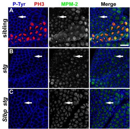
Embryonic MPM-2 foci depend on Cyclin E/Cdk2 activity
If the embryonic MPM-2 foci are related to the previously described MPM-2 foci in follicle cells, then their presence should depend on Cyclin E activity. To test this we characterized MPM-2 staining in stage 13 (cycle 17 in the epidermis) cyclin E mutant embryos relative to phenotypically wild type sibling controls. Wild type embryos at this developmental stage contain proliferating diploid cells in the central and peripheral nervous systems as well as endoreduplicating cells in various tissues (e.g. midgut, salivary gland, posterior spiracles). cyclin E mutant embryos develop normally until this stage because of maternal deposition of Cyclin E, and then arrest in G1 of cycle 17. Consequently, DNA synthesis in both proliferating neuronal cells and endoreduplicating cells is severely compromised in cyclin E mutants (Knoblich et al., 1994). We focused on endoreduplicating cells in the posterior spiracle primordium, since they are near the surface and relatively easy to image. In control embryos these cells contain robust MPM-2 foci (Figure MPM-2.MPM-2A-A’, arrow). In contrast, the posterior spiracle cells of stage matched
contain Lsm11 foci (Figure 2.2C-C’, arrow). These data indicate that Cyclin E is not required for maintenance of the HLB, but rather for the appearance of an MPM-2 epitope at the HLB. We also performed the reciprocal experiment of Cyclin E over-expression. Epidermal cells arrest in G1 of cycle 17, and contain Lsm11 foci but not MPM-2 foci (Fig. 2.2D). Ubiquitous expression of Cyclin E with a heat inducible promoter (hsp70::cyclin E) drives these G117 cells into S phase (Duronio and O'Farrell, 1995;
Figure 2.2: Embryonic MPM-2 foci depend on Cyclin E activity. (A-C) Images of the posterior
spiracles of germ band retracted (stage 13) embryos. (A) w1118 embryos stained with MPM-2 (A, green in
A’) and α-P-Tyr (blue in A’). (B,C) cycEAR95 homozygous mutant embryos stained with MPM-2 (B, green
in B’) or α-Lsm11 (C, red in C’) and α-P-Tyr (blue in B’ and C’). Arrows indicate a posterior spiracle cell.
w1118 control (D) or hsp70::cyclin E transgenic (E) embryos were given a 37oC heat shock for 30 minutes
MPM-2 foci co-localize with nascent histone transcripts
If MPM-2 recognizes an HLB phospho-protein involved in histone gene expression, then it should label sites of histone mRNA biosynthesis. We previously developed a cytological assay that detects nascent, chromatin-associated histone mRNA transcripts in intact Slbp mutant embryos (Lanzotti et al., 2002). Slbp mutants are defective in normal histone pre-mRNA processing, and accumulate inappropriately long, polyadenylated histone mRNAs (Sullivan et al., 2001). These aberrant mRNAs result from the use of cryptic polyadenylation signals located downstream of the normal pre-mRNA processing site in each of the 5 replication-associated histone genes (Lanzotti et
al., 2002). Because the 3’ UTR of these polyadenylated histone mRNAs contain
sequences not found in the wild type mRNA, we were able to develop a probe (H3-ds) that specifically recognizes the polyadenylated form of histone H3 mRNA. Cryptic polyadenylation is less efficient than the normal 3’ end processing, causing nascent pre-mRNA to accumulate on the chromatin template in Slbp mutants. These nascent transcripts can be visualized as a nuclear focus by fluorescent whole mount in situ hybridization with the H3-ds probe (Figure 2.3, A and B) (Lanzotti et al., 2002; Lanzotti et al., 2004). In addition, nascent histone transcripts appear soon after zygotic
transcription of the histone genes begins, because there is no maternal SLBP in the early embryo. We previously showed that nascent H3 transcripts arise in very late G214
sites of active histone mRNA biosynthesis in embryonic cells. MPM-2 also stains foci in cells in early G214 that lack nascent H3 transcripts but that are known to contain active
Cyclin E/Cdk2 (Figure 2.3A, C, arrowhead). This suggests that MPM-2 foci can be present without ongoing active histone gene transcription (see below, Fig. 2.6).
Figure 2.3: MPM-2 foci co-localize with the histone locus body in replicating cells. (A) An Slbp15
homozygous mutant post-blastoderm embryo was stained with α-P-Tyr (left panel in blue), MPM-2 (green
in merge), and hybridized with a fluorescent probe that recognizes misprocessed, polyadenylated H3
mRNA as nascent transcripts in the nucleus (H3-ds; left panel in red). Arrows indicate a cell in S15, which
contains an MPM-2 focus that co-localizes with nascent H3 transcripts. Arrowheads indicate an MPM-2
focus in a G214 cell that lacks nascent H3 transcripts. Anterior is to the top and ventral to the right. Bar: 20
µm. (B) S15 cell marked by arrows in (A). Bar: 2 µm. (C) G214 cell marked by arrowheads in (A). Bar: 2
The HLB disassembles during mitosis
Components of the mammalian Cajal body such as NPAT disassemble at the metaphase-anaphase transition and reassemble in the following interphase (Ma et al., 2000). To determine whether HLBs behave similarly, embryos were stained with MPM-2 or anti-Lsm11 antibodies and anti-phospho-histone H3 antibody (PH3) to mark mitotic cells. MPM-2 staining was examined in the early mitotic domains of cell cycle 14 in gastrulating embryos (Figure 2.4). Because the groups of cells comprising a mitotic domain do not enter mitosis synchronously, we could examine all stages of mitosis within a single domain. The nuclear MPM-2 foci present in G214 persist into early mitosis, and
all M14 prophase cells examined contained MPM-2 foci associated with condensing
chromosomes (n=125, Figure 2.4A, arrow). In contrast, only 77% of metaphase cells (n=106, Figure 2.4A, arrowhead) and none of the anaphase cells (n=102, Figure 2.4A, yellow arrowhead) contain detectable chromosome-associated MPM-2 foci. At the following interphase the nuclear MPM-2 foci reappear. In syncytial blastoderm embryos where progression through mitosis occurs synchronously, we also failed to detect MPM-2 foci during anaphase and some metaphase embryos (Figure 2.5). These data indicate that the MPM-2 phospho-epitope begins to decline during metaphase and becomes
lines (Ma et al., 2000; Zhao et al., 2000). Drosophila centrosomes have been reported to contain proteins with phospho-epitopes that are recognized by MPM-2 during mitosis (do Carmo Avides et al., 2001b; Lange et al., 2005a; Logarinho and Sunkel, 1998b). With the fixation conditions we used, MPM-2 staining increases throughout the cell during mitosis, but we did not observe specific staining of centrosomes.
arrowheads indicate anaphase cells. Bar: 20 µm. Single prophase, metaphase, and anaphase cells are shown
in (A’) and (B’), (A’’) and (B’’), and (A’’’) and (B’’’) respectively. Bars: 2 µm. Mitotic domain 5 is shown
in (A) and mitotic domain 1 is shown in (B). Note that the rat α-P-Tyr used here recognizes an antigen at
the HLB and the mouse α-PH3 used in (B) stains interphase cells weakly.
Shown are cycle 12 nuclei in (A) prophase, (B) metaphase, and (C) anaphase. (D) Cycle 13 interphase
nuclei. Bar: 10 µm.
MPM-2 foci assemble in the absence of the histone pre-mRNA processing factors
SLBP and U7 snRNP
Our analysis thus far suggests that MPM-2 recognizes a Cyclin E/Cdk2 substrate found in the HLB that could be involved in histone mRNA biosynthesis. One possibility is that one or more components of the histone pre-mRNA processing machinery are required for the formation of MPM-2 foci. To test this, we stained Slbp mutants with MPM2 antibodies. As shown above (Figure 2.3), MPM-2 foci are observed in Slbp mutant embryos, which are deficient in SLBP because there is no maternal store of SLBP protein (Lanzotti et al., 2002). This indicates that SLBP does not contain the primary MPM-2 epitope in the HLB, and suggests that components of the HLB assemble independently of SLBP. Consistent with this, Lsm11 foci are readily detected in Slbp mutant embryos, and co-localize with sites of nascent histone H3 transcription (data not shown).
Figure 2.6: MPM-2 foci form independently of the U7 snRNP. Fat bodies (A and B) and eye discs (C
and D) dissected from w1118 and U714 homozygous mutant third instar larvae were stained with MPM-2
(left panels, green in merge) and α-Lsm11 (middle panels, red in merge). Fat bodies were also stained with
DAPI (blue in merge). Arrows and arrowheads indicate nuclei with and without MPM-2 foci, respectively.
Note that in wild type Lsm11 foci are present in cells without MPM-2 (arrowhead in C) and that Lsm11
foci are absent in the U7 mutant cells. Bars: 20 µm.
Figure 2.7: MPM-2 foci are not dependent on Lsm11. Dmel-2 cells treated with either (A) control
dsRNA or (B) Lsm11 dsRNA stained with MPM-2 (left panels in green), α-Lsm11 (middle panels in red),
and DAPI (right panels in blue). Arrow in (A) indicates a cell with a single HLB. Arrow and arrowhead in
(B) indicates cells with two or three, respectively, MPM-2 foci and no Lsm11 foci. Bar:10 µm.
Embryonic MPM-2 foci do not require stringcdc25 or histone transcription
Cyclin E/Cdk2 phosphorylation of the protein containing the MPM-2 epitope may regulate, or even result from, the transcription of histone genes. To test this, we
examined MPM-2 staining in string mutant embryos. In late G214, histone gene
maternal string mRNA and protein (Figure 2.8, A and B) (Lanzotti et al., 2004). MPM-2 foci can be readily detected in string Slbp null mutant embryos that lack nascent histone transcripts (Figure 2.8, A and C). This result indicates that formation of MPM-2 foci does not require histone gene transcription or the function of mitotic Cdks activated by stringcdc25. Consistent with this interpretation, MPM-2 foci are present in all cells throughout interphase 14 (i.e. both S phase and G2 when Cyclin E/Cdk2 is active) at times when neither stringcdc25 is active nor histone transcription occurs (Figure 2.3C). These data indicate that the formation of MPM-2 foci occurs independently of stringcdc25 (and thus by inference Cdk1 activity) and histone transcription.
was used to distinguish stg mutants from siblings. Anterior is to the top and ventral to the right. Bar: 20
µm.
MPM-2 foci appear coincidentally with activation of zygotic histone gene expression
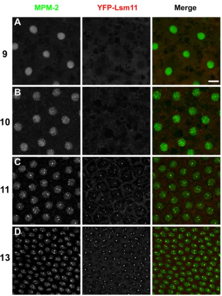
Most zygotic gene expression begins at the blastoderm stage during cycle 14 (Edgar and Schubiger, 1986). Histone genes are an exception, and become
transcriptionally active precisely in nuclear cycle 11 (Edgar and Schubiger, 1986). If the protein containing the MPM-2 epitope regulates some aspect(s) of histone mRNA biosynthesis, then we expect MPM-2 foci to be present once zygotic expression of the histone genes has begun. To determine whether there is a relationship between the onset of zygotic histone transcription, the assembly of the HLB, and the appearance of the MPM-2 epitope at the HLB, we carefully analyzed syncytial blastoderm embryos stained with MPM-2 and anti-Lsm11 antibodies. Nuclear density was used to accurately stage the embryos with respect to each nuclear cycle. Uniform MPM-2 staining throughout the nucleus was detectable in all syncytial blastoderm cycles, perhaps because maternal Cyclin E/Cdk2 is present throughout the early embryo. MPM-2 foci appear for the first time precisely during nuclear cycle 11, concomitantly with the onset of zygotic histone gene transcription (Figure 2.9). Similarly, nuclear Lsm11 staining is undetectable prior to cycle 11, and Lsm11 foci first appear during nuclear cycle 11 and co-localize with MPM-2 foci (Figure 2.9). The appearance of Lsm11 foci in cycle 11 is not the result of new, zygotic synthesis, since Lsm11 protein is present in 0-30 min embryos (data not shown), prior to the onset of zygotic gene expression. In addition, maternal expression of a YFP-Lsm11 fusion protein in da-GAL4/UAS-YFP-Lsm11 females results in
that the onset of zygotic histone gene transcription and formation of the MPM-2-positive HLB during early development are temporally and spatially coincident.
in merge), and α-P-Tyr (right panels in blue). Interphase of nuclear cycles 9 to 14 are shown, as indicated
on the left. Bar: 10 µm.
Figure 2.10: The histone locus body first forms and co-localizes with MPM-2 foci during nuclear cycle 11. Syncytial blastoderm embryos with a maternally provided supply of YFP-Lsm11 were stained
with MPM-2 (red) and α-GFP (YFP-Lsm11; green). Interphase of nuclear cycles 9, 10, 11 and 13 are
shown, as indicated on the left. Bar: 10 µm.
MPM-2 foci form independently of the histone locus
transcription may directly result in the nucleation of the HLB at the histone locus, or de
novo assembly of the HLB may trigger the initiation of histone transcription. To attempt
to distinguish between these possibilities, we stained embryos containing a homozygous deletion of the entire histone locus (Df(2L)Ds6) with MPM-2 and anti-Lsm11 antibodies. We hypothesized that chromatin at or near the histone locus is necessary for the assembly of the HLB, as it might function as a scaffold on which the HLB is assembled to carry out histone mRNA biosynthesis. Surprisingly, both MPM-2 and Lsm11 foci are detected in
Df(2L)Ds6 homozygous embryos lacking the histone genes (Figure 2.11, arrows), which
were distinguished from siblings containing histone genes by in situ hybridization of histone H3 mRNA (Figure 2.11, left panels). Moreover, the distribution of nuclei with one versus two MPM-2 foci is similar between wild type and Df(2L)Ds6 embryos, such that the percentage of cells containing one MPM-2 focus in wild type and Df(2L)Ds6 embryos is 89% and 84%, respectively, and the percentage of cells containing two MPM-2 foci in wild type and Df(MPM-2L)Ds6 embryos is 11% and 7.8%, respectively. This result indicates that the histone locus, and therefore histone transcription, is not required for the initial nucleation of the HLB. This is consistent with our analysis of string mutant embryos, which also indicated that histone transcription is not required to maintain MPM-2 foci.
or Df(2L)Ds6 embryos. Whereas in wild type embryos only 5% of interphase nuclei contained Lsm11 foci that did not co-localize with MPM-2, in Df(2L)Ds6 mutants the proportion of this class of nuclei increased to ~35% (Table 2.1). In addition, 8% of the nuclei contained more than one Lsm11 focus that did not co-localize with MPM-2 foci (Table 2.1). These data indicate that full association of the U7 snRNP and the protein containing the MPM-2 epitope requires the histone locus. Second, the HLBs in
Df(2L)Ds6 embryos are smaller than in controls as revealed by measuring the size (see
Materials and Methods) of the MPM-2 nuclear focus in 75 cells from two stage-matched, stage 11 embryos. The average size (defined as length plus width) of the MPM-2 focus in control and Df(2L)Ds6 embryos is 1.48 ± 0.37 and 1.06 ± 0.27 µm, respectively (p<0.001). Thus, while the histone locus is not absolutely essential for the formation of the HLB, as assessed by co-localization of MPM-2 and Lsm11 foci, full, stable assembly of the HLB requires the histone locus.
and DAPI (blue). Df(2L)Ds6 homozygous mutant embryos that lack the histone gene cluster (B) were
distinguished from control siblings (A) by the lack of zygotic histone H3 mRNA. Arrows indicate foci of
MPM-2 and α-Lsm11 that co-localize. Arrowheads indicate cells containing a focus of co-localizing
MPM-2 and α-Lsm11 as well as a focus of just α-Lsm11. Anterior is to the top. Bar: 20 µm.
Table 2.1 Characterization of the HLB in histone deletion embryos
1 ML* 2 ML
1 ML +
1 L 2 L 1 L
1 ML +
2 Ls 2 M Other
w1118 (n=191) 81.2 11.0 5.2 0 0.5 0 0 2.1
Ds6 (n=192) 54.7 4.2 20.3 3.1 3.6 7.8 0.5 5.7
*ML indicates a focus of co-localized MPM-2 and Lsm11 staining, L indicates a focus containing only
Lsm11, and M indicates a focus containing only MPM-2. “Other” refers to individual patterns of staining
that are not represented in the indicated categories and that each comprise less than 0.5% of cells.
Discussion
Using the MPM-2 monoclonal antibody as a tool, we have presented evidence that a Cyclin E/Cdk2 substrate localizes to the HLB, a recently described Drosophila nuclear organelle likely to be directly involved in histone mRNA biosynthesis (Liu et al. 2006). MPM-2 foci co-localize with the histone locus, nascent histone transcripts, and the U7 snRNP-specific protein Lsm11. We propose that the MPM-2 antibody recognizes an activator of histone mRNA biosynthesis that either stimulates histone transcription and/or pre-mRNA processing during S phase.
Miele et al., 2005; Wei et al., 2003; Zhao et al., 1998; Zhao et al., 2000; Zheng et al., 2003). The NH2-terminus of NPAT contains a LisH domain that is necessary for H4
transcription (Wei et al., 2003). Although there are fifteen predicted LisH-containing proteins in Drosophila, no ortholog of NPAT has yet been identified. Coilin, which functions to maintain the integrity of the Cajal body, can be phosphorylated by Cyclin E/Cdk2 in vitro (Hebert et al., 2001; Liu et al., 2000; Tucker et al., 2001). However, MPM-2 does not recognize Cajal bodies in HeLa cells (A.E.W. and R.J.D., unpublished), and the Drosophila Cajal body in Drosophila cells is distinct from the HLB (Liu et al., 2006). The orthologue of coilin has also not been reported in Drosophila.
HLB behavior during early Drosophila development
Although the HLB is capable of forming independently of the histone locus, the histone locus contributes to the structural integrity of the HLB. In histone deletion embryos, HLBs are smaller and some interphase nuclei contain Lsm11 foci that do not co-localize with MPM-2. The size of the nucleolus is determined by the amount of ribosomal gene transcription (Hernandez-Verdun, 2006). Thus, the size and overall composition of the HLB may be similarly dependent on transcription of the histone genes. Consistent with this, MPM-2 and Lsm11 foci are present at maximal size during prophase of cycle 14, just after the initiation of histone transcription in late G214.
Nascent histone transcripts are likely aborted during mitosis (Shermoen and O'Farrell, 1991), which correlates with the loss of MPM-2 and Lsm11 staining we observe in anaphase. Alternatively, reduced HLB size may be a secondary consequence of cell cycle arrest resulting from lack of histone biosynthesis (Smith et al., 1993).
In wild type embryos we observe either one or two MPM-2 or Lsm11 foci per cell at frequencies similar to the known pairing frequencies of the histone loci on homologous second chromosomes (Fung et al., 1998). This is also consistent with the association of the HLB with histone genes (Liu et al., 2006). Surprisingly, we often detect one or two MPM-2 foci in histone deletion embryos, suggesting that HLB number is not dictated solely by homologous pairing at the histone locus. HLB components may associate with another chromosomal locus in the absence of the histone genes.
Cell cycle regulation of the HLB
Histone mRNA is greatly depleted in cyclin E mutant embryos (Lanzotti et al., 2004). Since cyclin E mutant embryos arrest in G1 phase, it is difficult to know this observation is indicative of a direct involvement of cyclin E/Cdk2 in histone gene
expression, or arises as a secondary consequence of cell cycle arrest. Because interphase MPM-2 foci are coincident with the HLB, are present in cells where Cyclin E/Cdk2 is active, and are absent in cells that lack Cyclin E/Cdk2 activity, we favor the interpretation that Cyclin E/Cdk2 phosphorylates a protein directly involved in histone mRNA
biosynthesis. How Cyclin E/Cdk2 participates in this process is not known, but it is not required for recruitment of U7 snRNP to the HLB, since Lsm11 remains a stable
component of the HLB in wild type cells arrested in G1 and in cells of cyclin E mutant embryos.
During the early embryonic cell cycles that have constitutive Cyclin E/Cdk2 activity, MPM-2 foci disappear as cells progress through mitosis and are undetectable by anaphase. Focal Lsm11 staining is also lost during mitosis, suggesting the disassembly of HLB components into the cytoplasm subsequent to nuclear envelope breakdown rather than simply dephosphorylation or destruction of the MPM-2 target(s). The HLB rapidly reforms during the subsequent interphase. These behaviors are similar to the behavior of NPAT foci during mitosis in cultured mammalian fibroblasts (Ma et al., 2000; Zhao et
al., 2000). The nucleolus also disassembles during mitosis (Hernandez-Verdun, 2006),
Summary
The HLB is a dynamic structure capable of receiving input from both the histone locus and the cell cycle. Our analysis raises many questions, including how HLB assembly occurs at a specific time in a syncytial cytoplasm from abundant maternal components-even in the absence of a histone locus template, and how Cyclin E/Cdk2 regulates HLB function and histone mRNA biosynthesis. Determining the identity of the HLB protein(s) recognized by MPM-2 antigen will help answer such questions, and will provide a useful tool to examine how the regulation of such a fundamental process as histone mRNA biosynthesis is modulated by developmental programs.
Acknowledgements