Original Research Article
Keratometric changes after pterygium excision
Sharda Punjabi, Appurv Kachhwaha*
INTRODUCTION
Pterygium, a word derived from “pterygion” (ancient Greek for a wing) is a degenerative condition of the
subconjunctival tissue, which proliferates as a
vascularized granulation tissue to invade the cornea, destroying superficial layers of stroma and Bowman’s
membrane.1
All normal human eyes have mild degrees of spherocylindrical errors, consist of a bitoric optical system, i.e. have principal meridians of relatively higher and lower powers at right angles. A high degree of
astigmatism (3 D or more) is present in neonates and infants as the child grows astigmatism gradually disappears and transform into with the rule owing to eyelid pressure, apparently a part of normal eye maturation and emmetropisation. With aging, upper eyelid pressures on the cornea and the tone of orbicularis muscle decreases and hence, against-the-rule astigmatism is common over 40 years of age.
It is generally accepted that genetic factors have a significant role in determining ocular refractive status as well as astigmatism but many conditions and procedures such as surgery, suturing, wound healing, and ocular Department of Ophthalmology, Geetanjali Medical College and Hospital, Udaipur, Rajasthan, India
Received: 15 February 2020
Revised: 20 March 2020
Accepted: 01 April 2020
*Correspondence:
Dr. Appurv Kachhwaha, E-mail: [email protected]
Copyright: © the author(s), publisher and licensee Medip Academy. This is an open-access article distributed under the terms of the Creative Commons Attribution Non-Commercial License, which permits unrestricted non-commercial use, distribution, and reproduction in any medium, provided the original work is properly cited.
ABSTRACT
Background: A pterygium is a wing-shaped growth of conjunctiva and fibrovascular tissue on the superficial cornea. The pathogenesis of pterygia is strongly correlated with UV light exposure and environmental factors. The prevalence of pterygia increases steadily with proximity to the equator, and the condition is more common in men than women. It is well established fact that before entering the optical zone pterygium causes flattening of the cornea in horizontal meridian with the more normal side of the cornea usually temporally, resulting in with-the-rule astigmatism.
Methods: The study included 80 patients of primary pterygium who underwent pterygium excision with conjunctival autograft adhered by autologous blood surgery. After performing routine ocular examination which includes visual acuity without and with pinhole and pre-operative keratomery was assessed by autokeratorefractometer. Repeat examination was performed after 6 weeks of surgery. Patients with recurrent pterygium, pseudo-pterygium, and history of previous ocular surgery were excluded.
Results: The pre‑ and postoperative corneal astigmatism were compared after 6 weeks of surgery. The changes in corneal astigmatism were statistically significant p value <0.001. The preoperative mean corneal astigmatism of 3.41 D was reduced to 1.59 D (p value <0.001) 6 weeks after surgery, but maximum change in astigmatism was seen in Grade IV >Grade III >Grade II >Grade I.
Conclusions: Primary pterygium of all grades treated with well accepted technique pterygium excision with conjunctival autograft with autologous blood gives promising results in terms of improvement in corneal astigmatism and hence visual acuity as well.
Keywords: Conjunctival autograft, Corneal astigmatism, Keratometry, Pterygium
comorbidities like pterygium, limbal dermoid, OSSN, other ocular surface diseases modify the cylindrical status
of the eye.2-4 Pterygium induces with-the-rule
astigmatism in changing cornea in various ways:
• The mechanical traction exerted on cornea
• The size of pterygium, especially the double-headed pterygium
• The pooling of the tear film
This is measured by keratometry. Pterygium causes visual impairment by
• Occlusion of visual axis
• Inducing with the rule astigmatism
Surgery is the only effective treatment for pterygium, pterygium excision with conjunctival autograft remains the gold standard with less incidence of recurrence (5%-10%). The favoured site to harvest the autograft is superotemporal because this location in an oblique quadrant will avoid inadvertent injury to extraocular muscle, while its position under the upper lid will aid with patient comfort and healing of the donor site.5-9
As southern Rajasthan is a part of pterygium belt due to its geographical location and profession induced prolong outdoor activities which makes them vulnerable for pterygium.3-5
METHODS
This study was done between June 2018 to June 2019 at Geetanjali Medical College and Hospital, Udaipur. All patients of primary pterygium presented in Dept. of Ophthalmology were included in the study. Patients with recurrent pterygium, pseudo-pterygium, and history of previous ocular surgery were excluded. The study included 80 patients of primary pterygium who
underwent pterygium excision with conjunctival
autograft adhered by autologous blood surgery. After performing routine ocular examination which includes visual acuity without and with pinhole and pre-operative keratomery was assessed by autokeratorefractometer. Repeat examination which include visual acuity without and with pinhole and keratometry was performed after 6 weeks of surgery.
Pterygium grading
Pterygium was graded depending on the extent of corneal involvement as follows;
• Grade I : Just crossing the limbus (Figure 1)
• Grade II : Midway between limbus and pupil (Figure
2)
• Grade III: Reaching up to the pupillary margin (Figure 3)
• Grade IV: Crossing the pupillary margin (Figure 4)
Figure 1: Grade I pterigium “atrophic” as episcleral vessels clearly visible.
Figure 2: Grade II pterygium, episcleral vessels visible.
Figure 3: Grade III pterygium episcleral vessels partially visible.
the residual tissue was scraped from the corneal surface (Figure 5) after excision, autograft taken from superotemporal conjunctiva was placed over the bare sclera in its correct anatomical orientation and anchored to the limbus and peripherally to the surrounding conjunctiva (Figure 6).
Figure 4: Grade IV pterygium “fleshy, opaque” as vessels wholly obscured.
Figure 5: Pterygium excised.
Figure 6: conjunctival autograft adhered by autologous blood.
Statistical analysis
Statistical analysis was performed using SPSS for Windows (Version 16.0, 2007; SPSS Inc, Chicago, IL, USA). Paired and unpaired t‑tests were used to compare the variables. The probability level of 0.05 was set as the statistically significance value.
RESULTS
A total of 80 patients of primary pterygium were selected randomly, out of 80 patients, 44 were male (55%) and 36 were female (45%) (Figure 7). The mean age of patients was 47.07 years. Of the 80 patients, Grade I, II, III and IV pterygiums were present in 6(7.5%), 31(38.75%), 28(35%) and 15(18.75%) respectively (Figure 8).
Figure 7: Age and sex distribution of pterygium patients.
Figure 8: Number of patients according to grades of pterygium.
The pre‑ and postoperative corneal astigmatism were compared after 6 weeks of surgery. The changes in corneal astigmatism were statistically significant p value <0.001(Table 1 and Figure 9).
The amount of astigmatism varied with the grade of pterygium. The preoperative mean astigmatism was 1.16 D, 2.18 D, 3.87 D, and 6 D in eyes with grade I, II, III
0 2 4 6 8 10 12
20 to 30 31 to 40 41 to 50 51 t0 60 61 to 70 71 to 80
Sex Male Sex Female
0 5 10 15 20 25 30 35
and IV pterygiums, respectively (Table 2). The postoperative mean astigmatism at 6 weeks in eyes with grade I, II, III and IV pterygiums were 0.29 D (p value <0.001), 0.98 D (p value <0.001), 1.79 D (P value <0.001) and 3 D (p value <0.001) respectively (Table 2). The preoperative mean corneal astigmatism of 3.41 D
was reduced to 1.59 D (p value <0.001) 6 weeks after surgery (Table 1). Thus, a significant reduction in corneal astigmatism 6 weeks after surgery was observed for all the four grades of pterygium. But maximum change in astigmatism was seen in Grade IV >Grade III >Grade II >Grade I (Figure 10).
Table 1: Test of significant difference between pre and post op values.
Astigmatism N Mean Standard deviation Standard error of Mean T value p value
Pre Op 80 3.416 1.597 0.179
8.748 <0.001
(S)
Post Op 80 1.594 0.959 0.107
Table 2: Test of significant difference between pre and post op t values according to grade of Astigmatism.
Grade N PREOP POST OP T value p value
Mean SD SEM Mean SD SEM
I 6 1.167 0.342 0.139 0.292 0.246 0.100 5.088 <0.001 (S)
II 31 2.184 0.428 0.077 0.984 0.322 0.058 12.47 <0.001 (S)
III 28 3.875 0.323 0.061 1.795 0.596 0.108 16.82 <0.001 (S)
IV 15 6.000 0.807 0.208 3.000 0.762 0.197 10.47 <0.001 (S)
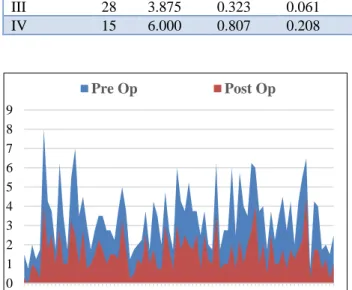
Figure 9: Comparison of pre and post op astigmatism in dioptres in all patients.
Figure 10: Change in astigmatism according to the grades to pterygium.
Visual acuity improvement seen in Grade II and Grade III pterygium which was 1-2 lines and 2-3 lines of Snellen’s Visual Acuity chart, respectively due to reduction in induced with the rule astigmatism and clearance of visual axis. In Grade I due to small pterygium size and little change in keratometric values 1.16 D±0.34 to 0.29 D±0.24 hence, improvement in visual acuity was not very significant. However in Grade IV though change in keratometric values was quite significant 6 D±0.80 to 3 D±0.76, but visual acuity improvement was not significant due scarring in the pupillary area (Figure11).
Figure 11: Post op corneal scarring in Grade IV pterygium.
DISCUSSION
A pterygium is a wing-shaped growth of conjunctiva and fibrovascular tissue on the superficial cornea. The
0 1 2 3 4 5 6 7 8 9
1 4 7 10 13 16 19 22 25 28 31 34 37 40 43 46 49 52 55 58 61 64 67 70 73 76 79
Pre Op Post Op
0.87
1.2
2.08
3
pathogenesis of pterygia is strongly correlated with UV light exposure, although environmental insults such as exposure to dust, wind, or other irritants causing chronic ocular inflammation may also be factors. The predominance of pterygia on the nasal side in the interpalpebral zone is results from light passing medially through the cornea, focusing on the nasal limbus area, while the shadow of the nose reduces the intensity of light transmitted to the temporal limbus. The prevalence of pterygia increases steadily with proximity to the equator, and the condition is more common in men than women, and in people who work outdoors. The histopathology of pterygia is basophilic degeneration of elastotic fibres and invasion of the superficial cornea, which is preceded by dissolution of the Bowman layer.1-5
It is well established fact that before entering the optical zone pterygium causes flattening of the cornea in horizontal meridian with the more normal side of the cornea usually temporally, resulting in with-the-rule astigmatism. An induced with-the-rule astigmatism was explained by several mechanisms: Pooling of the tear film at the leading edge of the pterygium, and mechanical traction exerted by the pterygium on the cornea. Postoperatively, a steepening of the cornea in the horizontal meridian was demonstrated, the magnitude of which was related to pterygium grade (p=0.0001).7-11
Many studies have proved that pterygium excision
surgery significantly reduces pterygium‑induced
astigmatism. In the study by Mohite et al, there was a significant reduction in mean pre-operative keratometric astigmatism from 3.046±1.20 D to mean post-operative 1.486±0.63 D (p<0.001) after pterygium surgery, they concluded that pterygium‑induced corneal astigmatism can be reduced by surgery.10 These results were comparable to this study as authors have also found significant reduction in mean corneal astigmatism after pterygium excision with conjunctival autograft with
autologous blood. The preoperative mean astigmatismof
3.41±1.59 D was significantly (p<0.0001) reduced to 1.59±0.95 D postoperatively after 6 weeks of surgery which can be attributed to the fact that the regularity and symmetry of corneal surface improved after pterygium surgery, thusreducing astigmatism.
Gumus et al, Seitz et al, concluded that there is a significant correlation between the size of pterygium and induced corneal astigmatism, the amount of induced corneal astigmatism increases with the increase in the size of pterygium.4-6 In this study, mean astigmatism was more in higher grades of pterygium. Preoperative mean astigmatism was minimum in Grade I 1.16 D±0.34 and maximum in Grade IV 6 D±0.80 while 2.18 D±0.42, 3.87 D±0.32, in grade II, III respectively. These results were comparable with results of the above‑mentioned studies.
The study by Maheswari et al, found significant improvements in visual acuity after pterygium excision surgery in all the grades of pterygium (p 0.05).5 But in
this study significant Visual acuity improvement was seen in Grade II and Grade III because of small pterygium size in Grade I and post op scarring in Grade IV.
CONCLUSION
Primary pterygium of all grades treated with well accepted technique pterygium excision with conjunctival autograft with autologous blood gives promising results in terms of improvement in corneal astigmatism and hence visual acuity as well.
Although a larger sample size would have helped in a better analysis and evaluation, this study has a small sample size (80).
Funding: No funding sources Conflict of interest: None declared
Ethical approval: The study was approved by the Institutional Ethics Committee
REFERENCES
1. Lin A, Stern G. Correlation between pterygium size
and induced corneal astigmatism. Cornea. 1998 Jan;17(1):28-30.
2. Fong KS, Balakrishnan V, Chee SP, Tan DT.
Refractive change following pterygium surgery. CLAO J: Office Publicat Contact Lens Assoc Ophthalmol, Inc. 1998 Apr;24(2):115-7.
3. Saw SM, Tan D. Pterygium: prevalence,
demography and risk factors. Ophthal Epidemiol. 1999 Jan 1;6(3):219-28.
4. Seitz B, Gütay A, Küchle M, Kus MM,
Langenbucher A. Impact of pterygium size on corneal topography and visual acuity-a prospective
clinical cross-sectional study. Klinische
Monatsblatter fur Augenheilkunde. 2001
Sep;218(9):609-15.
5. Maheshwari S. Effect of pterygium excision on
pterygium induced astigmatism. Ind J Ophthalmol. 2003 Jun 1;51(2):187-8.
6. Gumus K, Erkilic K, Topaktas D, Colin J. Effect of
pterygia on refractive indices, corneal topography, and ocular aberrations. Cornea. 2011;30(1):24-9. 7. Kheirkhah A, Safi H, Molaei S, Nazari R, Behrouz
MJ, Raju VK. Effects of pterygium surgery on front and back corneal astigmatism. Canadian J Ophthalmol. 2012 Oct 1;47(5):423-8.
8. Mohammadi SF, Tahvildari M, Z-Mehrjardi H.
Physiology of Astigmatism, Astigmatism - Optics, Physiology and Management. In: Goggin M. eds. ISBN: 978-953-51-0230-4, InTech; 2012:1-14. 9. Bhandari V, Rao CL, Ganesh S, Brar S. Visual
10. Mohite US, Dole NB, Jadhav SS. Effectiveness of pterygium surgery on corneal astigmatism. Med Pulse Int J Ophthalmol. 2017;3:12-7.
11. Pajic B, Aebersold DM, Eggspuehler A, Theler FR,
Studer HP. Biomechanical Modeling of Pterygium Radiation Surgery: A Retrospective Case Study. Sensors. 2017;17(6):1200.