EFFECT OF NF-κB HAPLOINSUFFICIENCY ON CALVARIAL BONE HEALING
Ruiwei Liang
A dissertation submitted to the faculty at the University of North Carolina at Chapel Hill in partial fulfillment of the requirements for the degree of Doctor of Philosophy in the
Curriculum in Oral Biology, School of Dentistry.
Chapel Hill 2015
Approved by: Lyndon Cooper Eric Everett Ching-Chang Ko
iii ABSTRACT
RUIWEI LIANG: EFFECT OF NF-κB HAPLOINSUFFICIENCY ON CRANIAL BONE HEALING (Under the direction of Lyndon F. Cooper)
Bone defect is a major and challenging health concern. The treatment for bone defect aims to enhance bone regeneration, which is highly regulated by many molecular signaling pathways. Growing evidence suggested that proper inflammatory signaling was crucial for bone regeneration. Previous study showed that treatment of MSCs with expression of NF-κB increased MSCs engraftment in damaged tissue. Previous work in our lab indicated a role of NF-κB on osteoblast differentiation during physiological bone development. The present study was designed to study role of NF-κB signaling in bone healing using genetically-modified mouse with haploinsufficiency of p65 in osteoblasts. Here, we showed that mice with osteoprogenitor-specific NF-κB haploinsufficiency displayed reduced calvarial defect bone repair manifested by micro-CT and histological analysis. The progenitor cells from p65 haploinsufficient mice demonstrated fewer CFU-OB colonies and decreased osteoblastic markers expression (Sp7, Alp and Bsp) in response to rhBMP2. Furthermore, rhBMP2 mediated Smad phosphorylation was disrupted in the absence of sufficient p65 signal. Therefore, we concluded that NF-κB haploinsufficiency impairs bone repair by
iv
vi
ACKNOWLEDGEMENTS
I would like to thank all those people who made this thesis possible and an unforgettable experience for me.
First and foremost I would like to express my sincerest gratitude to my advisor, Dr. Lyndon Cooper, who offered continuously support for my PhD study and thesis research with his encouragement, patience and immense knowledge. I would never have been able to complete my dissertation without his excellent guidance and caring help. I gratefully thank him for the systematic guidance and great efforts he put into training me in the scientific field.
I would like to express my sincere gratitude to Dr. Eric Everett, my committee member for his insightful comments at different stages of my research which incented me to widen my research from various perspectives. I gratefully thank my committee members, Dr. Ching-Chang Ko, Dr. Jenifer Webster-Cyriaque and Dr. Homa Zadeh for guiding my research with continuous support, help and those enlightening inspirations. Their practical advice and constructive criticisms help me understand and enrich my ideas in research.
I am grateful to my current and former fellow labmates in Dr. Cooper’s lab. It would not be such a lovely lab with an excellent atmosphere for doing research without any of you. I am thankful to Dr. Gustavo Mendonca, Dr. Daniela Mendonca, Dr. Sodsi
vii
supports on my research and precious friendship throughout these years. I also wish to thank Dr. Sheng Yang, Dr. Masako Nagasawa, Dr. Yoichiro Ogino and Dr. Kaori Eguchi for their technical assistance, insightful advice, caring and spiritual support. Working in this team gave me an invaluable and unforgettable experience over these five years.
I am indebted to the Oral biology PhD program in School of Dentistry of UNC. I give my sincere thanks to Dr. Patrick Flood for his selfless support, encouragement and
thoughtful inspiration. Gratitude also to Dr. Phillip Ceib and Cindy Blake for their help and generous caring that make me feel at home. I am grateful to Brittney Ciszek for her kindness helping me finish my thesis. I am also thankful to all my friends in the program for the time we shared with laughter and mutual encouragement.
viii
TABLE OF CONTENTS
LIST OF TABLES..……….xi
LIST OF FIGURES………..………xii
LIST OF ABBREVIATIONS……….……..….xiv
CHAPTER 1: INTRODUCTION………..1
BONE DEFECTS AND TISSU REGENERATION ………..…1
Phase 1: Inflammatory phase………2
Phase 2: Bone formation phase………..3
Phase 3: Bone remodeling phase………4
BONE DEVELOPMENT AND HOMEOSTASIS ………..….4
Runx2……….7
Osterix………...7
Alkaline phosphatase (ALP)……….8
Bone sialoprotein (BSP)……….9
Osteocalcin (OCN)……….9
ix
NF-κB PATHWAY AND ITS INVOLVEMENT IN BONE BIOLOGY………....…15
REFERENCES……….……..…………24
CHAPTER 2: EFFECT OF NF-κB HAPLOINSUFFICIENCY ON CRANIAL BONE HEALING……….……35
INTRODUCTION………..……….…………35
MATERIALS AND METHODS……….39
RESULTS………..……….………….48
Effects of NF-κB haploinsufficiency on bone healing in a critical size calvarial defect model……….………48
Micro-CT assessment of bone regeneration and image analysis……….48
Histological analysis of bone formation……….49
CFU-F and CFU-OB analysis……….…50
Effects of NF-κB haploinsufficiency on BMP2 mediated osteogenesis in vitro………51
Effects of NF-κB haploinsufficiency on BMP2 mediated Smads phosphorylation……….53
The effects of TNFα on BMP2- mediated Smads signaling in C2C12……….……….54
DISCUSSION……….56
CONCLUSION………..62
x
INTRODUCTION………..……….………84
MATERIALS AND METHODS……….87
RESULTS………..……….……….90
Microarray results………..………90
Gene oncology analysis……….90
DISCUSSION……….…93
REFERENCES………..…..104
CHAPTER 4: GENERAL DISCUSSION……….109
xi LIST OF TABLES
CHAPTER 2
Table 2.1 Primers used in PCR for genotyping………76 CHAPTER 3
Table 3.1 Gene downregulation observed in p65fl/+Osx-Cre mice at 4 weeks ……….96
Table 3.2 Gene upregulation observed in p65fl/+Osx-Cre mice at 4 weeks………..…..…97
Table 3.3 Gene Ontology Results for in p65fl/+Osx-Cre mice (Terms with
Fold Change > 3).………...98 Table 3.4 Gene Downregulation of the GO Regulation of Ossification
Category in p65fl/+Osx-Cre mice (Fold Change > 1.2)…….………..………....…99
Table 3.5 Gene Upregulation of the GO Ossification Category in
p65fl/+Osx-Cre mice (Fold Change > 1.2)…….……….…………...………100
Table 3.6 Gene Dysregulation of the GO Regulation of Biomineral
xii
LIST OF FIGURES CHAPTER 1
Figure 1.1 Differentiation of primitive mesenchymal stem cells into osteoblasts……….21 Figure 1.2 Signaling transduction of BMP2 pathway.………..……….….22 Figure 1.3 Canonical and non-canonical NF-κB signaling pathways in bone cells…….………….23 CHAPTER 2
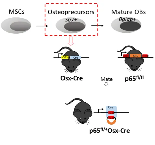
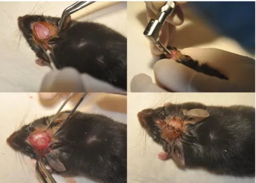
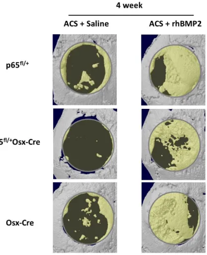
Figure 2.1 Schematic pictures describing the generation of transgenic mouse model………..63 Figure 2.2 Illustration of the critical-sized defect model and procedures in mouse……….……64 Figure 2.3 Micro-CT analysis of new bone formation in p65fl/+Osx-Cre,
p65fl/+ and Osx-Cre mice at 4 weeks following surgery.……….…….65
Figure 2.4 Micro-CT analysis of new bone formation in p65fl/+Osx-Cre
and p65fl/+ mice at 8 weeks following surgery.…..………..……….….………..66
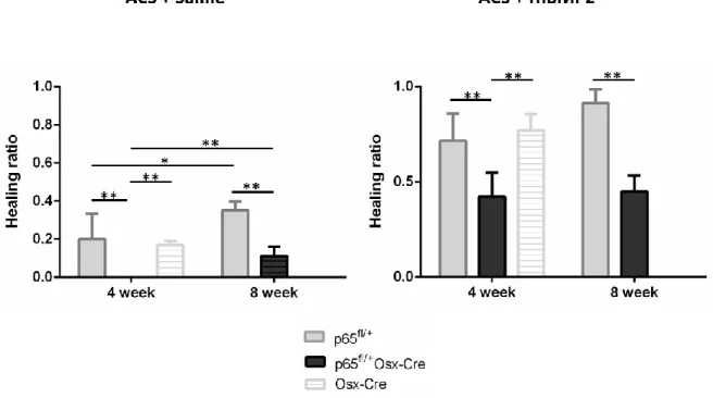
Figure 2.5 Micro-CT analysis of new bone formation in p65fl/+Osx-Cre
and p65fl/+ mice and Osx-Cre mice at 4 and 8 weeks following surgery.……….67
Figure 2.6 Histological evaluation of healing process in calvarial defects
in p65fl/+Osx-Cre and p65fl/+ mice.………..….…..68
Figure 2.7 Immunohistochemical assessment of bone sialoprotein
(BSP) expression in regenerated tissue of p65fl/+Osx-Cre and p65fl/+ mice.……….……..70
Figure 2.8 Colony-forming unit-fibroblasts (CFU-F) and colony-forming unit-osteoblasts (CFU-OB) were isolated and stained for p65fl/+Osx-Cre
and p65fl/+ mice………..……..71
Figure 2.9 Relative mRNA expression levels of osteogenic markers (Sp7, Alp, Bsp) of osteoprogenitors obtained from p65fl/+Osx-Cre
and p65fl/+ mice.………..……….….…….72
Figure 2.10 Relative mRNA expression levels of several osteogenic genes (Sp7, Alp, Bsp, Satb2) during the calvarial healing process
in p65fl/+Osx-Cre and p65fl/+ mice...………...73
xiii
by NF-κB signaling………..………..……….74 Figure 2.12 The BMP2 mediated Smads signaling pathway is affected
by NF-κB signaling……….……….75 CHAPTER 3
Figure 3.1 Sample quality control………..………..102 Figure 3.2 The BMP2 mediated Smad signaling pathway is affected
xiv
LIST OF ABBREVIATIONS
ACS Absorbable collagen sponge
ALP Alkaline phosphatase
BGLAP Bone gamma-carboxyglutamic acid-containing protein
BMD Bone mineral density
BMP Bone morphogenetic protein
BMP-R BMP receptor
BMSC Bone marrow stromal cell
BSA Bovine serum albumin
BSP Bone sialoprotein
C2C12 Mouse embryonic premyoblast cell line
C3H10T1/2 Mouse embryonic fibroblastic cell line
cDNA Complementary DNA
CFU-F Fibroblast colony forming unit
CFU-OB Osteoblast colony forming unit
COL1A1 Alpha chain of type 1 collagen
xv CRE Cre recombinase
Ct Threshold cycle
DLX5 Distal-less homeobox 5
DMEM Dulbecco’s modified Eagle’s medium
ECM Extracellular matrix
EDTA Ethylenediaminetetraacetic acid
FBS Fetal bovine serum
FDA Food and Drug Administration
FGF Fibroblast growth factor
GAPDH Glyceraldehyde-3-pohophate dehydrogenase
GFP Green fluorescent protein
GO Gene oncology
GPM6BA Glycoprotein m6b
H&E Hematoxylin and Eosin
IκB Protein inhibitor of NF-κB
IKK IκB kinase
xvi
LRP5 Low-density lipoprotein receptor-related protein 5
LOXP DNA sequence of locus of X-over P1 as a target of Cre
LPS Lipopolysaccharide
MAPK Mitogen-activated protein kinase
MC3T3 Preosteoblastic cell line from mouse calvaria
M-CSF Macrophage-colony-stimulating factor
MSC Mesenchymal stem cell
NFATC1 Nuclear factor of activated T cells, cytoplasmic 1
NF-κB Nuclear factor kappa B
NIK NF-κB inducing kinase
NSAIDs Non-steroidal anti-inflammatory drugs
OCN Osteocalcin
OPG Osteoprotegerin
OPN Osteopontin
OSX Osterix, Sp7
p65 fl/fl p65 gene flanked with loxP DNA sequence
xvii PDTC Pyrrolidine dithiocarbamate
PDVF Polyvinylidene fluoride
PGE2 Prostaglandin E2
PPARɣ Peroxisome proliferative activated receptor gamma
PRX-1 Paired-related homeobox
PTH Parathyroid hormone
RANK Receptor activator of NF-κB
RANKL Ligand of receptor activator of NF-κB
RHBMP2 Recombinant human BMP2
RHD Rel homology domain
RIN RNA integrity number
RIPA buffer Radioimmunoprecipitation assay buffer
RT-PCR Reverse transcriptase-polymerase chain reaction
RUNX2 Runt-related transcription factor 2
SATB2 Special AT-rich sequence-binding protein
SD Standard deviation
xviii
SIBLING Small integrin-binding ligand N-linked glycoprotein
SMADs Mothers against decapentaplegic (MAD) homolog
STAT1 Signal transducers and activators transcription factor 1
TGFβ Transforming growth factor beta
TNFα Tumor necrosis factor alpha
TNFR Receptor for tumor necrosis factor
TRAP Tartrate resistant acid phosphatase
TWIST1 Twist-related protein 1
WNT Wingless-type MMTV integration site family
1 CHAPTER 1
INTRODUCTION
Bone defects and tissue regeneration
Bony defect, a condition affecting the integrity of bone function, is a major and challenging problem for dental and orthopedic surgeons. Patients with bony defect suffer from detrimental impact to both physiological and psychological aspects of their lives. This condition represents a great economic burden, in the United States, as over 2.5 billion dollars are used for the treatment and care of bone defect patients each year. Moreover, this number is expected to double by 2020 (Amini et al., 2012; Baroli, 2009). Thus, there is high demand for affordable therapeutic options that can effectively treat this clinical condition. Current research aims to develop techniques that will enhance bone regeneration and promote bone repair.
2
delayed (Szpalski et al., 2010). In such cases, strategies to restore bone regeneration and function are required. Understanding the bone regeneration process may help to identify the key factors that control bone healing, so that they may be targeted in future clinical applications.
Bone healing is a complex but well-organized process, involving many cellular and molecular cascades. The process of bone healing resembles natural bone development. It has been described as three sequential and overlapping phases.
Phase 1: Inflammatory phase
The inflammation phase occurs immediately upon injury. This acute inflammatory response lasts for five to seven days in a limited, temporal manner. The onset of injury stimulates a series of signaling cascades, leading to a remarkable production of pro-inflammatory cytokines (Mountziaris and Mikos, 2008; Mountziaris et al., 2011) that are secreted by inflammatory cells, macrophages, and cells of mesenchymal origin on the periosteum (Kon et al., 2001). The tremendous induction of cytokines suggests that the contributing cells include not only the local residents within sites of trauma, but also recruited cells from other distinct locations. The expression levels of interleukin-1 (1), IL-6, and tumor necrosis factor α (TNFα) peak at 24 hours following skeletal injury and then return to baseline at 72 hours (Kon et al., 2001). Other inflammatory cytokines observed in the initial inflammation phase include IL-11, IL-18, osteoprotegerin (OPG), receptor
(Ai-3
Aql et al., 2008; Rundle et al., 2006). RANKL, OPG and M-CSF are vital regulators for osteocalstogenesis and osteoclast activity.
The inflammation phase of bone healing is crucial for bone regeneration as it is responsible for initiating the healing cascades (Schmidt-Bleek et al., 2012). The
pro-inflammatory cytokines help to recruit mesenchymal stem cells and promote angiogenesis (Schmidt-Bleek et al., 2012). The transforming growth factor-beta (TGF-β) and vascular endothelial growth factors (VEGFs) synthesized by recruited MSCs and osteoblasts further stimulate stem cell proliferation and differentiation (Ai-Aql et al., 2008).
The acute inflammation phase is critical for early bone repair and thus greatly affects the outcome of healing. Previous studies based on animal models suggest that the healing process is impaired in the absence of proper inflammatory signals (Gerstenfeld et al., 2003a; Katavic et al., 2003; Zhang et al., 2002). Anti-inflammatory medications, such as
nonsteroidal anti-inflammatory drugs (NSAIDs), increase the risk of nonunion, malunion, and infections in patients treated for long bone fracture (Bhattacharyya et al., 2005; Jeffcoach et al., 2014).
Phase 2: Bone formation phase
A combination of intramembranous ossification and endochondral ossification processes occur subsequently from 7-14 days following injury. This phase is associated with elevated level of TGFs and BMPs, which promote bone regeneration via the differentiation and growth of recruited progenitor cells. VEGFs rise in accordance with enhanced
4
lineage that produces bone matrix. Large amounts of extracellular matrix are deposited (Collagen, Aggrecan, OPN, BSP, etc.) at bone defects (Gerstenfeld et al., 2003b). The inflammatory response is absent during this phase.
Phase 3: Bone remodeling phase
Bone remodeling begins approximately 10 days, and can last anywhere from 28 days to 2 years, following injury (Ai-Aql et al., 2008; Khan et al., 2008; Kon et al., 2001). Pro-inflammatory cytokines (IL-1, IL-6, TNFα) produced primarily by osteoblasts and
hypertrophic chondrocytes reach a second peak. Along with factors that regulate osteoclast function (RANKL, OPG, M-CSF), these cytokines participate in replacing woven bone with mature lamellar bone.
The major goal for treatment of bony defects is to induce and accelerate bone regeneration. Successful bone regeneration is achieved through bone formation by osteoblasts and bone resorption by osteoclasts. The differentiation of primitive
mesenchymal stem cells into mature osteoblasts is referred to as osteoblastogenesis. It represents an essential component of bone healing and occurs in both bone development and post-natal bone repair. Bone regeneration shares a similar ossification mechanism with sequential expression of critical bone markers resembling the natural bone development. Understanding the process of bone development and identifying the signaling pathways involved in lineage specification of bone precursors will help to treat bone disorders that require bone regeneration.
5
Bone is one of the hardest tissues in the human body and provides mechanical support for the body and protection to the vital organs (brain, heart, etc.). In addition, it serves as a mineral reservoir for calcium and phosphorus. Bone is composed of organic components (bone matrix) including type I collagen and proteoglycans; and inorganic mineral composed of primarily hydroxyapatite (Allori et al., 2008b; Boskey and Posner, 1984). At the cellular level, bone formation is accomplished by highly regulated cooperation of bone forming cells (osteoblasts), bone resorbing cells (osteoclasts) and osteocytes
(Nakahama, 2010).
Bone development initiates during the embryonic state through two distinct mechanisms: endochondral and intramembranous ossification. Endochondral ossification starts with a cartilaginous template formed by chondrocytes followed by replacement with mineralized matrix synthesized by osteoblasts. The majority of bone tissue, including long bones of both the axial and appendicular skeleton, are derived from this process (Javed et al., 2010). During intramembranous ossification, mesenchymal stem cells (MSCs) from condensed mesenchyme differentiate into osteoprogenitors and then develop into mature osteoblasts. Craniofacial bones (the flat bones of the skull and mandible) primarily develop from this mechanism (Franz-Odendaal, 2011; Javed et al., 2010). The maintenance of bone mass evolves through bone modeling and remodeling controlled by strictly regulated
6
Bone undergoes continuous turnover, with a balance between bone formation by osteoblasts and bone resorption by osteoclasts. Osteoclasts are multi-nucleated cells that function in bone resorption. Osteoclasts are differentiated from hematopoietic cells of the monocyte/macrophage lineage. They differ from osteoblasts, which are developed from mesenchymal stem cells. Characterized by the expression of tartrate-resistant acid phosphatase (TRAP), calcitonin receptors, p60c-src and vitronectin receptors, mature osteoclasts have the capacity to degrade bone tissue(Suda et al., 1997). Previous evidence has shown that the maturation of osteoclasts from the progenitors requires osteoblasts (Owens et al., 1996). Further investigation found that several factors secreted by
osteoblasts are essential for osteoclast differentiation (Suda et al., 1999). These factors include M-CSF and RANKL. By association with their respective receptors on osteoclast precursors, M-CSF and RANKL promote osteoclastogenesis. This represents a positive regulatory role of osteoblast in osteoclast differentiation. In contrast, osteoblasts also produce OPG, a decoy receptor for RANK, thus inhibiting the RANKL-RANK binding (Hsu et al., 1999). In this way, osteoblast negatively regulates osteoclast differentiation. The
modulation of osteoclast differentiation by osteoblast indirectly affects bone resorption and homeostasis.
7
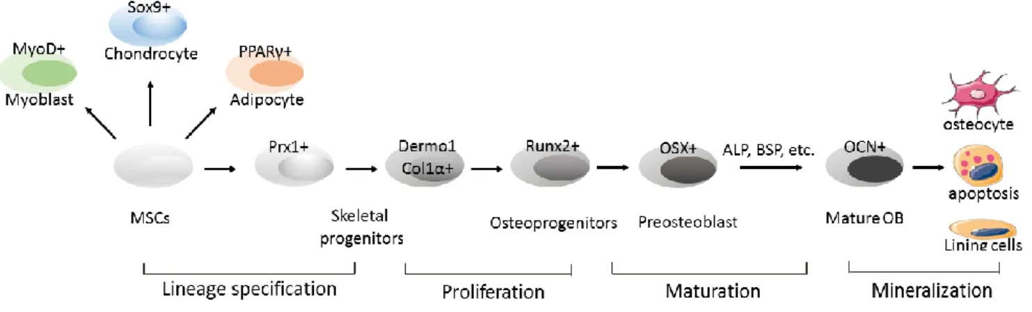
differentiate into osteochondroprogenitors that express Prx1 and Twist1 (ten Berge et al., 1998; Yu et al., 2003). These early skeletal progenitors have the capacity to develop into either chondrogenic or osteoblastic lineages. Sox9 is the key transcription factor that drives chondrogenic differentiation (Mori-Akiyama et al., 2003). On the other hand, runx2 and osterixprimarily direct the cell commitment into osteoblastic lineage (Harada and Rodan, 2003). The key transcription factors and several important osteogenic proteins involved in osteoblastogenesis are summarized below (Figure 1.1).
Runx2
Progressive osteoblast differentiation from early mesenchymal precursors requires tight modulation of transcription factors. Runx2 is the earliest transcriptional regulator, followed by osterix. Runx2 belongs to the Runx family, containing a runt DNA binding domain which is homologous with the Drosophila pair-rule gene runt (Komori, 2005). Runx2 is widely accepted as the master regulator for osteoblast differentiation and plays a pivotal role during bone development. Runx2-null mice exhibit complete absence of both
endochondral and intramembranous bone as a result of the lack of mature osteoblasts, and altered chondrocyte maturation as a result of hypertrophic chondrocytes. (Komori et al., 1997; Otto et al., 1997).
Runx2 expressing osteoprogenitors are able to differentiate into either osteoblasts or chondrocytes. Further differentiation from osteoprogenitors into functional mature osteoblasts that produce bone matrix requires osterix expression.
8
Osterix is a zinc finger-containing transcription factor belong to the Sp/XKLF family. Osterix functions immediately downstream of Runx2 (Javed et al., 2010). Previous literature demonstrates that osterix-deficient mice display normal Runx2 expression, while Runx2-null mice demonstrate no expression of osterix (Nakashima et al., 2002).
Osterix is also essential for osteoblast differentiation and is expressed in all the osteoblasts throughout the embryonic bone development. Sp7 -/- mice show a similar bone phenotype to the Runx2 -/- mice, with a lack of mature differentiated osteoblasts in either intramembranous or endochondral ossification. However, different from Runx2 -/- mice, the osterix-null mice develop normal cartilage formation as well as terminally differentiated hypertrophic chondrocytes (Nakashima et al., 2002). This observation highlights a specific role of osterix in osteoblastic differentiation.
Alkaline phosphatase (ALP)
Once committed into osteoblastic cell lineages, the osteoprogenitors then go through a proliferative period before they differentiate into osteoblasts that synthesize extracellular matrix. Alkaline phosphatase is a membrane-bound enzyme and is expressed immediately after the proliferation period by osteoblasts. ALP is an early marker for
osteoblasts during matrix maturation. Its expression is associated with maximal production of type I collagen (Javed et al., 2010).
9 Bone sialoprotein (BSP)
Osteoblasts are responsible for ECM production, maturation and mineralization. The ECM proteins in bone include mainly type I collagen, proteoglycans and other
non-collagenous proteins (bone sialoprotein, osteopontin, osteocalcin, etc.). These matrix proteins regulate osteoblast proliferation, differentiation and survival (Rodan and Noda, 1991). BSP is a glycoprotein that belongs to the SIBLING (Small Integrin-binding Ligand N-linked Glycoprotein) family. It is widely expressed by osteoblasts, hypertrophic
chondrocytes and osteoclasts.
BSP was shown to influence osteoblast differentiation and induce bone
mineralization (Gordon et al., 2007). Mice lacking BSP displayed delayed long bone growth, mineralization and a decrease of bone markers including osteopontin (OPN) and osteocalcin (OCN) (Bouleftour et al., 2014). BSP deficient mice also demonstrated impaired bone
healing, implicating a role for BSP in bone regeneration via bone formation and osteoclast activity (Malaval et al., 2009; Monfoulet et al., 2010).
Osteocalcin (OCN)
Osteocalcin, a vitamin K and vitamin D dependent non-collagenous protein, is also secreted by osteoblasts. It is not expressed in the early stages of osteoblast differentiation and, in fact, is the marker for the late phase of osteoblastogenesis (Bellows et al., 1999). Bgalap-expressing osteoblasts are usually recognized as mature osteoblasts.
10
overlapping expression of bone markers directs the commitment, proliferation and
differentiation of stem cells into osteoblasts. Evaluation of bone marker expression is often used to assess the differentiation stage of progenitor cells. The progression of stem cells into each differentiation stage requires many extracellular signals including growth factors (BMPs, FGFs, etc.), hormones (PTH, estrogen, etc.) and cytokines (interleukins) (Allori et al., 2008a). In this sense, these osteogenic signals function in a stage-specific manner with a defined temporal expression profile.
BMP2 in tissue engineering
For the past several decades, tremendous efforts have been taken by scientists to develop effective treatment for critical-sized defects. In such cases where a large amount of bone is missing, spontaneous bone repair cannot occur. Therefore, the goal for treatment is to restore function by providing a local environment favorable for bone regeneration.
11
allografts include the potential transmission of disease, immunogenic reaction and limited availability of cadaver bones. Due to the limitations of autografts and allografts, there is an increasing demand for the discovery of graft substitutes, which could replace, and resemble the form and properties of, natural bone (Giannoudis et al., 2005).
In recent decades, researchers have focused on developing synthetic bone graft substitutes. An optimal substitute material is expected to provide adequate strength in response to mechanical load, and should gradually be absorbed during the process of new bone formation. The key properties that will determine the regenerative outcome of graft substitutes are: 1) a biocompatible and degradable scaffold carrier that serves as a template for bone cells to adhere, proliferate and differentiate (osteoconduction); 2) growth factors that stimulate progenitor differentiation into osteoblastic lineages (osteoinduction); and 3) existence of host cells or implanted cells to assist with bone repair (osteogenesis). However, an optimal material that fulfills all of the above requirements has yet to be developed.
Due to this complexity of bone repair, an improved strategy using a combination of synthetic scaffolds, growth factors and cells holds the most promise as a tissue engineering application (Agarwal et al., 2009; Rahman et al., 2014). As researchers continue to gain a better understanding of the molecular pathways involved in bone healing, many relevant key factors have already been investigated in the lab and applied in clinical settings. Among the factors studied, bone morphogenetic proteins (BMPs), which belong to the transforming growth factor β (TGF-β) superfamily, have been heavily studied due to their strong
12
organogenesis, embryogenesis and wound healing (Attisano and Wrana, 2002; Miyazono, 2000a). BMPs greatly impact bone development and healing. In 1965, BMPs were first identified as osteoinductive agents in demineralized bone matrix, and were later termed bone morphogenetic proteins by Urist (Urist, 1965; Urist and Strates, 1971). Currently, at least 20 BMPs have been recognized and described in humans as well as other species (Carreira et al., 2014).
BMP2 transduces signals through direct binding to its serine-threonine kinase receptor complex (type I and type II receptors, BMPRI and BMPRII). This ligand-receptor interaction catalyzes the phosphorylation of downstream signaling transducers and
activates the intracellular cascades (Carreira et al., 2014). In the canonical Smads pathway, receptor specific Smad proteins (Smads, Smad 1/5/8) are phosphorylated. Activated R-Smads are then released from the cell membrane into the cytoplasm where they interact with Smads such as co-Smad and Smad 4. (Attisano and Wrana, 2002). The binding of R-Smads with Smad4 enables the exposure of nucleus-imported sequences, allowing for the nuclear translocation of the R-Smads-Smad 4 complex and initiating transcriptional regulation (Kawabata et al., 1999; Miyazono, 2000b) (Figure 1.2).
In the nucleus, the Smad heteromer modulates the transcription of major
13
development and regeneration. Moreover, BMP2 is also a central mediator that can control the expression of other several other BMPs. (Edgar et al., 2007).
Elevated BMP2 expression was observed as soon as 24 hours following bone injury, suggesting that it plays a role in the initiation of the bone healing process (Farhadieh et al., 2004; Khanal et al., 2008; Kugimiya et al., 2005). BMP2 is found actively involved
throughout the bone regeneration phase for at least 21 days after bone fracture (Bais et al., 2009). The Smad 1/5/8 complex shares a similar expression pattern and localization with BMP2, as shown by immunohistochemical staining after osteotomy (Khanal et al., 2008).
The importance of BMP2 in bone healing was addressed by the finding that mice with impaired BMP2 expression display normal bone development but defective bone healing (Tsuji et al., 2006). BMP2 is produced by osteoprogenitors, mesenchymal cells, chondrocytes and osteoblasts that either reside at, or are recruited to, the sites of injury. BMP2 induction upon bone injury was suggested to promote bone repair by: 1) initiating healing cascades and chemotaxis, and recruiting of mesenchymal stems cells; 2) activating other BMPs; and 3) activating many growth factors that control angiogenesis, cell
proliferation and differentiation (Dimitriou et al., 2005).
14
engineering applications in both experimental and clinical scenarios. For optimal effect, rhBMP2 is often delivered by carriers, which not only prolong protein retention, but also provide a platform for progenitor adhesion, proliferation and differentiation. Due to the desirable properties (biocompatibility, biodegradability, safety and being favorable for cell adhesion), absorbable collagen sponges (ACS) have become attractive carriers for tissue engineering.
Local delivery of BMP2 by absorbable collagen carriers enhances bone healing and regeneration in both experimental and clinical scenarios (Kim et al., 2005; Lin et al., 2013; Simpson et al., 2006). The beneficial effect of rhBMP2/ACS has been applied to many clinical cases and has been reported to bridge critical-sized defects, promote fracture healing, induce spinal fusion and promote dental and craniofacial reconstruction (Chenard et al., 2012; Geiger et al., 2003). Robust release of several pro-inflammatory cytokines (IL-1, IL-6, TNFα, etc.) is observed during both natural bone healing as well as rhBMP2 mediated bone regeneration (Gerstenfeld et al., 2003a; Lu et al., 2013). Interestingly, elevation of BMP2 expression occurs and peaks in parallel with pro-inflammatory cytokines, at 24 hours post injury, and is sustained throughout the acute inflammation phase (Kon et al., 2001; Mountziaris and Mikos, 2008). BMP2 enhances the early inflammation phase, acting as a chemoattractant for immune cells, such as lymphocytes and macrophages (Cunningham et al., 1992). Moreover, BMP2 induces the expression of inflammatory mediators in various cell types including preosteoblasts (Akeel et al., 2012; Helbing et al., 2011).
15
findings that the osteoinductive capability of BMP2 is impaired in the absence of proper inflammatory signals (Katavic et al., 2003; Zhang et al., 2002). In addition, proinflammatory factors are proven to stimulate osteogenic differentiation of stem cells by increasing BMP2 production (Fukui et al., 2003; Hess et al., 2009; Rifas, 2006). This evidence, taken together, implies a tight connection between the osteoinductivity of BMP2 and the inflammatory signaling pathway. On the other hand, this connection between BMP2 and inflammation is complex and not well understood, as many cases demonstrate a negative regenerative effect of rhBMP2. Excessive inflammation caused by systemic inflammatory disease or local infection impairs bone repair (Huang et al., 2014). High doses of rhBMP2 could cause adverse inflammatory responses such as soft tissue swelling, thereby diminishing the
efficacy of rhBMP2-mediated repair (Lee et al., 2011). The exact role of inflammation on the osteoinductive capability of BMP2 remains unclear. Further understanding of this
interaction will improve the current therapeutic strategy for skeletal repair.
Although promising bone regenerative effects have been achieved using the rhBMP2 based tissue engineering approach in the past several decades, an optimal healing process that mimics all of the healing cascades of natural bone repair has yet to be developed.
NF-κB pathway and its involvement in bone biology
16
responsible for dimerization and DNA binding. The five monomers can form homodimers or heterodimers that function in gene expression regulation. Among those transcriptionally active dimers, the p65-p50 heterodimer is the most common form of NF-κB (Oeckinghaus and Ghosh, 2009; Vallabhapurapu and Karin, 2009).
The NF-κB signaling is activated through two major pathways: the classical (canonical) pathway and alternative (non-canonical) pathway (Figure 1.3). These two pathways are different in many aspects including the participating NF-κB dimers activating signals as well as the biological functions. The classical pathway is induced by many
extracellular cues such as receptor activator of the NF-κB ligand (RANKL) and TNFα. In an unstimulated state, NF-κB complexes are sequestered in the cytoplasm via association with IκB proteins (IκBα, IκBβ, and IκBɛ). The classical NF-κB signaling pathway is initiated upon activation of the IκB kinase complex (IKKα, IKKβ, IKKγ). The IKK catalytic complex
phosphorylate IκB proteins for polyubiquitination and degradation in proteasomes, releasing the NF-κB dimer for nuclear translocation. p65-p50 and cRel-p50 heterodimers mainly signal through the classical pathway (Hayden and Ghosh, 2008; Oeckinghaus and Ghosh, 2009). On the other hand, the alternative pathway is stimulated by only a subset of the TNF superfamily and is mainly involved in lymphoid development. NF-κB inducing kinase (NIK) is the key regulatory point which activates IKKα, leading to the processing of p100 into p52. The p52-RelB heterodimer is the primary component in the alternative NF-κB signaling pathway (Bonizzi and Karin, 2004).
NF-κB signaling is essential for embryogenesis and has an important role in
17
related inflammatory bone disorders has been extensively investigated. Studies using a p50 and p52 double knockout animal model demonstrated that these animals display
osteopetrosis, with retarded growth and a significant decrease in osteoclast numbers (Iotsova et al., 1997). Other transgenic mice models with manipulated NF-κB elements also suggest the requirement of NF-κB signaling during osteoclastogenesis (Soysa and Alles, 2009). Further investigation has determined the importance of NF-κB in osteoclast differentiation, survival and function by transcriptional regulation of osteoclast master genes including c-Fos, NFATc1, tartrate-resistant acid phosphatase (TRAP) and cathepsin K (Novack, 2011; Soysa and Alles, 2009).
The role of NF-κB on osteoclasts is well defined. However, the effect of NF-κB on bone forming cells (osteoblasts) is relatively less explored. A role of NF-κB in osteoblast differentiation was suggested when Hess et al. reported that TNFα treatment/ p65
activation positively affects osteogenic differentiation in human mesenchymal stromal cells through regulation of BMP2 expression (Hess et al., 2009). As a potent NF-κB activator, TNFα was also reported to induce osteogenic differentiation of human dental pulp stem cells, mesenchymal stem cells and human stem cells from apical papilla respectively through NF-κB signaling pathway (Cho et al., 2010; Feng et al., 2013; Li et al., 2014). In addition, NF-κB/p65 signaling activation facilitates BMP2-mediated chondrogenic
18
2003). These findings indicate crosstalk between the NF-κB pathway and BMP2-mediated osteogenesis.
In an attempt to investigate the precise role of NF-κB in osteogenesis, a transgenic animal model characterized by manipulation of the NF-κB function has been employed. Global knockout of p65 (RelA) leads to embryonic lethality of mice due to liver degeneration at 15-16 days of gestation caused by liver cell apoptosis (Beg et al., 1995). To circumvent the early lethality, strategies to generate conditional NF-κB knockout animals enabled the further investigation of NF-κB function on bone in vivo. A Cre-LoxP system has been widely used for tissue-specific elimination of target gene expression in mouse models. In mice harboring both the Cre transgene and a floxed target gene, Cre-recombinase mediates the gene deletion by cleaving the two specific 34-base LoxP sites that flank the target gene. A genetically modified Cre-recombinase transgene under the control of a tissue-specific promoter allows for deletion of a target gene in a specific tissue (Chambers, 1994).
bglap2-IKK-DN mice with dominant negative IKKγ function in mature osteoblasts exhibit increased bone mineral density (BMD) and BV/TV at 2 and 4 weeks old (Chang et al., 2009). In another study, Coll2-IKK2ca mice, in which IKKβ is constitutively active in
19
bone formation (Caron et al., 2012; Cho et al., 2010; Feng et al., 2013; Hess et al., 2009; Li et al., 2014). The current findings regarding the function of NF-κB on osteogenesis are
controversial.
The discrepancy of the results may lie in the difference of approaches that have been applied. One issue that comes with the transgenic mouse with manipulated NF-κB regulators is the difficulty to interpret the results. Since many NF-κB subunits share the same regulators, the indirect effect from other NF-κB components may contribute to the observed outcome. The strategies employed in previous studies include the local
application of NF-κB inhibitors or activators and generation of virus expressing dominant negative or constitutive active NF-κB elements (Hess et al., 2009). Since NF-κB is a major inflammatory mediator, expressed by many cell types and not limited to bone cells, the treatment of NF-κB inhibitors and/or activators may affect other cells, indirectly influencing the outcome. An improved approach for investigating the direct role of NF-κB on
osteogenesis will greatly improve the current knowledge and enhance accuracy and interpretation of future experiments.
20
need to elucidate the precise function of NF-κB in bone homeostasis and healing,
21
Figure 1.1 Differentiation of primitive mesenchymal stem cells into osteoblasts.
23
24 REFERNCES
Agarwal, R., Williams, K., Umscheid, C.A., and Welch, W.C. (2009). Osteoinductive bone graft substitutes for lumbar fusion: a systematic review. Journal of neurosurgery Spine 11, 729-740.
Aghaloo, T., Cowan, C.M., Zhang, X., Freymiller, E., Soo, C., Wu, B., Ting, K., and Zhang, Z. (2010). The effect of NELL1 and bone morphogenetic protein-2 on calvarial bone
regeneration. Journal of oral and maxillofacial surgery : official journal of the American Association of Oral and Maxillofacial Surgeons 68, 300-308.
Ai-Aql, Z.S., Alagl, A.S., Graves, D.T., Gerstenfeld, L.C., and Einhorn, T.A. (2008). Molecular mechanisms controlling bone formation during fracture healing and distraction
osteogenesis. Journal of dental research 87, 107-118.
Akeel, S., El-Awady, A., Hussein, K., El-Refaey, M., Elsalanty, M., Sharawy, M., and Al-Shabrawey, M. (2012). Recombinant bone morphogenetic protein-2 induces up-regulation of vascular endothelial growth factor and interleukin 6 in human pre-osteoblasts: role of reactive oxygen species. Archives of oral biology 57, 445-452.
Allori, A.C., Sailon, A.M., and Warren, S.M. (2008a). Biological basis of bone formation, remodeling, and repair-part I: biochemical signaling molecules. Tissue engineering Part B, Reviews 14, 259-273.
Allori, A.C., Sailon, A.M., and Warren, S.M. (2008b). Biological basis of bone formation, remodeling, and repair-part II: extracellular matrix. Tissue engineering Part B, Reviews 14, 275-283.
Amini, A.R., Laurencin, C.T., and Nukavarapu, S.P. (2012). Bone tissue engineering: recent advances and challenges. Critical reviews in biomedical engineering 40, 363-408.
Attisano, L., and Wrana, J.L. (2002). Signal transduction by the TGF-beta superfamily. Science 296, 1646-1647.
25
Baroli, B. (2009). From natural bone grafts to tissue engineering therapeutics: Brainstorming on pharmaceutical formulative requirements and challenges. Journal of pharmaceutical sciences 98, 1317-1375.
Beg, A.A., Sha, W.C., Bronson, R.T., Ghosh, S., and Baltimore, D. (1995). Embryonic lethality and liver degeneration in mice lacking the RelA component of NF-kappa B. Nature 376, 167-170.
Bellows, C.G., Reimers, S.M., and Heersche, J.N. (1999). Expression of mRNAs for type-I collagen, bone sialoprotein, osteocalcin, and osteopontin at different stages of osteoblastic differentiation and their regulation by 1,25 dihydroxyvitamin D3. Cell and tissue research 297, 249-259.
Bhattacharyya, T., Levin, R., Vrahas, M.S., and Solomon, D.H. (2005). Nonsteroidal
antiinflammatory drugs and nonunion of humeral shaft fractures. Arthritis and rheumatism 53, 364-367.
Blokhuis, T.J., Calori, G.M., and Schmidmaier, G. (2013). Autograft versus BMPs for the treatment of non-unions: what is the evidence? Injury 44 Suppl 1, S40-42.
Bonizzi, G., and Karin, M. (2004). The two NF-kappaB activation pathways and their role in innate and adaptive immunity. Trends in immunology 25, 280-288.
Boskey, A.L., and Posner, A.S. (1984). Bone structure, composition, and mineralization. The Orthopedic clinics of North America 15, 597-612.
Bouleftour, W., Boudiffa, M., Wade-Gueye, N.M., Bouet, G., Cardelli, M., Laroche, N., Vanden-Bossche, A., Thomas, M., Bonnelye, E., Aubin, J.E., et al. (2014). Skeletal
development of mice lacking bone sialoprotein (BSP)--impairment of long bone growth and progressive establishment of high trabecular bone mass. PloS one 9, e95144.
Caron, M.M., Emans, P.J., Surtel, D.A., Cremers, A., Voncken, J.W., Welting, T.J., and van Rhijn, L.W. (2012). Activation of NF-kappaB/p65 facilitates early chondrogenic
differentiation during endochondral ossification. PloS one 7, e33467.
26
Chambers, C.A. (1994). TKO'ed: lox, stock and barrel. BioEssays : news and reviews in molecular, cellular and developmental biology 16, 865-868.
Chang, J., Wang, Z., Tang, E., Fan, Z., McCauley, L., Franceschi, R., Guan, K., Krebsbach, P.H., and Wang, C.Y. (2009). Inhibition of osteoblastic bone formation by nuclear factor-kappaB. Nature medicine 15, 682-689.
Chen, G., Deng, C., and Li, Y.P. (2012). TGF-beta and BMP signaling in osteoblast
differentiation and bone formation. International journal of biological sciences 8, 272-288.
Chenard, K.E., Teven, C.M., He, T.C., and Reid, R.R. (2012). Bone morphogenetic proteins in craniofacial surgery: current techniques, clinical experiences, and the future of personalized stem cell therapy. Journal of biomedicine & biotechnology 2012, 601549.
Cho, H.H., Shin, K.K., Kim, Y.J., Song, J.S., Kim, J.M., Bae, Y.C., Kim, C.D., and Jung, J.S. (2010). NF-kappaB activation stimulates osteogenic differentiation of mesenchymal stem cells derived from human adipose tissue by increasing TAZ expression. Journal of cellular physiology 223, 168-177.
Clohisy, J.C., Hirayama, T., Frazier, E., Han, S.K., and Abu-Amer, Y. (2004). NF-kB signaling blockade abolishes implant particle-induced osteoclastogenesis. Journal of orthopaedic research : official publication of the Orthopaedic Research Society 22, 13-20.
Cunningham, N.S., Paralkar, V., and Reddi, A.H. (1992). Osteogenin and recombinant bone morphogenetic protein 2B are chemotactic for human monocytes and stimulate
transforming growth factor beta 1 mRNA expression. Proceedings of the National Academy of Sciences of the United States of America 89, 11740-11744.
Dimitriou, R., Jones, E., McGonagle, D., and Giannoudis, P.V. (2011). Bone regeneration: current concepts and future directions. BMC medicine 9, 66.
Dimitriou, R., Tsiridis, E., and Giannoudis, P.V. (2005). Current concepts of molecular aspects of bone healing. Injury 36, 1392-1404.
27
Einhorn, T.A. (1998). The cell and molecular biology of fracture healing. Clinical orthopaedics and related research, S7-21.
Farhadieh, R.D., Gianoutsos, M.P., Yu, Y., and Walsh, W.R. (2004). The role of bone
morphogenetic proteins BMP-2 and BMP-4 and their related postreceptor signaling system (Smads) in distraction osteogenesis of the mandible. The Journal of craniofacial surgery 15, 714-718.
Feng, J.Q., Xing, L., Zhang, J.H., Zhao, M., Horn, D., Chan, J., Boyce, B.F., Harris, S.E., Mundy, G.R., and Chen, D. (2003). NF-kappaB specifically activates BMP-2 gene expression in growth plate chondrocytes in vivo and in a chondrocyte cell line in vitro. The Journal of biological chemistry 278, 29130-29135.
Feng, X., Feng, G., Xing, J., Shen, B., Li, L., Tan, W., Xu, Y., Liu, S., Liu, H., Jiang, J., et al. (2013). TNF-alpha triggers osteogenic differentiation of human dental pulp stem cells via the NF-kappaB signalling pathway. Cell biology international 37, 1267-1275.
Franz-Odendaal, T.A. (2011). Induction and patterning of intramembranous bone. Frontiers in bioscience 16, 2734-2746.
Fukui, N., Zhu, Y., Maloney, W.J., Clohisy, J., and Sandell, L.J. (2003). Stimulation of BMP-2 expression by pro-inflammatory cytokines IL-1 and TNF-alpha in normal and osteoarthritic chondrocytes. The Journal of bone and joint surgery American volume 85-A Suppl 3, 59-66.
Geiger, M., Li, R.H., and Friess, W. (2003). Collagen sponges for bone regeneration with rhBMP-2. Advanced drug delivery reviews 55, 1613-1629.
Gerstenfeld, L.C., Cho, T.J., Kon, T., Aizawa, T., Tsay, A., Fitch, J., Barnes, G.L., Graves, D.T., and Einhorn, T.A. (2003a). Impaired fracture healing in the absence of TNF-alpha signaling: the role of TNF-alpha in endochondral cartilage resorption. Journal of bone and mineral research : the official journal of the American Society for Bone and Mineral Research 18, 1584-1592.
28
Giannoudis, P.V., Dinopoulos, H., and Tsiridis, E. (2005). Bone substitutes: an update. Injury 36 Suppl 3, S20-27.
Gordon, J.A., Tye, C.E., Sampaio, A.V., Underhill, T.M., Hunter, G.K., and Goldberg, H.A. (2007). Bone sialoprotein expression enhances osteoblast differentiation and matrix mineralization in vitro. Bone 41, 462-473.
Harada, S., and Rodan, G.A. (2003). Control of osteoblast function and regulation of bone mass. Nature 423, 349-355.
Hayden, M.S., and Ghosh, S. (2008). Shared principles in NF-kappaB signaling. Cell 132, 344-362.
Helbing, T., Rothweiler, R., Ketterer, E., Goetz, L., Heinke, J., Grundmann, S., Duerschmied, D., Patterson, C., Bode, C., and Moser, M. (2011). BMP activity controlled by BMPER regulates the proinflammatory phenotype of endothelium. Blood 118, 5040-5049.
Hess, K., Ushmorov, A., Fiedler, J., Brenner, R.E., and Wirth, T. (2009). TNFalpha promotes osteogenic differentiation of human mesenchymal stem cells by triggering the NF-kappaB signaling pathway. Bone 45, 367-376.
Hsu, H., Lacey, D.L., Dunstan, C.R., Solovyev, I., Colombero, A., Timms, E., Tan, H.L., Elliott, G., Kelley, M.J., Sarosi, I., et al. (1999). Tumor necrosis factor receptor family member RANK mediates osteoclast differentiation and activation induced by osteoprotegerin ligand. Proceedings of the National Academy of Sciences of the United States of America 96, 3540-3545.
Huang, R.L., Yuan, Y., Zou, G.M., Liu, G., Tu, J., and Li, Q. (2014). LPS-stimulated
inflammatory environment inhibits BMP-2-induced osteoblastic differentiation through crosstalk between TLR4/MyD88/NF-kappaB and BMP/Smad signaling. Stem cells and development 23, 277-289.
Iotsova, V., Caamano, J., Loy, J., Yang, Y., Lewin, A., and Bravo, R. (1997). Osteopetrosis in mice lacking NF-kappaB1 and NF-kappaB2. Nature medicine 3, 1285-1289.
29
Jeffcoach, D.R., Sams, V.G., Lawson, C.M., Enderson, B.L., Smith, S.T., Kline, H., Barlow, P.B., Wylie, D.R., Krumenacker, L.A., McMillen, J.C., et al. (2014). Nonsteroidal anti-inflammatory drugs' impact on nonunion and infection rates in long-bone fractures. The journal of trauma and acute care surgery 76, 779-783.
Katavic, V., Grcevic, D., Lukic, I.K., Vucenik, V., Kovacic, N., Kalajzic, I., and Marusic, A. (2003). Non-functional Fas ligand increases the formation of cartilage early in the endochondral bone induction by rhBMP-2. Life sciences 74, 13-28.
Kawabata, M., Imamura, T., Inoue, H., Hanai, J., Nishihara, A., Hanyu, A., Takase, M., Ishidou, Y., Udagawa, Y., Oeda, E., et al. (1999). Intracellular signaling of the TGF-beta superfamily by Smad proteins. Annals of the New York Academy of Sciences 886, 73-82.
Khan, S.N., Solaris, J., Ramsey, K.E., Yang, X., Bostrom, M.P., Stephan, D., and Daluiski, A. (2008). Identification of novel gene expression in healing fracture callus tissue by DNA microarray. HSS journal : the musculoskeletal journal of Hospital for Special Surgery 4, 149-160.
Khanal, A., Yoshioka, I., Tominaga, K., Furuta, N., Habu, M., and Fukuda, J. (2008). The BMP signaling and its Smads in mandibular distraction osteogenesis. Oral diseases 14, 347-355.
Kim, C.S., Kim, J.I., Kim, J., Choi, S.H., Chai, J.K., Kim, C.K., and Cho, K.S. (2005). Ectopic bone formation associated with recombinant human bone morphogenetic proteins-2 using absorbable collagen sponge and beta tricalcium phosphate as carriers. Biomaterials 26, 2501-2507.
Komori, T. (2005). Regulation of skeletal development by the Runx family of transcription factors. Journal of cellular biochemistry 95, 445-453.
Komori, T., Yagi, H., Nomura, S., Yamaguchi, A., Sasaki, K., Deguchi, K., Shimizu, Y., Bronson, R.T., Gao, Y.H., Inada, M., et al. (1997). Targeted disruption of Cbfa1 results in a complete lack of bone formation owing to maturational arrest of osteoblasts. Cell 89, 755-764.
30
Kugimiya, F., Kawaguchi, H., Kamekura, S., Chikuda, H., Ohba, S., Yano, F., Ogata, N., Katagiri, T., Harada, Y., Azuma, Y., et al. (2005). Involvement of endogenous bone morphogenetic protein (BMP) 2 and BMP6 in bone formation. The Journal of biological chemistry 280, 35704-35712.
Lee, K.B., Murray, S.S., Taghavi, C.E., Song, K.J., Brochmann, E.J., Johnson, J.S., Keorochana, G., Liao, J.C., and Wang, J.C. (2011). Bone morphogenetic protein-binding peptide reduces the inflammatory response to recombinant human bone morphogenetic protein-2 and recombinant human bone morphogenetic protein-7 in a rodent model of soft-tissue inflammation. The spine journal : official journal of the North American Spine Society 11, 568-576.
Lee, M.H., Kim, Y.J., Kim, H.J., Park, H.D., Kang, A.R., Kyung, H.M., Sung, J.H., Wozney, J.M., Kim, H.J., and Ryoo, H.M. (2003). BMP-2-induced Runx2 expression is mediated by Dlx5, and TGF-beta 1 opposes the BMP-2-induced osteoblast differentiation by suppression of Dlx5 expression. The Journal of biological chemistry 278, 34387-34394.
Li, J., Yan, M., Wang, Z., Jing, S., Li, Y., Liu, G., Yu, J., and Fan, Z. (2014). Effects of canonical NF-kappaB signaling pathway on the proliferation and odonto/osteogenic differentiation of human stem cells from apical papilla. BioMed research international 2014, 319651.
Lin, C.Y., Chang, Y.H., Li, K.C., Lu, C.H., Sung, L.Y., Yeh, C.L., Lin, K.J., Huang, S.F., Yen, T.C., and Hu, Y.C. (2013). The use of ASCs engineered to express BMP2 or TGF-beta3 within scaffold constructs to promote calvarial bone repair. Biomaterials 34, 9401-9412.
Lu, Z., Wang, G., Dunstan, C.R., Chen, Y., Lu, W.Y., Davies, B., and Zreiqat, H. (2013). Activation and promotion of adipose stem cells by tumour necrosis factor-alpha preconditioning for bone regeneration. Journal of cellular physiology 228, 1737-1744.
Luu, H.H., Song, W.X., Luo, X., Manning, D., Luo, J., Deng, Z.L., Sharff, K.A., Montag, A.G., Haydon, R.C., and He, T.C. (2007). Distinct roles of bone morphogenetic proteins in osteogenic differentiation of mesenchymal stem cells. Journal of orthopaedic research : official publication of the Orthopaedic Research Society 25, 665-677.
31
Miyazono, K. (2000a). Positive and negative regulation of TGF-beta signaling. Journal of cell science 113 ( Pt 7), 1101-1109.
Miyazono, K. (2000b). TGF-beta signaling by Smad proteins. Cytokine & growth factor reviews 11, 15-22.
Monfoulet, L., Malaval, L., Aubin, J.E., Rittling, S.R., Gadeau, A.P., Fricain, J.C., and Chassande, O. (2010). Bone sialoprotein, but not osteopontin, deficiency impairs the mineralization of regenerating bone during cortical defect healing. Bone 46, 447-452.
Mori-Akiyama, Y., Akiyama, H., Rowitch, D.H., and de Crombrugghe, B. (2003). Sox9 is required for determination of the chondrogenic cell lineage in the cranial neural crest. Proceedings of the National Academy of Sciences of the United States of America 100, 9360-9365.
Mountziaris, P.M., and Mikos, A.G. (2008). Modulation of the inflammatory response for enhanced bone tissue regeneration. Tissue engineering Part B, Reviews 14, 179-186.
Mountziaris, P.M., Spicer, P.P., Kasper, F.K., and Mikos, A.G. (2011). Harnessing and modulating inflammation in strategies for bone regeneration. Tissue engineering Part B, Reviews 17, 393-402.
Nakahama, K. (2010). Cellular communications in bone homeostasis and repair. Cellular and molecular life sciences : CMLS 67, 4001-4009.
Nakashima, K., Zhou, X., Kunkel, G., Zhang, Z., Deng, J.M., Behringer, R.R., and de Crombrugghe, B. (2002). The novel zinc finger-containing transcription factor osterix is required for osteoblast differentiation and bone formation. Cell 108, 17-29.
Novack, D.V. (2011). Role of NF-kappaB in the skeleton. Cell research 21, 169-182.
Oeckinghaus, A., and Ghosh, S. (2009). The NF-kappaB family of transcription factors and its regulation. Cold Spring Harbor perspectives in biology 1, a000034.
32
cleidocranial dysplasia syndrome, is essential for osteoblast differentiation and bone development. Cell 89, 765-771.
Owens, J.M., Gallagher, A.C., and Chambers, T.J. (1996). Bone cells required for osteoclastic resorption but not for osteoclastic differentiation. Biochemical and biophysical research communications 222, 225-229.
Peng, Y., Kang, Q., Cheng, H., Li, X., Sun, M.H., Jiang, W., Luu, H.H., Park, J.Y., Haydon, R.C., and He, T.C. (2003). Transcriptional characterization of bone morphogenetic proteins (BMPs)-mediated osteogenic signaling. Journal of cellular biochemistry 90, 1149-1165.
Rahman, C.V., Ben-David, D., Dhillon, A., Kuhn, G., Gould, T.W., Muller, R., Rose, F.R.,
Shakesheff, K.M., and Livne, E. (2014). Controlled release of BMP-2 from a sintered polymer scaffold enhances bone repair in a mouse calvarial defect model. Journal of tissue
engineering and regenerative medicine 8, 59-66.
Rawadi, G., Vayssiere, B., Dunn, F., Baron, R., and Roman-Roman, S. (2003). BMP-2 controls alkaline phosphatase expression and osteoblast mineralization by a Wnt autocrine loop. Journal of bone and mineral research : the official journal of the American Society for Bone and Mineral Research 18, 1842-1853.
Rifas, L. (2006). T-cell cytokine induction of BMP-2 regulates human mesenchymal stromal cell differentiation and mineralization. Journal of cellular biochemistry 98, 706-714.
Rodan, G.A., and Noda, M. (1991). Gene expression in osteoblastic cells. Critical reviews in eukaryotic gene expression 1, 85-98.
Rundle, C.H., Wang, H., Yu, H., Chadwick, R.B., Davis, E.I., Wergedal, J.E., Lau, K.H., Mohan, S., Ryaby, J.T., and Baylink, D.J. (2006). Microarray analysis of gene expression during the inflammation and endochondral bone formation stages of rat femur fracture repair. Bone 38, 521-529.
33
Simpson, A.H., Mills, L., and Noble, B. (2006). The role of growth factors and related agents in accelerating fracture healing. The Journal of bone and joint surgery British volume 88, 701-705.
Soysa, N.S., and Alles, N. (2009). NF-kappaB functions in osteoclasts. Biochemical and biophysical research communications 378, 1-5.
Stein, G.S., Lian, J.B., and Owen, T.A. (1990). Relationship of cell growth to the regulation of tissue-specific gene expression during osteoblast differentiation. FASEB journal : official publication of the Federation of American Societies for Experimental Biology 4, 3111-3123.
Suda, T., Nakamura, I., Jimi, E., and Takahashi, N. (1997). Regulation of osteoclast function. Journal of bone and mineral research : the official journal of the American Society for Bone and Mineral Research 12, 869-879.
Suda, T., Takahashi, N., Udagawa, N., Jimi, E., Gillespie, M.T., and Martin, T.J. (1999). Modulation of osteoclast differentiation and function by the new members of the tumor necrosis factor receptor and ligand families. Endocrine reviews 20, 345-357.
Swarnkar, G., Zhang, K., Mbalaviele, G., Long, F., and Abu-Amer, Y. (2014). Constitutive activation of IKK2/NF-kappaB impairs osteogenesis and skeletal development. PloS one 9, e91421.
Szpalski, C., Barr, J., Wetterau, M., Saadeh, P.B., and Warren, S.M. (2010). Cranial bone defects: current and future strategies. Neurosurgical focus 29, E8.
ten Berge, D., Brouwer, A., Korving, J., Martin, J.F., and Meijlink, F. (1998). Prx1 and Prx2 in skeletogenesis: roles in the craniofacial region, inner ear and limbs. Development 125, 3831-3842.
Tsuji, K., Bandyopadhyay, A., Harfe, B.D., Cox, K., Kakar, S., Gerstenfeld, L., Einhorn, T., Tabin, C.J., and Rosen, V. (2006). BMP2 activity, although dispensable for bone formation, is required for the initiation of fracture healing. Nature genetics 38, 1424-1429.
34
Urist, M.R. (1965). Bone: formation by autoinduction. Science 150, 893-899.
Urist, M.R., and Strates, B.S. (1971). Bone morphogenetic protein. Journal of dental research 50, 1392-1406.
Vallabhapurapu, S., and Karin, M. (2009). Regulation and function of NF-kappaB
transcription factors in the immune system. Annual review of immunology 27, 693-733.
Wang, E.A., Rosen, V., Cordes, P., Hewick, R.M., Kriz, M.J., Luxenberg, D.P., Sibley, B.S., and Wozney, J.M. (1988). Purification and characterization of other distinct bone-inducing factors. Proceedings of the National Academy of Sciences of the United States of America 85, 9484-9488.
Yu, K., Xu, J., Liu, Z., Sosic, D., Shao, J., Olson, E.N., Towler, D.A., and Ornitz, D.M. (2003). Conditional inactivation of FGF receptor 2 reveals an essential role for FGF signaling in the regulation of osteoblast function and bone growth. Development 130, 3063-3074.
35 CHAPTER 2
EFFECT OF NF-κB HAPLOINSUFFICIENCY ON CALVARIAL BONE HEALING
INTRODUCTION
Critical sized bony defect caused by congenital disorders, trauma, tumors or infection remains a challenge for dental and orthopedic surgeons and represents a great economic burden to health resources (Amini et al., 2012; Baroli, 2009). The treatment for such clinical conditions requires enhancement and acceleration of bone regeneration with the application of bone grafts (Pneumaticos et al., 2010; Tevlin et al., 2014). To avoid the intrinsic shortcoming of natural bone grafts (i.e donor site morbidity, immunogenic property, pain, etc.), synthetic bone graft substitutes using biocompatible scaffold carrier, with osteoinductive agents including BMPs and other growth factors, have been developed (Amini et al., 2012; Chenard et al., 2012).
Bone morphogenic protein 2 (BMP2) is a growth factor that belongs to the transforming growth factor-β superfamily. At the cellular level, BMP2 triggers the
36
research experiments using animal models, showed improved bone formation and healing following local delivery of BMP2 by an absorbable collagen carrier (Kim et al., 2005; Lin et al., 2013; Simpson et al., 2006). BMP2-mediated bone regeneration involves in an initial inflammatory response associated with the production of cytokines such as tumor necrosis factor-α (TNFα) and interleukin-6 (IL-6) (Gerstenfeld et al., 2003). In vitro studies revealed that BMP2 induces expression of inflammatory mediators in various cell types including preosteoblasts (Akeel et al., 2012; Helbing et al., 2011). Inflammatory signals are crucial for the recruitment and subsequent differentiation of stem and progenitor cells, reflecting a signaling mechanism through which BMP2 promotes bone regeneration. Furthermore, the osteoinductive capability of BMP2 is impaired in the absence of proper inflammatory signals (Katavic et al., 2003; Zhang et al., 2002).
Nuclear factor kappa B (NF-κB) is recognized as a master regulator for the immune system and inflammatory response. The NF-κB family is comprised of transcription factors that mediate many biological activities including cell proliferation, survival and apoptosis (Li and Verma, 2002). The NF-κB family consists of 5 proteins in mammalian cells: RelA (p65), RelB, c-Rel, p50 and p52. These members all share an N-terminal Rel homology domain which is responsible for dimerization and DNA binding. The p65-p50 heterodimer is the most common form of NF-κB in mammals (Oeckinghaus and Ghosh, 2009; Vallabhapurapu and Karin, 2009). In unstimulated cells, inactive NF-κB complexes remain in the cytoplasm in association with IκB proteins (IκBα, IκBβ and IκBɛ). Upon activation, IκB proteins are
37
activator of NF-κB ligand (RANKL) and TNFα are able to induce NF-κB activation through the classical pathway, which requires the p65-p50 and cRel-p50 heterodimers. (Hayden and Ghosh, 2008; Oeckinghaus and Ghosh, 2009).
NF-κB signaling is required for bone development. Its interaction with bone resorbing cells (osteoclasts) and role in relevant inflammatory bone disorders has been extensively investigated (Abu-Amer, 2013). Growing evidence suggests that NF-κB, as a vital inflammatory mediator, is vital for osteoblastic regulation and osteogenesis (Caron et al., 2012; Cho et al., 2010; Hess et al., 2009). NF-κB/p65 signaling activation facilitates BMP2-mediated chondrogenic differentiation during endochondral ossification (Caron et al., 2012). Moreover, TNFα treatment and p65 activation positively influences osteogenic differentiation of human mesenchymal stromal cells (hMSCs) via the regulation of BMP2 expression (Hess et al., 2009). These findings indicate an interaction between NF-κB mediated inflammatory signals and BMP2-induced bone formation and repair. The precise role of NF-κB in the context of bone forming cells (osteoblasts), however, remains unclear. Moreover, little is known regarding the role of the NF-κB signaling pathway in the
osteoinductivity of BMP2 during bone healing.
In the present study, we sought to investigate the interplay between NF-κB signaling and BMP2 mediated bone repair in a critical size calvarial defect model using a genetically modified mouse model. Given the fact that the global deletion of p65, a major NF-κB
Osx-38
Cre mice, we obtained mice with a p65 haploinsufficiency in osteoblastic cell lineages. p65 expression was regulated by the Sp7 promoter.
39 Materials and Methods
Mice
p65-floxed mice were crossed with Osterix-Cre mice (Osx-Cre) in order to generate mice with an osteoblast-restricted p65 ablation (p65fl/+Osx-Cre) (see Figure 2.1). p65 floxed
mice (p65fl/fl) in which exons 5-8 of p65 were flanked with loxp constructs were provided by
Dr. Albert S Baldwin, University of North Carolina at Chapel Hill (Steinbrecher et al., 2008). Osterix-Cre mice (Osx-Cre) which express a GFP fused Cre recombinase gene downstream of the Sp7 promoter were purchased from the Jackson Laboratory (Bar Harbor, ME). The phenotype of Osx-Cre mice has been described previously (Rodda and McMahon, 2006). All animals were on the C57BL/6 background.
Genotyping of mice was performed using PCR of genomic DNA, which was isolated from tail samples taken from mice. Primer sequences used for genotyping are listed in Table 2.1. The animals were housed in a pathogen-free environment with controlled temperature and humidity. Mice were maintained on a 12-hour light/dark cycle. Routine husbandry procedures included cage cleaning, feeding and watering and were conducted every other day. All procedures were performed in accordance with the animal protocol approved by the Institutional Animal Care and Use Committees of the University of North Carolina (IACUC).
Calvarial Defect Model and Surgery procedures
40
the head was shaved, skin was sterilized using 70% ethanol and betadine. A midline incision parallel to the sagittal suture was made using a scalpel in order to expose the parietal bones of the animal. A trephine bur attached to a dental handpiece was then used to create a defect with a diameter of 3.5 mm (Figure 2.2). Great caution was taken to minimize injury to the dura mater. An absorbable collagen sponge (diameter ≈ 4 mm, thickness ≈ 1 mm) (DuraGen, Integra, Plainsboro, NJ) containing saline or 500 ng rhBMP2 (Medtronic,
Minneapolis, MN) was placed over the defect. The collagen sponge was molded into a disk shape (diameter ≈4 mm, thickness ≈1 mm) using a tissue biopsy punch. The skin was sutured using absorbable 4 or 5-0 chromic gut sutures (Ethicon, Somerville, NJ). Animals were allowed to recover on a heating pad before being transferred to clean recovery cages with soft bedding. Pain was managed using ketoprofen (5mg/kg, s.c) following surgery. Full time veterinarians were available at all times in the facility to ensure the health and care of the animals. All surgical procedures were approved by the Institutional Animal Care and Use Committees of the University of North Carolina (IACUC).
Micro-CT imaging and analysis
Separate groups of mice were euthanized 4 and 8 weeks following surgery, using CO2 asphyxiation followed by cervical dislocation. The heads were harvested and
41
software (Skyscan, Aartselaar, Belgium). Representative 3-Dementional images were created using CTAn and CTVol software provided by Skyscan.
In order to compare the level of the healing between p65fl/+ and p65fl/+Osx-Cre mice,
a parameter “healing ratio” was introduced into the analysis. This was defined as follows: Healing ratio= (1-[Current Volume of defect – Original Volume of defect]) X 100 percent. The volume of the calvarial defect was determined using ITK-SNAP (www.itksnap.org).
Histology analysis
For separate groups of animals, heads were harvested 4 and 8 weeks following surgeries. The tissue was immediately immersed into 4% paraformaldehyde for 3 days and then decalcified in 10% Ethylenediamine-tetraacetic acid (EDTA, Sigma, St. Louis, MO, USA) at pH 7.4 at 4 oC for 2 weeks. The samples were then dehydrated through a serial dilution of
ethanol before paraffin embedding. The embedded samples were cut at the center of defect into 5 µm thick sections. Paraffin sections were then stained for Hematoxylin and eosin as well as Masson’s trichrome staining according to the manufacturer’s instructions (Sigma-Aldrich, St. Louis, MO). The stained sections were examined and photographed using a light microscope (Nikon Eclipse50i, Nikon, Melville, NY).
Immunohistochemical analysis
42
(50%-100%). Antigen was retrieved by incubating specimens in 10 mM sodium citrate buffer (PH=6.0) for 20 minutes at 70 oC. Endogenous peroxidase was quenched using 3% hydrogen
peroxide in methanol. The slides were then incubated with primary antibody overnight at 4
oC after blocking with 2.5% normal goat serum in 0.1% bovine serum albumin (BSA) for 30
minutes at room temperature. The Avidin/Biotin Blocking Kit (SP-2001, Vector Laboratories, Burlingame, CA) was used to block all endogenous biotin and avidin binding sites present in tissues. A biotinylated secondary antibody was applied to the slides for 45 minutes at room temperature. The immunoreaction was amplified using RTU ABC Elite reagents (Vector Laboratories, Burlingame, CA). Finally, the slides were detected using DAB substrate kit (Vector Laboratories, Burlingame, CA) and then counterstained with hematoxylin (Sigma-Aldrich, St. Louis, MO) and mounted. Non-specific rabbit IgG was used in place of a primary antibody for the negative control slides. The primary antibody was used at a 1:400 dilution and the secondary antibody was performed at 1:800 dilution in 2.5% goat serum in 0.1% BSA. Photographs of stained sections were taken using a Nikon Eclipse 50i microscope (Nikon, Melville, NY).
CFU-F and CFU-OB assay