Melanoma central nervous system metastases: current
approaches, challenges, and opportunities
Justine V. Cohen1, Hussain Tawbi2, Kim A. Margolin3, Ravi Amravadi4, Marcus Bosenberg1, Priscilla K. Brastianos5, Veronica L. Chiang1, John de Groot6, Isabella C. Glitza2, Meenhard Herlyn7, Sheri L. Holmen8, Lucia B. Jilaveanu1, Andrew Lassman9, Stergios Moschos10, Michael A. Postow11, Reena Thomas12, John A. Tsiouris13, Patrick Wen14, Richard M. White15, Timothy Turnham16, Michael A. Davies2,†, and Harriet M. Kluger1,†
1Yale Cancer Center, Yale School of Medicine, New Haven, CT, USA
2Department of Melanoma, Medical Oncology, University of Texas MD Anderson Cancer Center,
Houston, TX, USA
3Department of Medical Oncology & Therapeutics Research, City of Hope Cancer Center, Duarte,
CA, USA
4Abramson Cancer Center, University of Pennsylvania, Philadelphia, PA, USA 5Division of Neuro-Oncology, Massachusetts General Hospital, Boston, MA, USA 6Division of Neuro-Oncology, MD Anderson Cancer Center, Houston, TX, USA 7Melanoma Research Center, The Wistar Institute, Philadelphia, PA, USA
8Huntsman Cancer Institute, University of Utah Health Sciences Center, Salt Lake City, UT, USA 9Department of Neurology & Herbert Irving Comprehensive, Cancer Center, Columbia University
Medical Center, New York, NY, USA
10UNC Lineberger Comprehensive Cancer Center, University of North Carolina at Chapel Hill,
Chapel Hill, NC, USA
11Department of Oncology, Memorial Sloan Kettering Cancer Center and Weill Cornell Medicine,
New York, NY, USA
12Division of Neuro-Oncology, Department of Neurology, Stanford University, Stanford, CA, USA 13Department of Radiology, New York-Presbyterian Hospital – Weill Cornell Medicine, New York,
NY, USA
14Department of Neuro-Oncology, Dana-Farber Cancer Institute, Boston, MA, USA
15Department of Cancer Biology & Genetics, Memorial Sloan Kettering Cancer Center and Weill
Cornell Medicine, New York, NY, USA
16Melanoma Research Foundation, Washington, DC, USA
HHS Public Access
Author manuscript
Pigment Cell Melanoma Res
. Author manuscript; available in PMC 2017 April 20.Published in final edited form as:
Pigment Cell Melanoma Res. 2016 November ; 29(6): 627–642. doi:10.1111/pcmr.12538.
A
uthor Man
uscr
ipt
A
uthor Man
uscr
ipt
A
uthor Man
uscr
ipt
A
uthor Man
uscr
Summary
Melanoma central nervous system metastases are increasing, and the challenges presented by this patient population remain complex. In December 2015, the Melanoma Research Foundation and the Wistar Institute hosted the First Summit on Melanoma Central Nervous System (CNS) Metastases in Philadelphia, Pennsylvania. Here, we provide a review of the current status of the field of melanoma brain metastasis research; identify key challenges and opportunities for improving the outcomes in patients with melanoma brain metastases; and set a framework to optimize future research in this critical area.
Keywords
brain metastasis; leptomeningeal disease; tumor microenvironment; clinical trials; experimental models
Introduction: clinical features and outcomes of melanoma central nervous
system (CNS) metastases
Metastases to the brain far outnumber primary brain tumors (Maher et al., 2009). The most common sources of brain metastases are lung cancer, breast cancer, and melanoma. While lung cancer and breast cancer are much more prevalent, melanoma has the highest risk of spread to the CNS among all common cancer types. Previous studies have demonstrated that 40–60% of patients with metastatic melanoma develop brain metastases at some point in the course of their disease, and autopsy series identified CNS involvement in up to 80% of patients with metastatic melanoma (Glitza et al., 2016). Several previous large studies reported the median survival of ~4 months from the diagnosis of melanoma CNS metastases, and melanoma brain metastases (MBM) are a leading cause of death from this disease (Davies et al., 2011; Fife et al., 2004; Raizer et al., 2008; Sampson et al., 1998; Skibber et al., 1996).
Clinical features associated with shorter survival in patients with MBMs in previous studies included the presence of active non-CNS disease, >3 MBMs, poor performance status (PS), and leptomeningeal involvement (reviewed in Glitza et al., 2016). Notably, the
overwhelming majority of studies reporting the incidence and outcomes of MBMs were conducted prior to the development of contemporary immunotherapies and BRAF/MEK-targeted therapies that have dramatically improved the survival in patients with metastatic melanoma. As these treatments achieve higher response rates and increasingly durable control of non-CNS disease, it is possible that the incidence of MBMs will increase, similar to the trajectory of breast cancer therapies. Alternatively, depending on blood–brain barrier penetrance and other relevant factors, the incidence of MBMs might actually decrease. The CNS has been identified as a common first site of systemic treatment failure in patients with melanoma treated with FDA-approved targeted therapies, but has yet to be characterized in depth in patients treated with immunotherapies (Frenard et al., 2015; Kim et al., 2011a). In addition, it is probable that these new therapies will impact the outcomes in patients with MBM, based on the results from initial prospective clinical studies (Long et al., 2012;
A
uthor Man
uscr
ipt
A
uthor Man
uscr
ipt
A
uthor Man
uscr
ipt
A
uthor Man
uscr
Margolin et al., 2012). The increasing availability and use of stereotactic radiosurgery (SRS), which was relatively limited in several historical studies, provides further rationale to re-evaluate contemporary outcomes in patients with MBM. Notably, technological advances in SRS now make it feasible for many small brain metastases to be treated in a single session, which may impact the prognostic significance of MBM number.
Updating information about the prevalence and outcomes of CNS metastases will provide an important resource for both clinicians and patients fighting this disease. Perhaps more importantly, this information will further highlight the critical unmet clinical need for more clinical trials for patients with MBM. Over the last decade, many key randomized clinical trials were conducted that ultimately resulted in the approval by the US FDA of 10 new treatment regimens for patients with stage IV melanoma from 2011 to the end of 2015 (Table 1; Chapman et al., 2011; Hauschild et al., 2012; Hodi et al., 2010; Larkin et al., 2015; Long et al., 2015; Ribas et al., 2015; Robert et al., 2015a, b, 2011; Weber et al., 2015). All of these landmark trials, which in total included over 6000 patients with metastatic melanoma, excluded the participation of patients with active MBMs, irrespective of size and number, given the presumption that the prognosis of MBM is uniformly dismal. Small
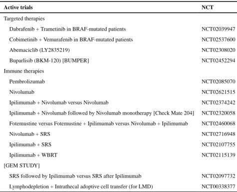
nonrandomized trials testing several of these agents in patients with MBM were opened after those landmark studies completed accrual. These studies included 234 patients and thus represented only 4.1% of the patients enrolled in the trials (Di Giacomo et al., 2012; Long et al., 2012; Margolin et al., 2012). However, based at least in part on the promising clinical activity that was observed in those clinical trials, an increasing number of trials are now becoming available for metastatic melanoma patients with active brain metastases (Table 2). An improved understanding of the factors that are prognostic in contemporary MBM patients will be important for appropriately interpreting the results of those trials. Ultimately, the availability of such information will be critical to the appropriate design of future prospective randomized clinical trials in patients with MBM, which are clearly needed.
Improving outcomes in MBMs will also be accelerated by developing improved
understanding of the pathophysiology and therapeutic resistance of these tumors. There is growing evidence in multiple tumor types, including melanoma, that while many molecular features can be shared, brain metastases often have key, distinct features compared with both primary tumors and metastases to other organs, even in the same patient (Brastianos et al., 2015; Chen and Davies, 2012). These findings support the need for focused preclinical studies of MBMs to identify the critical factors and therapeutic targets for MBMs. Notably, existing data support the rationale to characterize both tumor cells and the unique tumor microenvironment (TME) of the CNS. In addition, and in parallel with the multidisciplinary clinical approach, there is strong rationale to incorporate multiple approaches to the study of MBMs, including but not limited to ‘omics’ (i.e., mutational, transcriptional, proteomic, epigenetic), immunology, and metabolism.
Clinical investigations
Targeted therapy
Temozolomide, a chemotherapy agent that crosses the blood–brain barrier (BBB), has been used frequently in patients with MBM for decades despite the clinical trials demonstrating
A
uthor Man
uscr
ipt
A
uthor Man
uscr
ipt
A
uthor Man
uscr
ipt
A
uthor Man
uscr
intracranial clinical response rates (ICRR) of 3–7% (Agarwala et al., 2004). Similar to the experience in patients without brain metastases, much more impressive results have been observed with BRAF inhibitors (BRAFi). The initial signal of CNS activity came from the phase I trial of dabrafenib, which included 10 patients with BRAFV600-mutant melanoma with untreated or progressing MBMs. Unconfirmed clinical responses were observed in eight patients, and the intracranial disease control rate (IDCR) was 100% (Falchook et al., 2012). The subsequent BREAK-MB study, a phase II trial of dabrafenib in patients with MBM, remains to date the largest clinical trial conducted in this patient population (n = 172). The trial included distinct cohorts of patients who had not received any previous local treatment for brain metastases and those with disease progression in the brain after surgery, whole-brain radiotherapy (WBRT), or SRS. Stable or tapering doses of corticosteroids were permitted. In patients with previously untreated MBMs, the ICRR was 39.2%, and the IDCR was 81.1%; the similar results were observed in patients with progressive MBMs after prior CNS surgery or radiation (ICRR 30.8%, IDCR 89.2%; Long et al., 2012). Median overall survival was 33.1 and 31.4 weeks for the two cohorts, respectively. Notably, neither trial required testing of tissue from the brain metastases for BRAF mutation testing, based on data supporting a very high concordance of BRAFV600 mutation status between brain metastases and other extracranial metastatic sites, a finding consistent with the clinical activity observed in the trial (Chen et al., 2014; Colombino et al., 2012). As initial preclinical studies indicated that dabrafenib did not cross the intact BBB significantly, the results also support that the BBB is compromised by brain metastases. A smaller phase II study of vemurafenib in melanoma patients with symptomatic brain metastases reported an ICRR of only 16.1% (Dummer et al., 2014). Treatment was generally well tolerated with rare cases of adverse events (neurologic or systemic) requiring discontinuation of the study drug. While there is some preclinical evidence to suggest that vemurafenib penetrates brain tissue less efficiently than dabrafenib (Mittapalli et al., 2013), the presence of neurologic symptoms has been identified as a negative prognostic factor in previous studies of patients with MBM, suggesting that this was a cohort of patients with particularly aggressive disease (Glitza et al., 2016). Additional retrospective studies also support a significant activity for vemurafenib in MBMs (Dzienis and Atkinson, 2014).
Randomized clinical trials in BRAFV600-mutant metastatic melanoma patients without CNS metastases demonstrated the superiority of combined BRAF and MEK inhibition to BRAFi monotherapy (Larkin et al., 2014; Long et al., 2015; Robert et al., 2015a). These results led to the regulatory approval of the dabrafenib + trametinib (D+T) regimen in 2014 and for vemurafenib + cobimetinib (V+C) in 2015. Clinical trials are currently ongoing to evaluate the safety and efficacy of each of these combinations in patients with previously untreated or progressing MBMs [NCT02039947 for D+T; NCT02537600 and NCT02230306 for V+C]. Dabrafenib is also being evaluated in combination with SRS [NCT01721603]. In addition to MAPK pathway inhibitors, a limited number of novel targeted therapies against other pathways are being explored in patients with MBM. Examples include a trial of abemaciclib (LY2835219, CDK4/6 inhibitor) for brain metastases from breast cancer, non-small-cell lung cancer (NSCLC), and melanoma [NCT 02308020], and a trial of buparlisib (BKM-120, pan-PI3K inhibitor) specifically in patients with MBM [NCT02452294]. Abemaciclib penetrates
A
uthor Man
uscr
ipt
A
uthor Man
uscr
ipt
A
uthor Man
uscr
ipt
A
uthor Man
uscr
the BBB as does buparlisib (Koul et al., 2012; Raub et al., 2015). Other potential novel approaches include inhibitors of apoptosis, autophagy, and metabolism.
While MAPK pathway inhibitors achieve high rates of disease control in MBMs, the overwhelming majority of patients have short progression-free survival. In contrast to extracranial tumors, currently there is very limited understanding of the mechanisms of resistance to MAPK pathway inhibitors in MBMs. While it is very possible that the same alterations mediate resistance in MBMs as in extracranial tumors, there are also reasons to consider additional mechanisms. For example, analysis of biopsies collected in the phase I trial of vemurafenib demonstrated a linear relationship between the degree of MAPK pathway inhibition achieved and the amount of tumor regression observed (Bollag et al., 2010). Subsequent studies have demonstrated that the approved doses of both vemurafenib and dabrafenib produce a marked (>90%) inhibition of MAPK pathway activation in biopsies of non-CNS metastases (Sosman et al., 2012). However, as many agents achieve significantly lower levels in the CSF and brain compared with the blood, it is possible that suboptimal MAPK pathway inhibition could cause the diminished activity in patients with MBM. At this time, there are no data available about the degree of MAPK pathway inhibition achieved in melanoma brain metastases with the approved doses of vemurafenib or dabrafenib, but ‘neoadjuvant’ or precraniotomy window trials in patients with surgically resectable MBMs could address this gap in understanding (i.e., NCT01978236). The tolerability of BRAF inhibitors, and particularly of BRAF/MEK combination regimens, supports the feasibility of evaluating higher doses in patients with MBM if suboptimal pathway inhibition is detected. Indeed, a case report in a patient with progressing brain metastases from NSCLC with an activating EGFR mutation demonstrated that increasing dosing of the EGFR inhibitor gefitinib above the standard levels achieved higher drug exposure in the CSF, along with radiographic and symptomatic improvement of CNS lesions (Jackman et al., 2006). Interestingly, DNA sequencing on progressing tumors collected at autopsy in that patient demonstrated the presence of a known resistance mutation in EGFR (T790M) in all sampled non-extracranial lesions, but it was not detected in the progressing CNS disease. Similar findings were demonstrated in a second NSCLC case report (Balak et al., 2006; Jackman et al., 2006).
In addition to pharmacodynamics, the differences in tumor biology and genetics could also contribute to resistance in MBMs. Whole-exome sequencing of brain metastases from patients with multiple tumor types, including a small number of melanomas, demonstrated that although the brain metastases and primary tumors share a common genetic ancestor, the brain metastases harbor additional oncogenic drivers not detected in the primary tumor (Brastianos et al., 2015). Somatic mutations affecting the CDK, MAPK, and the PI3K-AKT pathways were frequently detected in the brain metastases that were not detectable or that were only present in a small subpopulation of the DNA of the primaries. Two different melanoma-specific protein-based analyses, one using immunohistochemistry and the other using quantitative reverse-phase protein arrays (RPPA), also demonstrated evidence of increased activation of the PI3K-AKT pathway, but not in the MAPK pathway, in MBMs compared with extracranial metastases from the same patients (Chen et al., 2014; Niessner et al., 2013). Recent studies suggest that the PI3K-AKT pathway may be activated in tumor cells growing in the brain due to the transmission of microRNAs (miRNAs) that
A
uthor Man
uscr
ipt
A
uthor Man
uscr
ipt
A
uthor Man
uscr
ipt
A
uthor Man
uscr
downregulate PTEN expression by exosomes released by astrocytes in the tumor
microenvironment (Niessner et al., 2013; Zhang et al., 2015). However, the loss of PTEN in patients with stage III melanoma has also been shown to correlate with significantly increased risk of MBM (Bucheit et al., 2014). Another recent study suggests that factors in the cerebrospinal fluid (CSF) may activate the PI3K-AKT pathway in melanoma cells (Seifert et al., 2016). Together, these findings, along with preclinical studies that
demonstrated increased antitumor activity and survival (Chen et al., 2014; Niessner et al., 2016; Seifert et al., 2016), support the rationale for clinical testing of combinatorial approaches targeting the PI3K-AKT and MAPK pathways in patients with MBM. Notably, recent data demonstrate that the loss of PTEN can promote resistance to immunotherapy as well, suggesting additional combinatorial approaches (Dong et al., 2013; Peng et al., 2016).
Immune therapy
Immune therapies have dramatically changed the treatment landscape for metastatic melanoma. Similar to the experience with targeted therapy, clinical trial results with contemporary immunotherapies are relatively limited (Hong et al., 2010) in MBMs as the CNS was thought to be a relatively immune-privileged site. Furthermore, immune checkpoint inhibitors are antibodies, and concerns about minimal antibody passage across the blood– brain barrier, much like experiences with monoclonal antibody therapies in other cancers metastatic to the brain, resulted in the initial exclusion of patients with active MBMs from all clinical trials. Finally, some patients with MBM require steroids to reduce
perilesional edema, which might curtail T-cell activation by immune therapies.
Results thus far from early trials, however, support the potential for clinical activity in patients with MBM, albeit with some unique challenges. In a subset analysis of a phase II trial of ipilimumab in patients with advanced melanoma, five of 12 patients with untreated MBMs responded to therapy (Weber et al., 2009, 2011). This was the first report of immune checkpoint inhibitors for patients with active MBMs and it led to a phase II trial of
ipilimumab specifically for patients with MBM, which accrued 72 patients with asymptomatic MBMs or patients requiring corticosteroids (Margolin et al., 2012). Intracranial disease control was seen in 24% of the first group and 10% of the group requiring steroids. An expanded access protocol of ipilimumab allowed patients with stable asymptomatic MBMs with similar results; one-year overall survival was 20% among 165 patients with MBM (Heller et al., 2011). The Italian Network for Tumor Biotherapy (NIBIT) conducted a phase II trial of ipilimumab and a nitrosourea alkylating agent, fotemustine (NIBIT-M1). Partial responses or stable diseases were seen in 25%, while 25% had a complete response in the brain (Di Giacomo et al., 2012, 2015). This led to the ongoing NIBIT-M2 trial for patients with untreated MBMs comparing fotemustine monotherapy, fotemustine plus ipilimumab, and ipilimumab plus nivolumab [NCT02460068].
A phase II trial of pembrolizumab for patients with metastatic melanoma or NSCLC with untreated brain metastases is also ongoing [NCT02085070], and the preliminary results are published (Goldberg et al., 2016). In the melanoma cohort, four of the first 18 patients were not evaluable due to the rapid extracerebral progression or hemorrhage. Four achieved partial intracranial response (22% of all patients, 29% of evaluable patients), three had stable
A
uthor Man
uscr
ipt
A
uthor Man
uscr
ipt
A
uthor Man
uscr
ipt
A
uthor Man
uscr
disease, and seven had disease progression; two had mixed responses, and one had histologically demonstrated pseudoprogression (Cohen et al., 2016). Response in the body was largely concordant with the CNS and responses were prolonged. It remains critical to recognize the possible effects of immunotherapy on MBMs, such as worsening edema and pseudo-progression, in order to allow for the appropriate treatment and interpretation of response.
A number of current and planned trials for patients with MBM will test the combinations of immune checkpoint inhibitors, and combinations with radiation, including ipilimumab plus nivolumab versus nivolumab monotherapy [NCT02320058 and NCT02374242], and ipilimumab plus radiation [NCT01703507, NCT01950195, and NCT02097732]. Details of radiation studies are discussed below. Tumor infiltrating lymphocyte (TIL) therapy has been used in the MBM population, and CNS responses have been observed (Hong et al., 2010). However, patients with untreated or progressing MBMs are generally excluded from trials, as the time required to produce TIL may be unacceptable for such patients.
Despite the challenges noted in treating MBM patients with immune-based therapy, durable CNS responses can be achieved, similar to responses in extracerebral sites. While these results are very promising, randomized trials are still lacking.
Intrathecal therapy for leptomeningeal disease (LMD)
The prognosis for LMD is dismal with a median overall survival of 4–6 weeks (Davies et al., 2011; Groves, 2011; Oechsle et al., 2010; Raizer et al., 2008). Treatment options are very limited due to the generally diffuse pattern of involvement, and these patients have been excluded from almost all clinical trials for patients with advanced melanoma (including those for patients with MBMs). There is minimal evidence of clinical benefit from any intervention, although there are case reports of individual patients achieving good outcomes with various therapies (Hottinger et al., 2011; Kim et al., 2015; Pape et al., 2012; Salmaggi et al., 2002; Schaefer et al., 2011; Wilgenhof and Neyns, 2014).
One unique approach for patients with LMD that has largely been explored in other cancers such as breast cancer and lymphoma is direct intrathecal (IT) administration of therapies (Perissinotti and Reeves, 2010). Small trials of patients with LMD treated with IT
chemotherapy showed very little benefit (Pape et al., 2012; Segura et al., 2012). As approved BRAF and MEK inhibitors do not have intravenous formulations, there are no data about the safety and efficacy of IT administration of targeted therapies for melanoma. Initial
evaluation of IT immunotherapies supports the IT approach. While minimal activity was observed with IT interferon alpha-2b (Chamberlain, 2002; Dorval et al., 1992), a report of 43 melanoma patients with LMD treated with IT interleukin-2 (IT IL-2) demonstrated 1-, 2-, and 5-year survival rates of 36%, 26%, and 13%, respectively, with some patients surviving and receiving treatment for >10 yr (Glitza et al., 2015b). However, IT IL-2 was associated with significant toxicities related to an increased intracranial pressure. Case reports have documented individual patients treated with IT cytotoxic T cells and IT TILs in combination with IT IL-2 (Clemons-Miller et al., 2001; Glitza et al., 2015a; Papadopoulos et al., 2002; Shonka et al., 2014). This supports the feasibility of such treatments, and a prospective trial
A
uthor Man
uscr
ipt
A
uthor Man
uscr
ipt
A
uthor Man
uscr
ipt
A
uthor Man
uscr
to determine the safety and efficacy of IT TIL in patients with LMD was recently activated (NCT00338377).
Radiation therapy
Much of what is known of the efficacy of radiation in MBM is based on studies including multiple cancer types. What is clear from randomized trials about the management of brain metastases is that (i) surgical resection of a large single symptomatic brain metastasis in patients with good systemic control and good functional status results in better neurologic outcome and survival (9.2 versus 3.5 months, P = 0.01) than WBRT (Patchell et al., 1990) and (ii) surgical resection followed by WBRT results in better local control and survival than surgical resection alone (Patchell et al., 1998).
For multiple brain metastases, no single paradigm has demonstrated superiority with regard to survival. WBRT has therefore played a central role for decades in the treatment for MBMs. WBRT was reported to improve neurologic symptoms as early as the 1950s (Chao et al., 1954). While survival is not prolonged, a subset analysis of patients with symptomatic brain metastases showed that the clinical benefit is limited to this population. However, WBRT fails to provide the long-term disease control, as most patients develop recurrent brain metastases. In addition, melanoma is relatively radiation resistant; melanoma cells were shown in early in vitro studies to have a low responsiveness to radiation, which corresponded with the documented low efficacies of WBRT in clinical use (Fertil and Malaise, 1985). Patients with MBM who underwent WBRT in the early 2000s had a median survival of 3.4 months compared with 2.1 months if provided with supportive care (Davies et al., 2011; De La Fuente et al., 2014; Morris et al., 2004; Sampson et al., 1998). WBRT is therefore generally limited to MBM patients with no surgical options, symptomatic diffuse disease, large-volume single lesions, or LMD.
In contrast, SRS has emerged as a highly effective local therapy for MBM. Since studies performed by Patchell et al. (Patchell et al., 1990, 1998) in the 1990s, the overall management of brain metastases has changed significantly. Not only has there been
recognition that screening for brain metastases using high-resolution MR imaging allows for the detection of small asymptomatic lesions that can be treated prior to the onset of
symptoms, but also that treating small lesions minimizes the treatment risks and achieves better outcomes. Radiosurgery involves the treatment of individual brain metastases using single-fraction high-dose radiation while sparing the surrounding normal brain. The added value of WBRT to SRS is questionable; a randomized trial involving multiple tumor types showed that intracranial metastatic control following WBRT + SRS was not different than after SRS alone, and OS was equivalent (Andrews et al., 2004). Response of brain metastases was independent of cancer type, even for cancers previously designated radioresistant (Yaeh et al., 2015). A small study comparing WBRT alone to WBRT + SRS was stopped early due to a one-year local failure rate of 100% in those undergoing WBRT alone versus 89% in those undergoing WBRT + SRS (Kondziolka et al., 1999). SRS alone has therefore become the standard treatment for the majority of patients with limited MBMs. While there is still no prospective randomized MBM-specific study looking at SRS and its effect on survival, multiple single institution studies have demonstrated one-year intracranial
A
uthor Man
uscr
ipt
A
uthor Man
uscr
ipt
A
uthor Man
uscr
ipt
A
uthor Man
uscr
control rates of >80% and median survival of 5–11 months, with improved OS, especially for patients with controlled extracranial disease (Ajithkumar et al., 2015). Current recommendations are for use of SRS for patients with ≤ four brain metastases (single and oligometastatic disease) that are ≤3 cm in diameter. While many retrospective studies have also reported excellent outcomes following SRS for >4 lesions, no prospective or
randomized trial data exist at this time (Flanigan et al., 2013). There are no studies of newer fractionated radiation techniques such as hippocampal sparing WBRT to preserve cognitive function in patients with MBM (Gondi et al., 2014). Similarly, no randomized studies exist comparing the outcome of WBRT consolidation versus SRS alone following the surgical resection of metastases, although retrospective series reports the favorable results using SRS alone for postoperative consolidation (Christ et al., 2015). Current radiosurgery delivery systems such as the Gamma Knife Perfexion are capable of treating multiple (i.e., >4) metastases in a single session, and patients can be treated repeatedly for new emerging metastases.
While there is strong rationale to combine treatment modalities, there are a paucity of prospectively generated data on the safety of combining WBRT or SRS with contemporary targeted and immune therapies for MBMs. Patients receiving radiation concurrently with vemurafenib have increased radiation-induced toxicity, and current practice is to interrupt BRAFi use during radiation therapy (Boussemart et al., 2013). A prospective study is ongoing to investigate the safety and activity of dabrafenib + SRS [NCT01721603]. One retrospective single institution review of patients with MBM treated with ipilimumab and SRS reported the improved outcomes compared with patients receiving SRS alone, but these findings were not corroborated by other retrospective single institution studies (Knisely et al., 2012; Mathew et al., 2013; Patel et al., 2015). It is currently unknown whether the improved control of CNS disease allows patients receiving immunotherapy to benefit maximally from immunotherapy or whether radiation-induced cell death results in an antitumor immune response that enhances immunotherapy (Postow et al., 2012). The safety and activity of ipilimumab and SRS or WBRT is being investigated (NCT02115139, NCT02097732, NCT02107755), and retrospective (Qian et al., 2016) and prospective studies of radiation and PD-1 inhibitors are underway.
CNS imaging and response assessment
Standardization of the neuroimaging protocol utilized in identifying and tracking brain metastases is of paramount importance to any clinical practice or experimental trial design. The current standard of care in imaging brain metastases relies on closed high-field 1.5- to 4-Tesla MRI units. Open/low-field magnets are strongly discouraged due to their poor signal-to-noise resolution and therefore decreased lesion conspicuity. All follow-up lesion tracking should be performed at the same magnet field strength if possible, because metastases will be less conspicuous at lower field strengths. The type, dose (usually 0.1 mmol/kg), and timing of the gadolinium-based contrast agent should be kept constant between studies. Single-dose gadolinium is preferred due to the concern for nephrogenic systemic fibrosis (NSF) in end-stage renal failure patients (Perez-Rodriguez et al., 2009; Prince et al., 2008) and the recent reports of gadolinium accumulation in patient’s brains after multiple MRI scans, which is of unknown significance (Kanda et al., 2014, 2015;
A
uthor Man
uscr
ipt
A
uthor Man
uscr
ipt
A
uthor Man
uscr
ipt
A
uthor Man
uscr
Mcdonald et al., 2015). The MR imaging protocol should utilize volumetric imaging techniques whenever possible with a maximum isotropic voxel size of 1.2 × 1.2 × 1.2 mm. 2D sequences acquired should be performed at a maximum slice thickness of 5 mm with no gap. T2* gradient recall echo (GRE) or susceptibility weighted imaging (SWI) is
recommended due to the high rate of hemorrhage in melanoma metastases. Interposing the T2-weighted sequences between contrast administration and the post-contrast T1-weighted sequences will increase the contrast delay time and therefore the enhancement of metastases. There is also supportive literature for acquiring the T2-weighted FLAIR sequence after contrast administration due to higher sensitivity for LMD than for T1W gradient or spin echo sequences (Fukuoka et al., 2010). Table 3 outlines the key sequences to be performed as part of any brain metastases imaging protocol. Specific parameters are not outlined due to the immense variability between MRI scanner hardware and software at individual
institutions. Finally, it is well established in the literature and in practice that MRI is far more accurate and precise for assessing brain metastases than CT (Kanda et al., 2015; Mcdonald et al., 2015). CT should only be performed in the setting of an absolute MRI contraindication or in centers without MRI access. If performed, pre- and post-contrast CT should be collimated at 2.5 mm or less. Mixing modalities at different time points is strongly discouraged, as comparing metastases becomes highly inaccurate.
Until recently, there were no standardized response criteria or endpoints for clinical trials involving patients with brain metastases. Different studies have variably used
one-dimensional, two-one-dimensional, or volumetric measurements, and the specific thresholds for defining response and progression have also varied considerably between trials, making comparison very difficult. The International Response Assessment in Neuro-Oncology (RANO) Working Group has summarized the challenges in designing brain metastasis clinical trials (Lin et al., 2013a, b) and has recently proposed response criteria for brain metastases (RANO-BM; Lin et al., 2015). These criteria used one-dimensional
measurements (RECIST) for assessing tumor response in both systemic tumors and brain metastases. However, the brain compartment is separated from the rest of the body, and clinical status and corticosteroid doses are taken into consideration. To address the effects of immunotherapies on tumor response, and the potential for the initial tumor flair, the RANO group recently published the iRANO criteria (Okada et al., 2015). These criteria suggest that patients ‘progressing’ in the first 6 months of immunotherapy, but clinically stable, should continue on therapy and have a repeat MRI to confirm the progression. Both the RANO-BM and iRANO criteria are being incorporated into brain metastases clinical trials and will require the prospective validation.
Trial design for CNS metastasis patients
The systemic drug development paradigm to date remains to establish the clinical safety and therapeutic efficacy in clinical trials designed for patients with extracerebral disease, excluding patients with brain involvement. Elucidating the specific pathways for melanoma brain metastasis and gaining further insights into the microenvironment and tumor–host interactions will lend support for the development of needed MBM-specific clinical trials. This is most significant for the development of drugs whose mechanism of action targets MBM-specific pathways. For example, historically, the use of a PI3K inhibitor would have
A
uthor Man
uscr
ipt
A
uthor Man
uscr
ipt
A
uthor Man
uscr
ipt
A
uthor Man
uscr
to be explored first in patients with extracerebral disease prior to patients with brain metastases. However, there are data to suggest that the PI3K pathway may be more important in cerebral than in extracerebral metastases, thus supporting the evaluation of inhibitors against this pathway specifically in the MBM patient population (Chen et al., 2014; Davies et al., 2009; Seifert et al., 2016). An equally efficient approach is to allow the inclusion of melanoma brain metastases in clinical trials designed to bring the most promising therapies to patients with extracerebral metastases. As noted in the development of dabrafenib, phase I trials can systematically include a few patients with active melanoma brain metastases, perhaps in dedicated dose expansion cohorts, to provide an early safety and activity signal. A frequently cited challenge to include patients with MBM in later-phase studies in parallel with patients with extracerebral disease is their overall worse prognosis. However, this issue could be easily addressed in randomized settings where the presence of active untreated brain metastases in melanoma could be utilized as a stratification factor for number, and size of intracranial lesions as well as the presence/absence of neurologic symptoms, thereby isolating the impact of this population on the overall outcome of studies. Notably, the comparison of data from trials testing the same drug regimen in extracranial-only versus active brain metastasis-specific disease setting supports that agents that demonstrate the efficacy in extracranial disease generally also show the activity in MBMs, lessening concerns about issues related to the penetration of the BBB (Azer et al., 2014; Goldberg et al., 2016; Margolin et al., 2012). Indeed, existing data support that brain metastases significantly compromise the BBB to a greater degree than primary brain tumors (Gerstner and Fine, 2007).
While there is a strong rationale to develop more clinical trials for patients with MBMs, such trials will be strengthened by attention to key aspects of their design. For example,
standardization of the annotation of characteristics of patient with MBM in trials will facilitate meaningful comparisons of outcomes between studies. Consensus regarding key inclusion and exclusion criteria may also be helpful, particularly regarding the number and size of brain metastases and the use of prior radiation. Notably, the use of steroids is highly dependent on whether immunotherapy is utilized or not. Generally, steroids are not allowed at the time of initiation of immunotherapy based on the principle that they could inhibit early immune responses triggered by these agents.
Radiographic assessment of response of intracranial metastatic disease remains the most established primary endpoint for MBM trials, yet how these measurements should best be performed and interpreted remains controversial as discussed above (Quant and Wen, 2011). The ability to decrease the size of MBM may have a direct impact on the quality of life of patients, as even small changes in tumor size in critical areas of the brain might dramatically improve symptoms. Thus, response-related criteria are particularly meaningful in brain metastasis studies. However, standardization of response criteria is needed to facilitate the comparison between studies. The issue of standardization of response criteria is being addressed in part by the development of novel, brain-specific criteria such as RANO-BM and iRANO. Those approaches are critically needed, and the incorporation of the use of steroids into RANO-BM is a testament not only to the complexity of radiographic assessments in the brain, but also to the flexibility of response criteria to capture more realistically the clinical situation. Despite those efforts, there is an inherent variability in
A
uthor Man
uscr
ipt
A
uthor Man
uscr
ipt
A
uthor Man
uscr
ipt
A
uthor Man
uscr
MRI-based assessments related to multiple factors, including the high incidence of
hemorrhage in MBM, the paramagnetic properties of melanin, and vasogenic edema, which can be induced both by tumor progression and by an immune response to therapy. Therefore, while radiographic endpoints may offer efficient primary endpoints for study designs, landmark survival endpoints should be considered to allow proper comparisons across clinical trials notwithstanding the mechanism of action of agents used (immune, targeted, or both). A meaningful and ‘hard’ endpoint is the one-year OS rate. This endpoint was very relevant in the development of the first wave of contemporary therapies. While it is quickly receding in favor of two-year OS with the advent of increasingly effective and durable therapies, one-year OS in the MBM population remains dismal and is a reasonable initial standard as we try to emulate the control of extracranial disease. Progression-free survival at 6 months remains less than 50% in published studies and may represent another key benchmark, with the caveat that progression is still largely defined radiographically and thus will be subject to the challenges noted above. Other meaningful endpoints include time-to-distant brain metastases failure, particularly in studies incorporating SRS in the treatment algorithm. Finally, the use of validated instruments of neurocognitive function and/or health-related quality of life (HR-QoL) measures that have specifically been developed for patients with brain metastases, such as MDASI-BT and FACT-Br, will provide additional important information about the clinical impact of therapies for MBMs (Armstrong et al., 2006; Thavarajah et al., 2014).
Biology of brain metastases
Molecular determinants of brain metastases
An improved understanding of the pathogenesis of MBMs will facilitate the development and prioritization of rational therapeutic approaches for patients. There is growing evidence in multiple cancer types that brain metastases may harbor unique features. Such features may reflect advantages that support metastasis to the brain. As noted previously, one study of patients with stage III melanoma identified a strong association between the loss of expression of PTEN, which results in an increased activation of the PI3K-AKT pathway, and risk of MBM (Bucheit et al., 2014). This association is also supported by recent studies in genetically engineered mouse model, which will be described in more detail below (Cho et al., 2015). An integrated approach utilizing both cerebrotropic cell lines and clinical specimens identified PLEKHA5, a gene involved in brain development, as another possible promoter of cerebral metastasis (Jilaveanu et al., 2015). In vitro studies suggest that PLEKHA5 may promote transmigration across the BBB, while additional unpublished data indicate a possible interplay between PLEKHA5 and PI3K-AKT signaling.
Other possible links have been documented between PI3K pathway activation and expression or activity of a number of molecules previously implicated in brain metastasis, including vascular endothelial growth factor-A (VEGF-A), which causes BBB
hyperpermeability and the rapid growth of MBMs; heparanase (HSPE), which enhances the invasiveness of melanoma cells to the brain; and connexins, which can mediate early events in brain metastasis, such as tumor cell extravasation and blood vessel co-option (Gingis-Velitski et al., 2004; Huang et al., 2008; Kusters et al., 2002; Murry et al., 2006; Park et al.,
A
uthor Man
uscr
ipt
A
uthor Man
uscr
ipt
A
uthor Man
uscr
ipt
A
uthor Man
uscr
2007; Vogt and Hart, 2011; Xie et al., 2006). In addition to the PI3K-AKT pathway, other studies support a role for JAK-STAT signaling to promote MBM, although an increased metastatic potential to other organ sites was also observed in those studies (Huang et al., 2008; Xie et al., 2006). A retrospective study analyzing genome-wide and targeted miRNA expression profiling of primary melanoma tumors identified a miRNA-based signature to predict the development of brain metastasis (Hanniford et al., 2015). The expression of a group of four miRNAs in primary melanoma correlated with time to brain metastasis, and mechanistic studies are underway to further understand the basis of this correlation.
In addition to selective pressures, there is also evidence that the interactions of tumor cells within the TME of the brain may influence the pathogenesis and molecular biology of MBMs. Xenograft studies performed in mice demonstrated that implanting tumors cells in the brain results in reprogramming of a large (>1000) number of genes, regardless of the type of tumor cell that was implanted (Park et al., 2011). Gene expression patterns of brain metastases from different tumor types were more similar to each other than xenografts of the same tumor type growing in other metastatic sites. Interestingly, the gene expression pattern that characterized the brain metastases was similar to that seen in primary brain tumors. Many of these TME-induced gene network changes could be recapitulated in vitro by co-culturing tumor cells with astrocytes, which also induced a marked resistance to
chemotherapy (Kim et al., 2011b). In contrast, co-culturing of cancer cells with pulmonary fibroblasts had minimal effects. The interaction between the tumor cells and the astrocytes resulted in the increased production of the growth factor endothelin-1 (ET-1) by the
astrocytes (Kim et al., 2014). Subsequent experiments showed that the treatment with a dual small-molecule inhibitor of endothelin receptor A and B markedly sensitized the tumor cells to chemotherapy, including in vivo models of breast and lung cancer brain metastasis (Lee et al., 2016). As noted previously, recent studies have also demonstrated that exosomes containing miRNAs released by astrocytes in the brain microenvironment cause
downregulation of PTEN expression in breast cancer and melanoma brain metastases (Zhang et al., 2015). Recent data also suggest that the PI3K-AKT pathway may be activated in melanoma brain metastases by factors in the CSF (Seifert et al., 2016).
At this time, there are relatively limited data about the immunologic features of MBMs (Berghoff et al., 2015; Harter et al., 2015; Kluger et al., 2015). An IHC-based analysis of 139 MBM craniotomy specimens from two academic institutions revealed that a high density of TILs and a low degree of intratumoral hemorrhage were associated with the prolonged overall survival. Furthermore, a high percentage of CD8+ effector T cells was a favorable prognostic factor, whereas the density of mature (CD31+/aSMA+), immature (CD31+/aSMA−), or sprouting (CD31+/Ang2+) blood vessels was neither prognostic nor associated with intratumoral hemorrhage. PD-L1, which could be an unfavorable prognostic marker, but may also be an important therapeutic target, was expressed not only in
melanoma and immune cells, but also in reactive glial cells, suggesting that the brain tumor microenvironment is unique and actively contributes to local immunoregulatory
mechanisms.
Together, these studies support the need for additional characterization of MBMs. Challenges to histopathologic assessment of craniotomy specimens are that only a
A
uthor Man
uscr
ipt
A
uthor Man
uscr
ipt
A
uthor Man
uscr
ipt
A
uthor Man
uscr
proportion of patients currently undergo craniotomies, unless the progress through noninvasive approaches, such as stereotactic radiosurgery and systemic treatments. This implies that less craniotomy specimens will come from untreated patients and more from patients who have received the prior treatment. Comparison of MBMs with other metastases, particularly from the same patients, will provide particularly important information about the shared and unique features of these tumors, which is critical to the development of rational therapeutic approaches. Building upon the studies described above, there is a rationale and need to evaluate other features of MBMs, such as metabolism and autophagy, which may identify the additional rational therapeutic strategies (Haq et al., 2013; Rebecca and Amaravadi, 2016). As noted in the discussion of clinical investigations, there is also a clear need to understand how contemporary therapies (targeted therapies, immunotherapies, radiation therapy) affect MBM biology. Due to the practical limitations of tissue acquisition from the CNS in the living subjects, the evaluation of noninvasive correlates of markers, including circulating markers in the blood or CSF, will likely facilitate translation and clinical application of discoveries. Another approach to overcome the limited tissue material in living subjects is to collect tumor specimens from patients with MBM shortly after death (warm autopsies).
Animal models of CNS metastasis
The development of more effective therapies for MBMs also critically depends upon the availability of clinically relevant models for functional testing. Several sophisticated mouse models of melanoma have recently been generated featuring relevant genetic alterations (Table 4; Mckinney and Holmen, 2011). While these models have provided a wealth of information, a major obstacle in studying MBM has been the lack of animal models that mimic the pattern of metastasis observed in human disease. To circumvent this limitation, several groups have employed experimental models in which human cells have been introduced into the brain via direct intracranial injection or into the CNS circulation via intracarotid or intracardiac injection (Gaziel-Sovran et al., 2013). Interestingly, data exist that melanoma cell lines may form different patterns of CNS metastases following such procedures. These models are useful for studying later stages of metastasis, but are limited in their ability to model earlier steps in the metastatic process. Such models have also been used for in vivo selection to isolate subclones with increased proclivity to establish brain metastases, which have been used to identify candidate features associated with MBM.
Xenograft animal models that could fully interrogate the development of MBMs would allow for testing of targeted agents that could prevent the development of CNS disease. Cruz-Munoz et al. (Cruz-Munoz et al., 2008) described a model engineered by subdermal injection of severe combined immune-deficient (SCID) mice with metastatic human melanoma cells. This model develops spontaneous brain metastases but with relatively long latency and low incidence. Recently, Cho et al. (Cho et al., 2015). demonstrated that the expression of activated AKT1 is sufficient to drive spontaneous lung and brain metastases in a non-metastatic autochthonous mouse model of melanoma driven by mutant BRAF and Ink4a/Arf loss. When also combined with PTEN loss, metastases developed with high penetrance after a relatively short latency and lung and brain metastases were observed in 70% and 80% of the mice, respectively. This model not only allows further study into the
A
uthor Man
uscr
ipt
A
uthor Man
uscr
ipt
A
uthor Man
uscr
ipt
A
uthor Man
uscr
biology of melanoma metastasis but also enables testing of rational targeted strategies. In addition to these approaches, multiple laboratories have active programs to establish and propagate patient-derived xenografts (PDX). Data in other cancer types suggest that such models may reflect the molecular features of clinical disease better than the cell lines propagated in tissue culture. Investigations are underway in several laboratories to identify PDX models that can metastasize to the brain from subcutaneous tumors. Overall, these xenografts better recapitulate human MBM. However, currently, they do not allow studies of host immune response. Realizing the full potential of PDX models will require the
development of humanized mice bearing patient-derived xenografts along with hematopoietic system reconstituted from CD34+ cells derived from the same patient to allow for interrogation of immunotherapeutic strategies in immunocompetent mouse (Werner-Klein et al., 2014). Notably, there also remains a largely unmet need to establish the robust models of LMD. Existing models generally utilize rats, not mice, and are generally technically challenging to establish, thus limiting their use by investigators (Cranmer et al., 2005).
The zebrafish has emerged as a powerful preclinical model for melanoma due to its capacity for genetic manipulation, small-molecule screens, and in vivo imaging. Expression of either
BRAFV600E or NRASQ61K under the mitf promoter leads to a 100% penetrant cutaneous
melanoma in a p53−/− background. From these animals, Heilmann et al. (Heilmann et al., 2015) developed green/red fluorescent protein (GFP/RFP)-tagged zebrafish melanoma cell lines, which can be transplanted into the optically transparent casper recipients. This allows for real-time in vivo imaging of metastatic progression at single-cell resolution. While the
BRAFV600E model is characterized by a low rate of spontaneous brain metastases, brain
metastases can be achieved through the direct implantation, suggesting that additional genetic, epigenetic, or microenvironmental factors can promote brain metastasis in the zebrafish. Efficient screening for such factors in an unbiased manner is a major strength of the zebrafish and can be achieved using genetic or small-molecule screening approaches. The zebrafish melanoma model is an ideal platform in which to discover candidate new pathways that can be validated in other models and therapeutically targeted.
Conclusions
Improving outcomes in melanoma CNS metastases: a path forward
To improve the outcomes for patients with MBM, both clinical and preclinical efforts focused on this disease population and the unique challenges posed by the CNS tumor microenvironment and associated toxicities are necessary. Key aspects of research and patient care include the following:
• Multidisciplinary care. Perhaps more than any other metastatic site, the clinical management of patients with MBMs requires multidisciplinary expertise and approaches. Collaborative teams optimally involve medical oncologists, neurosurgeons, neurooncologists, neuroradiologists, neuropathologists, and radiation oncologists to make treatment decisions and to evaluate and manage treatment responses and complications. Notably, as outcomes improve in patients
A
uthor Man
uscr
ipt
A
uthor Man
uscr
ipt
A
uthor Man
uscr
ipt
A
uthor Man
uscr
with MBM, there is a growing role/need for neurologists to evaluate the effects and sequelae of treatments on normal brain tissue and function/cognition.
• Clinical trials. Trials specifically tailored to this patient population are needed, and the inclusion of patients with MBM in clinical trials early in the drug development process will accelerate access to active therapies. This might include brain metastasis cohorts in early-phase trials of drugs that have
preclinical activity in melanoma or brain metastasis-specific studies. While there are clearly overlapping molecular features in melanoma metastases to the brain compared with other sites, in which case brain metastasis cohorts on general melanoma trials is appropriate, it appears that certain pathways (i.e., PI3K) may be more activated in brain metastasis, and studies of pathways more activated in brain metastases might be tailored to patients whose disease course is driven by CNS lesions.
• Standardization of trial endpoints. As the number of clinical trials for this patient population increases, there is great need to standardize imaging response criteria and to tailor interventions (local and systemic) to different clinical settings based on the number, location, and size of brain metastases. Although trial design and eligibility criteria might differ depending on the intervention, standardized descriptions of patient characteristics and treatment outcomes will facilitate meaningful comparison between regimens prior to conducting randomized trials.
• Leptomeningeal disease. LMD remains a unique challenge. Inclusion of cohorts of patients with LMD in MBM trials, or separate trials for these patients, will be important moving forward. Clinical experience in melanoma and other cancers supports the rationale to evaluate intrathecal therapies in this population. There is also a need to develop preclinical models of LMD to expedite rational
therapeutic development.
• Preclinical studies of the biology of melanoma brain metastases. Oncogenic pathways activated in brain metastases and extracerebral metastases frequently differ, as do the TME and immune response in the CNS. Basic science and functional preclinical studies focused specifically on brain metastases will likely result in further improvements in systemic therapies for this population.
Continued development, and expansion of the repertoire, availability, and functional testing, of relevant preclinical models will be critical to future investigations and progress.
• Establishment of multi-institutional collaborative specimen banks. Given the practical limitations to obtaining high-quality clinical samples of MBMs, there is a strong rationale to support the pooling of MBM tissue resources among melanoma research centers. The availability of such a resource could accelerate research, validation, and discovery. The expansion of warm autopsy efforts may provide further important resources for MBM research, as will the evaluation of potential surrogates (i.e., blood, CSF) of the features of MBMs.
A
uthor Man
uscr
ipt
A
uthor Man
uscr
ipt
A
uthor Man
uscr
ipt
A
uthor Man
uscr
• Collaborations with physicians and scientists working on other CNS tumors. Primary CNS tumors and brain metastases from other histologic types often share molecular and immune features with MBMs. Interdisciplinary collaborations may help to provide new insights into the key molecular and immunologic dependences of brain tumors, and expedite the development of more effective therapies.
Results from these concerted efforts will be presented at future workshops, with the ultimate goal of improving treatments and outcomes for melanoma patients with brain metastases.
Competing interests
HT receives research funding from BMS, Novartis, and Merck and has consulted for BMS, Novartis, and Genen-tech/Roche. PKB has consulted for Genentech and has received Speaker’s Honoraria for Genentech and Merck. SM has received a travel grant from Novartis and research funding from Merck. MAP has received honoraria from BMS and Merck and receives research grant support from BMS. He sits on the advisory board of BMS. PW has research support from Agios, Angiochem, Astra Zeneca, BMS, Exelixis, Genentech/Roche, GlaxosmithKline, Karyopharm, Novartis, ORIC Pharmaceuticals, Sanofi-Aventis,
Regeneron Pharmaceuticals Inc., Vascular Biogenics. He sits on the advisory boards of AbbVie, Cavion, Celldex, Genentech/Roche, Midatech, Momenta, Novartis, Novocure, SigmaTau, and Vascular Biogenics and on the speaker’s bureau of Merck. MAD has served on advisory boards for Novartis, Glaxosmithkline, Roche/Genentech, and Sanofi-Aventis and has received research funding from Glaxosmithkline, Roche/Genentech, Sanofi-Aventis, Myriad, and Oncothyreon. HMK has received research funding from Merck. She has consulted for Regeneron, Alexion, and Prometheus.
References
Agarwala SS, Kirkwood JM, Gore M, Dreno B, Thatcher N, Czarnetski B, Atkins M, Buzaid A, Skarlos D, Rankin EM. Temozolomide for the treatment of brain metastases associated with metastatic melanoma: a phase II study. J Clin Oncol. 2004; 22:2101–2107. [PubMed: 15169796] Ajithkumar T, Parkinson C, Fife K, Corrie P, Jefferies S. Evolving treatment options for melanoma
brain metastases. Lancet Oncol. 2015; 16:e486–e497. [PubMed: 26433822]
Andrews DW, Scott CB, Sperduto PW, et al. Whole brain radiation therapy with or without stereotactic radiosurgery boost for patients with one to three brain metastases: phase III results of the RTOG 9508 randomised trial. Lancet. 2004; 363:1665–1672. [PubMed: 15158627]
Armstrong TS, Mendoza T, Gning I, Coco C, Cohen MZ, Eriksen L, Hsu MA, Gilbert MR, Cleeland C. Validation of the M.D. Anderson Symptom Inventory Brain Tumor Module (MDASI-BT). J Neurooncol. 2006; 80:27–35. [PubMed: 16598415]
Azer MW, Menzies AM, Haydu LE, Kefford RF, Long GV. Patterns of response and progression in patients with BRAF-mutant melanoma metastatic to the brain who were treated with dabrafenib. Cancer. 2014; 120:530–536. [PubMed: 24496868]
Balak MN, Gong Y, Riely GJ, et al. Novel D761Y and common secondary T790M mutations in epidermal growth factor receptor-mutant lung adenocarcinomas with acquired resistance to kinase inhibitors. Clin Cancer Res. 2006; 12:6494–6501. [PubMed: 17085664]
Berghoff AS, Ricken G, Widhalm G, Rajky O, Dieckmann K, Birner P, Bartsch R, Holler C, Preusser M. Tumour-infiltrating lymphocytesand expression of programmed death ligand 1 (PD-L1) in melanoma brain metastases. Histopathology. 2015; 66:289–299. [PubMed: 25314639]
A
uthor Man
uscr
ipt
A
uthor Man
uscr
ipt
A
uthor Man
uscr
ipt
A
uthor Man
uscr
Bollag G, Hirth P, Tsai J, et al. Clinical efficacy of a RAF inhibitor needs broad target blockade in BRAF-mutant melanoma. Nature. 2010; 467:596–599. [PubMed: 20823850]
Boussemart L, Boivin C, Claveau J, Tao YG, Tomasic G, Routier E, Mateus C, Deutsch E, Robert C. Vemurafenib and radiosensitization. JAMA Dermatol. 2013; 149:855–857. [PubMed: 23699661] Brastianos PK, Carter SL, Santagata S, et al. Genomic characterization of brain metastases reveals
branched evolution and potential therapeutic targets. Cancer Discov. 2015; 5:1164–1177. [PubMed: 26410082]
Bucheit AD, Chen G, Siroy A, et al. Complete loss of PTEN protein expression correlates with shorter time to brain metastasis and survival in stage IIIB/C melanoma patients with BRAFV600 mutations. Clin Cancer Res. 2014; 20:5527–5536. [PubMed: 25165098]
Busch C, Krochmann J, Drews U. Human melanoma cells in the rhombencephalon of the chick embryo: a novel model for brain metastasis. Exp Dermatol. 2012; 21:944–947. [PubMed: 23171456]
Chamberlain MC. A phase II trial of intra-cerebrospinal fluid alpha interferon in the treatment of neoplastic meningitis. Cancer. 2002; 94:2675–2680. [PubMed: 12173336]
Chao JH, Phillips R, Nickson JJ. Roentgen-ray therapy of cerebral metastases. Cancer. 1954; 7:682– 689. [PubMed: 13172684]
Chapman PB, Hauschild A, Robert C, et al. Improved survival with vemurafenib in melanoma with BRAF V600E mutation. N Engl J Med. 2011; 364:2507–2516. [PubMed: 21639808]
Chen G, Davies MA. Emerging insights into the molecular biology of brain metastases. Biochem Pharmacol. 2012; 83:305–314. [PubMed: 21946085]
Chen G, Chakravarti N, Aardalen K, et al. Molecular profiling of patient-matched brain and extracranial melanoma metastases implicates the PI3K pathway as a therapeutic target. Clin Cancer Res. 2014; 20:5337–5346.
Cho JH, Robinson JP, Arave RA, et al. AKT1 activation promotes development of melanoma metastases. Cell Rep. 2015; 13:898–905. [PubMed: 26565903]
Christ SM, Mahadevan A, Floyd SR, Lam FC, Chen CC, Wong ET, Kasper EM. Stereotactic
radiosurgery for brain metastases from malignant melanoma. Surg Neurol Int. 2015; 6:S355–S365. [PubMed: 26392919]
Clemons-Miller AR, Chatta GS, Hutchins L, Angtuaco EJ, Ravaggi A, Santin AD, Cannon MJ. Intrathecal cytotoxic T-cell immunotherapy for metastatic leptomeningeal melanoma. Clin Cancer Res. 2001; 7:917s–924s. [PubMed: 11300492]
Cohen JV, Alomari AK, Vortmeyer AO, Jilaveanu LB, Gold-berg SB, Mahajan A, Chiang VL, Kluger HM. Melanoma brain metastasis pseudoprogression after pembrolizumab treatment. Cancer Immunol Res. 2016; 4:179–182. [PubMed: 26701266]
Colombino M, Capone M, Lissia A, et al. BRAF/NRAS mutation frequencies among primary tumors and metastases in patients with melanoma. J Clin Oncol. 2012; 30:2522–2529. [PubMed: 22614978]
Cranmer LD, Trevor KT, Bandlamuri S, Hersh EM. Rodent models of brain metastasis in melanoma. Melanoma Res. 2005; 15:325–356. [PubMed: 16179861]
Cruz-Munoz W, Man S, Xu P, Kerbel RS. Development of a preclinical model of spontaneous human melanoma central nervous system metastasis. Cancer Res. 2008; 68:4500–4505. [PubMed: 18559492]
Davies MA, Stemke-Hale K, Lin E, et al. Integrated molecular and clinical analysis of AKT activation in metastatic melanoma. Clin Cancer Res. 2009; 15:7538–7546. [PubMed: 19996208]
Davies MA, Liu P, Mcintyre S, Kim KB, Papadopoulos N, Hwu WJ, Hwu P, Bedikian A. Prognostic factors for survival in melanoma patients with brain metastases. Cancer. 2011; 117:1687–1696. [PubMed: 20960525]
De La Fuente M, Beal K, Carvajal R, Kaley TJ. Whole-brain radiotherapy in patients with brain metastases from melanoma. CNS Oncol. 2014; 3:401–406. [PubMed: 25438811]
Di Giacomo AM, Ascierto PA, Pilla L, et al. Ipilimumab and fotemustine in patients with advanced melanoma (NIBIT-M1): an open-label, single-arm phase 2 trial. Lancet Oncol. 2012; 13:879–886. [PubMed: 22894884]
A
uthor Man
uscr
ipt
A
uthor Man
uscr
ipt
A
uthor Man
uscr
ipt
A
uthor Man
uscr
Di Giacomo AM, Ascierto PA, Queirolo P, et al. Three-year follow-up of advanced melanoma patients who received ipilimumab plus fotemustine in the Italian Network for Tumor Biotherapy (NIBIT)-M1 phase II study. Ann Oncol. 2015; 26:798–803. [PubMed: 25538176]
Dong Y, Richards JA, Gupta R, Aung PP, Emley A, Kluger Y, Dogra SK, Mahalingam M, Wajapeyee N. PTEN functions as a melanoma tumor suppressor by promoting host immune response. Oncogene. 2013; 33:4632–4642. [PubMed: 24141770]
Dorval T, Beuzeboc P, Garcia-Giralt E, Jouve M, Palangie T, Pouillart P. Malignant melanoma: treatment of metastatic meningitis with intrathecal interferon alpha-2b. Eur J Cancer. 1992; 28:244–245. [PubMed: 1567672]
Dummer R, Goldinger SM, Turtschi CP, Eggmann NB, Michielin O, Mitchell L, Veronese L, Hilfiker PR, Felderer L, Rinderknecht JD. Vemurafenib in patients with BRAF(V600) mutation-positive melanoma with symptomatic brain metastases: final results of an open-label pilot study. Eur J Cancer. 2014; 50:611–621. [PubMed: 24295639]
Dzienis MR, Atkinson VG. Response rate to vemurafenib in patients with B-RAF-positive melanoma brain metastases: a retrospective review. Melanoma Res. 2014; 24:349–353. [PubMed: 24709889] Einarsdottir BO, Bagge RO, Bhadury J, et al. Melanoma patient-derived xenografts accurately model
the disease and develop fast enough to guide treatment decisions. Oncotarget. 2014; 5:9609–9618. [PubMed: 25228592]
Falchook GS, Long GV, Kurzrock R, et al. Dabrafenib in patients with melanoma, untreated brain metastases, and other solid tumours: a phase 1 dose-escalation trial. Lancet. 2012; 379:1893–1901. [PubMed: 22608338]
Fertil B, Malaise EP. Intrinsic radiosensitivity of human cell lines is correlated with
radioresponsiveness of human tumors: analysis of 101 published survival curves. Int J Radiat Oncol Biol Phys. 1985; 11:1699–1707. [PubMed: 4030437]
Fife KM, Colman MH, Stevens GN, et al. Determinants of outcome in melanoma patients with cerebral metastases. J Clin Oncol. 2004; 22:1293–1300. [PubMed: 15051777]
Flanigan JC, Jilaveanu LB, Chiang VL, Kluger HM. Advances in therapy for melanoma brain metastases. Clin Dermatol. 2013; 31:264–281. [PubMed: 23608446]
Frenard C, Peuvrel L, Jean MS, Brocard A, Knol AC, Nguyen JM, Khammari A, Quereux G, Dreno B. Development of brain metastases in patients with metastatic melanoma while receiving
ipilimumab. J Neurooncol. 2015; 126:355–360. [PubMed: 26511495]
Fujimaki T, Price JE, Fan D, Bucana CD, Itoh K, Kirino T, Fidler IJ. Selective growth of human melanoma cells in the brain parenchyma of nude mice. Melanoma Res. 1996; 6:363–371. [PubMed: 8908596]
Fukuoka H, Hirai T, Okuda T, Shigematsu Y, Sasao A, Kimura E, Hirano T, Yano S, Murakami R, Yamashita Y. Comparison of the added value of contrast-enhanced 3D fluid- attenuated inversion recovery and magnetization-prepared rapid acquisition of gradient echo sequences in relation to conventional postcontrast T1-weighted images for the evaluation of leptomeningeal diseases at 3T. AJNR Am J Neuroradiol. 2010; 31:868–873. [PubMed: 20037130]
Gaziel-Sovran A, Osman I, Hernando E. In vivo modeling and molecular characterization: a path toward targeted therapy of melanoma brain metastasis. Front Oncol. 2013; 3:127. [PubMed: 23750336]
Gerstner ER, Fine RL. Increased permeability of the blood-brain barrier to chemotherapy in metastatic brain tumors: establishing a treatment paradigm. J Clin Oncol. 2007; 25:2306–2312. [PubMed: 17538177]
Gingis-Velitski S, Zetser A, Flugelman MY, Vlodavsky I, Ilan N. Heparanase induces endothelial cell migration via protein kinase B/Akt activation. J Biol Chem. 2004; 279:23536–23541. [PubMed: 15044433]
Glitza IC, Haymaker C, Bernatchez C, et al. Intrathecal administration of tumor-infiltrating
lymphocytes is well tolerated in a patient with leptomeningeal disease from metastatic melanoma: a case report. Cancer Immunol Res. 2015a; 11:1201–1206.
Glitza, IC., Rohlfs, M., Bassett, R., et al. Therapeutic outcomes of intrathecal interleukin-2 in metastatic melanoma patients with leptomeningeal disease (LMD). Society for Neuro-Oncology Annual Meeting; San Antonio, TX. 2015b.