125
potential anticancer activity of curcumin analogs containing
sulfone on human cancer cells
Qiuyan Zhang1,2, Dongli Li,1,2, Yue Liu,2, Hui Wang1, Changyuan Zhang1, Huarong Huang1, Yan He1, Xuan Chen1, Zhiyun Du1,* and Xi Zheng1,2,*
1 Allan H. Conney Laboratory for Cancer Research, Guangdong University of Technology, Guangzhou 510006, P.R. China 2 Susan Lehman Cullman Laboratory for Cancer Research, Department of Chemical Biology, Ernest Mario School of
Pharmacy, Rutgers, The State University of New Jersey, Piscataway, NJ 08854, USA
*corresponding authors: *[email protected]; [email protected]
received: March 23, 2015; revised: October 8, 2015; accepted: November 2, 2015, 2015; published online: December 21, 2015
abstract:Three curcumin analogs (S1-S3) containing sulfone were investigated for their effects on human prostate cancer PC-3, colon cancer HT-29, lung cancer H1299 and pancreatic cancer BxPC-3 cells. The three compounds were approxi-mately 16- to 96-fold more active than curcumin in these cell lines as determined by the MTT assay. The effects of these compounds on cell growth were further studied in prostate cancer PC-3 cells in both two dimensional (2D) and three dimensional (3D) cultures. S1-S3 strongly inhibited the growth and induced cell death in PC-3 cells, and the effects of these compounds were associated with suppression of nuclear factor kappa B (NF-κB) transcriptional activity. Moreover, treatment of PC-3 cells with all three compounds caused a decrease in the level of phosphorylated signal transducer and activator of transcription-3 (p-STAT3) (Tyr705), but not p-STAT3 (Ser727). Only S1 and S2 decreased the presence of phosphorylated Akt (p-Akt) in PC-3 cells. These curcumin analogs warrant further in vivo studies for anticancer activities in suitable animal models.
Key words: anticancer; curcumin analogs; NF-κB; sulfone; 3D cell culture
introDuction
Numerous studies have shown that curcumin, a yel-low natural compound, which is isolated from the rhizomes of the plant Curcuma longa (Mehta et al., 2014),can be used for preventing and treating vari-ous human diseases because of its multiple biological properties,including antioxidant (Ruby et al., 1995; Trujillo et al., 2013), anti-inflammatory (Ramsewak et al., 2000; Aggarwal et al., 2013), antiviral, antibacte-rial, antifungal and antitumor activities (Moghadam-tousi et al., 2014; Prasad et al., 2014; Aggarwal et al., 2003). The effectiveness and safety of curcumin have been proven, but bioavailability of curcumin remains a major concern, as a relative high concentration of curcumin is needed to achieve its antitumor effects (Chen et al., 2012; Tuorkey, 2014; Li et al., 2014). The weakness of the pharmacokinetic profile of curcumin
in vivo significantly inhibits its clinical application (Chuah et al., 2014). Much effort has been devoted to developing useful curcumin analogs to not only circumvent curcumin’s low bioavailability but also to enhance its pharmacological capability. Various syn-thetic curcumin derivatives and curcumin analogs had been designed and evaluated for their biological activities (Wei et al., 2012; Zhou et al., 2013; Zhang et al., 2014).
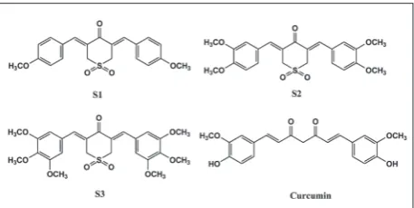
the first report investigating the effects of three cur-cumin analogs containing sulfone including (3Z,5Z)- 3,5-bis-(4-methoxybenzylidene)-dihydro-2H-thio-pyran-4-(3H)-one-1,1-di-oxide (S1) (Rovnyak et al., 1982), (3Z,5Z)-3,5-bis(3,4-dimethoxy-benzylidene) dihydro-2H-thiopyran-4(3H)-one-1,1-dioxide (S2) and (3Z,5Z)-3,5-bis-(3,4,5-trimethoxybenzyli-dene)-dihydro-2H-thiopyran-4(3H)-one-1,1-dioxide (S3) (Tan et al., 2014) (Fig. 1) against several cancer cell lines including prostate cancer PC-3 cells, colon can-cer HT-29 cells, pancreatic cancan-cer BxPC-3 cells and lung cancer H1299 cells.
We also employed a three dimensional (3D) cul-ture model to observe the morphology and effects of curcumin and curcumin analogs on prostate cancer PC-3 cells. To further investigate the mechanism of these compounds, we determined the effect of these compounds on the activity of nuclear factor kappa B (NF-κB) in PC-3 cells using a luciferase reporter assay, as well as the levels of phosphorylated protein kinase B (p-Akt), phosphorylated signal transducer and activator of transcription-3 (STAT3) (Tyr705 and Ser727) by Western blotting analysis.
materials anD methoDs cell culture and reagents
Human cancer PC-3, colon cancer HT-29, lung can-cer H1299 and pancreatic cancan-cer BxPC-3 cells were obtained from the American Type Culture Collection (ATCC, Rockville, MD, USA). The cell lines were all maintained in RPMI-1640 medium (Gibco, USA), which was supplemented with 10% fetal bovine se-rum (Gibco, Grand Island, NY, USA) and penicillin (100 units/mL)-streptomycin (100 μg/mL). The cells were incubated at 37ºC in a humidified atmosphere containing 5% CO2. Curcumin and curcumin analogs were dissolved in DMSO, and the final concentration of DMSO in all experiments was 0.1%. Primary anti-bodies included Actin (C-2: sc-8432, Santa Cruz Bio-technology), p-Akt antibody (Cat. #9271, Cell Signal-ing), anti-phospho-STAT3 (Tyr705) clone EP2147Y, rabbit monoclonal (Cat. #04-1059, Millipore) and
fig. 1. Structure of curcumin and curcumin analogs containing sulfone.
phospho-STAT3 (Ser727) antibodies (Cat. #9134, Cell Signaling). Secondary antibodies, all purchased from Santa Cruz Biotechnology, included goat anti-mouse IgG-HRP 2055 and goat anti-rabbit IgG-HRP sc-2004.
mtt assay
PC-3, HT-29, H1299 and BxPC-3 human cancer cells were seeded at a density of 2.5×10⁴ cells/mL (200 μL/ well) in 96-well microplates and incubated for 24 h at 37°C. The cells were treated with various concen-trations of different compounds. After 72 h of incu-bation, the medium was removed, and 3-[4,5-dime- thylthiazol-2-yl]-2,5-diphenyltetrazolium bromide (MTT) solution (5.5 mg/mL, 100 μL/well) was added to the wells. After 4 h of incubation, the MTT solu-tion was removed and 100 μL of DMSO was added to each well. Plates were then swirled gently to facilitate formazan crystal solubilization. Finally, the absor-bance was measured at 570 nm using a microplate reader (Tecan Infinite M200 Pro, Switzerland).
trypan blue assay
not absorb dye were counted as live cells. The cells, including dead and alive cells, were then examined under a light microscope (Nikon Optiphot, Japan).
three dimensional (3D) cell culture
PC-3 cells were mixed with Matrigel (Collaborative Research, Bedford, MA) on ice at a density of 0.5×10⁵ cells/mL The Matrigel containing PC-3 cells was then placed in a 12-well plate (1.0 mL/well) and incubated at 37°C for 2 h to let the Matrigel solidify. Afterward, RPMI-1640 medium was added to each well on top of the gel. The cells were incubated for 24 h and then treated with curcumin or curcumin analogs once every other day. At day 10, the 3D cultures were ex-amined under a microscope (Nikon Eclipse TE200, Japan) for the formation of tissue-like structures.
nf-κB dependent reporter gene expression assay
The NF-κB-luciferase construct (Lenti NF-κB Re-porter, Qiagen, Valencia, CA) was transfected into PC-3 cells by LipofectamineTM 2000 (Invitrogen Life Tech, Grand Island, NY) following the manufacturer’s instructions, and a single stable clone, PC-3/N, was used in the present study. The cells were seeded at a density of 2.5×10⁴ cells/mL of medium in 12-well plates (1.0 mL/well) and incubated for 24 h. The cells were then treated with the different concentrations of curcumin or curcumin analogs for 24 h, and the NF-κB luciferase activities were determined by luciferase reporter assay kits (E1500, Promega Madsion WI) ac-cording the manufacturer’s instructions. The protein concentrations of cell lysates were determined using a Bio-Rad protein assay kit (Bio-Rad, Hercules, CA, USA) according to the manufacturer’s instructions. The NF-κB transcriptional activities were normalized against protein concentrations and expressed as the percentage of luciferase activity relative to the control cells.
Western blotting
PC-3 cells were seeded at a density of 1.0×10⁵ cells/ mL in a 100-mm dish (10.0 mL/dish) and incubated
at 37°C for 24 h. The cells were treated with curcumin or curcumin analogs for 24 h. Thereafter, cells were washed with ice-cold PBS and lysed in 200 μL of ly-sis buffer (10 mM Tris-HCl, pH 7.4, 50 mM sodium chloride, 30 mM sodium pyrophosphate, 50 mM so-dium fluoride, 100 μM soso-dium orthovanadate, 2 mM iodoacetic acid, 5 mM zinc chloride, 1 mM phenyl-methylsulfonyl fluoride and 0.5% Triton X-100). The lysates were centrifuged at 13000×g for 15 min at 4°C. The protein concentration of whole cell lysates was de-termined with a Bio-Rad protein assay kit (Bio-Rad). Equal amounts (22.5 μg) of protein were subjected to sodium dodecyl sulfate polyacrylamide gel electro-phoresis (SDS-PAGE) and transferred onto polyvinyli-dene fluoride (PVDF) membrane. The membrane was washed three times with Tris-buffered saline (TBS) for 10 min and once with TBS containing 0.05% Tween 20 (TBST) for 10 min. The membrane was incubated in blocking buffer for 1 h. Then the membrane was cut and separately incubated at 4°C overnight with specific primary antibody against actin (C-2), p-Akt and p-STAT3 (Tyr705 and Ser727) (all primary anti-bodies were diluted 1:1000). Following removal of the primary antibody, the membranes were washed three times with TBS and once with TBST at room tempera-ture. The membranes were subjected to fluorochrome-conjugated secondary antibody at a dilution of 1:5000 for 1 h at room temperature without light. Membranes were washed four times with TBST. Immunoreactivity was detected using SuperSignal West Femto Luminol/ Enhancer solution (Thermo Scientific) and analyzed by Quantity One software (Bio-Rad). Relative quan-tification of protein markers compared to actin was analyzed using Image Studio Lite Ver 4.0 software.
statistical analysis
results
effects of curcumin and curcumin analogs on different human cancer cell lines
The effects of curcumin and its analogs (S1-S3) on the growth of PC-3, HT-29, H1299 and BxPC-3 cells were determined using MTT assay. The resulting IC50 values are presented in Table. 1. The IC50 values of S1-S3 ranged from 0.72 μM to 1.73 μM on PC-3 cells, 0.19 μM to 0.38 μM on HT-29 cells, 0.46 μM to 1.24 μM on H1299 cells and 0.29 μM to 1.01 μM on BxPC-3 cells. The results showed that S1-SBxPC-3 were approxi-mately 16- to 96-fold more active than curcumin. Both inhibition of proliferation and cytotoxicity can result in a decrease in the MTT assay. Therefore, we assessed the number of viable cells and dead cells using the trypan blue exclusion assay. Since our laboratory has an experimental system for the prostate cancer PC-3 cells, we used this cell line for further studies.
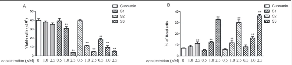
In experiments using the trypan blue exclusion assays, we found that treatment of PC-3 cells with S1-S3 decreased the number of viable cells in a con-centration-dependent manner (Fig. 2A). Treatment of PC-3 cells with the compounds also resulted in a concentration-dependent increase in the number of dead cells (Fig. 2B). All compounds were much more active than curcumin, and S3 showed the strongest activity.
effects of curcumin and curcumin analogs on pc-3 cells in 2D and 3D cultures
The 3D cell culture model was used to determine the effects of curcumin and curcumin analogs on the growth and formation of 3D structures of PC-3 cells. The morphology of PC-3 cells treated with curcumin or curcumin analogs in the 3D culture system were examined and compared with conventional 2D mono-layer culture. As shown in Fig. 3, S1-S3 showed strong inhibitory effects at a concentration of 1.0 μM (Fig. 3C, E and G) and 2.5 μM (Fig. 3D, F and H) in 2D culture, compared to control PC-3 cells or treatment with curcumin (Fig. 3A, B). As for the 3D culture, control PC-3 cells or treatment with curcumin formed a tissue-like morphology in extra cellular matrix gel (Fig. 4A, B), while treatment with S1-S3 at a concen-tration of 2.5 μM showed inhibitory effects on the growth of tissue-like structures (Fig. 4D, F and H). However, these compounds had no obvious effects at the concentration of 1.0 μM except for S3 (Fig. 4C, E and G).
table 1. Inhibitory effect of curcumin and curcumin analogs on the growth of different cells.
compounds ic50 (μm)
ht-29 h1299 pc-3 Bxpc-3
Curcumin S1 S2 S3
18.39±0.35 0.19±0.14** 0.38±0.15** 0.29±0.09**
19.87±0.94
1.24±0.08**
0.58±0.04** 0.46±0.01**
21.64±1.83
1.73±0.26**
0.85±0.10**
0.72±0.17**
18.25±1.27
1.01±0.11**
0.32±0.08**
0.29±0.09**
HT-29, H1299, PC-3 and BxPC-3 cells were seeded at a density of 2.5×10⁴
cells/mL in 96-well plates and incubated 24 h, respectively, after 72 h with various concentrations of test samples. MTT solution (100 μL, 5 mg/mL) was added to the wells and further incubated for 4 h at 37ºC. Thereafter, the medium was removed and DMSO was added. Measured with micro-plate reader at a wavelength of 570 nm. Data are shown as means±SEM of three independent experiments. Significantly different from the curcumin *p<0.05, **p<0.01.
effects of curcumin and curcumin analogs on nf-κB activation
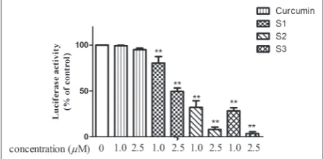
The effects of curcumin and curcumin analogs on activation of NF-κB were determined by a luciferase reporter gene expression assay. As shown in Fig. 5, treatments of PC-3 cells with curcumin analogs S1-S3 caused a significant inhibitory effect on the luciferase activity at the concentration of 1.0 μM, resulting in 19.41%, 67.87% and 71.57% inhibition of the lucifer-ase activity, respectively. With a higher concentration of 2.5 μM, S1-S3 caused 50.4%, 91.95% and 96.37%
in-hibitions of the luciferase activity, respectively. There were good correlations between the inhibition of NF-κB activity and cell growth inhibition of PC-3 cells at concentrations of both 1.0 μM and 2.5 μM.
effects of curcumin and curcumin analogs on levels of p-akt, and p-stat3 on pc-3 cells
Expressions of p-Akt, p-STAT3 were determined by the Western blotting analysis. In these experiments, PC-3 cells were treated with curcumin or curcumin an-alogs at a concentration of 1.0 μM for 24 h. The levels fig. 3. Morphology of PC-3 cells treated with curcumin or curcumin analogs in the 2D cells culture system.
PC-3 cells were treated with curcumin or curcumin analogs for 48 h, and then checked the morphology of the cultures in a microscope. (A) control; (B) curcumin; (C) s1-1.0 μM; (D) s1-2.5 μM; (E) s2-1.0 μM; (F) s2-2.5 μM; (G) s3-1.0 μM; (H) s3-2.5 μM.
of p-Akt, p-STAT3 (Tyr705 and Ser727) were analyzed by optical density measurements and normalized for actin. As shown in Fig. 6, the three curcumin analogs all decreased the expression of p-STAT3 (Tyr705) but not p-STAT3 (Ser727), while only S1 and S2 decreased the expression of p-Akt on PC-3 cells.
Discussion
Earlier studies synthesized and characterized cur-cumin analogs containing sulfone s1-s3 (Rovnyak et al., 1982; Tan et al., 2014). Although a recent study
fig. 5. Effects of curcumin and curcumin analogs on NF-κB tran-scriptional activity in PC-3/N cells. PC-3/N cells were treated with curcumin or curcumin analogs for 24 h. NF-κB transcriptional activity was determined by a luciferase reporter gene assay. Data are shown as means±SEM of three independent experiments. Significance of results compared to the control groups *p<0.05, **p<0.01.
fig. 6. Effect of curcumin or curcumin analogs on Ser727) pro-tein expression in p-Akt, p-Stat3 (Tyr705 and Ser727) PC-3 cells. PC-3 cells were treated with curcumin or curcumin analogs for 24 h. The levels of p-Akt and p-Stat3 were determined by Western blot analysis.
showed that this class of curcumin analogs induced apoptotic cell death in acute promyelocytic leukemic cells, their activities on prostate and other cancer cells were not reported. In the present study, we investi-gated the effects of three curcumin analogs (s1-s3; Fig. 1) against human prostate cancer PC-3, colon cancer HT-29, lung cancer H1299 and pancreatic cancer BxPC-3 cells. Results from the MTT assay indicated that these compounds were approximately 16- to 96-fold more active than curcumin. Further studies in PC-3 cells using the trypan blue exclusion assay showed that S1-S3decreased the number of viable cells and increased the number of dead cells. This result indicated that these compounds had both growth inhibition and cytotoxic effects on the cells. It is reasonable to assume that the effects of these com-pounds on PC-3, HT-29, H1299 and BxPC-3 cells as determined by the MTT assay were from both growth inhibition and cytotoxicity. Future studies are needed to determine the cytotoxicity of these compounds in normal cells and animals.
Compared to a conventional 2D monolayer cell culture, the 3D culture system had advantages in that it can mimic the structural architecture and differ-ent function of the tumor tissues (Weiswald et al., 2015; Rothan et al., 2014). It is well known that cell-cell interactions and cell-cell-matrix interactions within a 3D environment are important to the physiological function and response of cancer cells to anticancer agents (Liu et al., 2011; Tsunoda et al., 2014; Akeda et al., 2009). In the present study, we determined the effects of curcumin and S1-S3 on PC-3 cells both in 2D and 3D cell cultures. We found that PC-3 cells formed 3D tissue-like morphology in extra cellular matrix Matrigel. Compared to the control group, the tissue-like structure did not change much when the cells treated with a single compound at the concentra-tion of 1.0 μM, except for S3. When treated with S1-S3 at a higher concentration (2.5 μM), the tissue-like structures dispersed. This result indicates that S1-S3 may inhibit the formation of tumor structures and have useful anticancer activities.
inflamma-tion (Terlizzi et al., 2014; Jing et al., 2014; Marusawa et al., 2014). NF-κB not only regulates the expression of most anti-apoptotic gene products associated with the survival of the tumor, but also regulates the gene products linked with the proliferation, invasion, an-giogenesis and metastasis of tumors (Liu et al., 2011; Zhang et al., 2014). Extensive research showed that curcumin exerts a wide range of antitumor effects through modulation of significant signaling pathways, including the NF-κB pathway (Oyagbemi et al., 2009; Chen et al., 2008; Kunnumakkara et al., 2008). To fur-ther determine the mechanism of growth inhibition on PC-3 cells, we also investigated the effect of S1-S3 on the activation of NF-κB through an NF-κB-luciferase reporter gene expression assay. We found that treat-ment with S1-S3 strongly inhibited NF-κB activity at concentrations of both 1.0 μM and 2.5 μM in compari-son to curcumin. This result suggests that inhibition of NF-κB activity may contribute to the inhibitory effect on cell growth induced by S1-S3.
Previous studies have demonstrated that STAT3 activation was associated with cell proliferation, inhi-bition of apoptosis and cellular transformation (Ka-mran et al., 2013; Yu et al., 2014; Kluge et al., 2011), and Akt was shown to play a major role in a variety of tumors because Akt activation not only inhibits apoptosis of cells and is thus involved in cell survival pathways, but induces protein synthesis pathways (Li-ang et al., 2015; Mollazadeh et al., 2015; Almhanna et al., 2011). In addition, curcumin can suppress NF-κB activation by an Akt-dependent or Akt-independent inhibition of IKK (Lin et al., 2007; Aggarwal et al., 2006). In the present study, our results indicated that the effects on PC-3 cells of curcumin analogs contain-ing sulfone were associated with a decrease in p-Akt and p-STAT3. S1-S3 can decrease the expression of p-STAT3 (Tyr705) but not p-STAT3 (Ser727). In ad-dition, S1 and S2 can decrease the expression of p-Akt, but S3 had no effects. Possibly S3 exerts its antitumor effects through other pathways.
In summary, we found that curcumin analogs S1-S3 containing sulfone strongly inhibited the growth of human prostate cancer PC-3, colon cancer HT-29, lung cancer H1299 and pancreatic cancer BxPC-3
cells. In addition, the compounds S1-S3 had strong effects on growth inhibition of PC-3 cells not only in 2D culture but also in 3D culture system. We further found that the potent effects of S1-S3 on PC-3 cells were associated with their inhibition of NF-κB tran-scriptional activity, indicating that the NF-κB pathway may be involved in the growth inhibition induced by these compounds. Moreover, our results showed that S1-S3 can decrease the level of p-STAT3 (Tyr705) but not p-STAT3 (Ser727). This result suggests the phos-phorylation in tyrosine 705 is important for the effect of S1-S3 in PC-3 cells. Decreases in the amount of p-Akt in PC-3 cells treated with S1 and S2 indicate that suppression of Akt was involved in the effects of S1 and S2 but not in S3. Our studies indicate that these curcumin analogs warrant further in vivo studies for anticancer activities in suitable animal models.
acknowledgments: The present studywas supported by grants from projects of Guangzhou Science & Technology Interna-tional Collaboration (2013J4500014), Guangdong Province (2012B091000170& 2012B091100342) and Nation (21272043), and a Guangdong Province Leadership Grant, the Rutgers Cancer Institute of New Jersey (CCSG P30-CA072720 RSD).
authors’ contribution: Conception and design: XZ, ZD, QZ; experiments: DL, YL, HW, CZ; analysis and interpretation: QZ, HH, YH, data collection: CZ, XC; writing the article: QZ, XZ, ZD.
conflict of interest disclosure: The authors declare that there is no conflict of interest regarding the publication of this paper.
references
Aggarwal, B.B., Gupta, S.C. and B. Sung (2013). Curcumin: an
orally bioavailable blocker of TNF and other pro-inflam-matory biomarkers. Br. J. Pharmacol. 169, 1672-1692.
Aggarwal, B.B., Kumar, A. and A.C. Bharti (2003). Anticancer
potential of curcumin: preclinical and clinical studies.
Anti-cancer Res.23, 363-398.
Aggarwal, B.B. and S. Shishodia (2006). Molecular targets of
dietary agents for prevention and therapy of cancer.
Bio-chem. Pharmacol. 71, 1397-1421.
Akeda, K., Nishimura, A., Satonaka, H., Shintani, K., Kusuzaki,
K., Matsumine, A., Kasai, Y., Masuda, K. and A. Uchida
(2009). Three-dimensional alginate spheroid culture sys-tem of murine osteosarcoma. Oncol. Rep. 22, 997-1003.
Almhanna, K., Strosberg, J. and M. Malafa (2011). Targeting AKT
Chen, A. and S. Zheng (2008). Curcumin inhibits connective tissue growth factor gene expression in activated hepatic stellate cells in vitro by blocking NF-kappaB and ERK sig-nalling. Br. J. Pharmacol. 153, 557-567.
Chen, M.B., Wu, X.Y., Tao, G.Q., Liu, C.Y., Chen, J., Wang, L.Q.
and P.H. Lu (2012). Perifosine sensitizes curcumin-induced
anti-colorectal cancer effects by targeting multiple signal-ing pathways both in vivo and in vitro. Int. J. Cancer.131,
2487-2498.
Chuah, A.M., Jacob, B., Jie, Z., Ramesh, S., Mandal, S., Puthan, J.K., Deshpande, P., Vaidyanathan, W., Gelling, R.W., Patel,
G., Das, T. and S. Shreeram (2014). Enhanced
bioavailabil-ity and bioefficacy of an amorphous solid dispersion of curcumin. Food Chem.156, 227-233.
Jing, H. and S. Lee (2014). NF-κB in cellular senescence and
can-cer treatment. Mol Cell., 37, 189-195.
Kamran, M.Z., Patil, P. and R.P. Gude (2013). Role of STAT3 in
cancer metastasis and translational advances. Biomed. Res. Int. 2013, 421821.
Kluge, A., Dabir, S., Vlassenbroeck, I., Eisenberg, R. and A. Dowlati
(2011). Protein inhibitor of activated STAT3 expression in lung cancer. Mol. Oncol. 5, 256-264.
Kunnumakkara, A.B., Anand, P. and B.B. Aggarwal (2008).
Cur-cumin inhibits proliferation, invasion, angiogenesis and metastasis of different cancers through interaction with multiple cell signaling proteins. Cancer Lett. 269, 199-225.
Liang, F., Yue, J., Wang, J., Zhang, L., Fan, R., Zhang, H. and Q.
Zhang (2015). GPCR48/LGR4 promotes tumorigenesis
of prostate cancer via PI3K/Akt signaling pathway. Med. Oncol. 32, 486.
Lin, Y.G., Kunnumakkara, A.B., Nair, A., Merritt, W.M., Han, L.Y., Armaiz-Pena, G.N., Kamat, A.A., Spannuth, W.A.,
Gerhen-son, D.M., Lutgendorf, S.K., Aggarwal, B.B. and A.K. Sood
(2007). Curcumin inhibits tumor growth and angiogenesis in ovarian carcinoma by targeting the nuclear factor-kap-paB pathway. Clin. Cancer Res. 13, 3423-3430.
Liu, L., Sun, B., Pedersen, J.N., Aw Yong, K.M., Getzenberg, R.H.,
Stone, H.A. and R.H. Austin (2011). Probing the
invasive-ness of prostate cancer cells in a 3D microfabricated land-scape. Proc. Nati. Acad. Sci. U.S.A. 108, 6853-6586.
Liu, S., Wang, Z., Hu, Z., Zeng, X., Li, Y., Su, Y., Zhang, C. and
Z. Ye (2011). Anti-tumor activity of curcumin against
androgen-independent prostate cancer cells via inhibition of NF-κB and AP-1 pathway in vitro. J. Huazhong. Univ. Sci.
Technolog. Med. Sci. 31, 530-534.
Li, Y. and T. Zhang (2014). Targeting cancer stem cells by
cur-cumin and clinical applications. Cancer Lett. 346, 197-205.
Marusawa, H. and B.J. Jenkins (2014). Inflammation and
gastro-intestinal cancer: an overview. Cancer Lett. 345, 153-156.
Mehta, H.J., Patel, V. and R.T.Sadikot (2014). Curcumin and lung
cancer-a review. Target Oncol. 9, 295-310.
Moghadamtousi, S.Z., Kadir, H.A., Hassandarvish, P., Tajik, H.,
Abubakar, S. and K. Zandi (2014). A review on
antibacte-rial, antiviral, and antifungal activity of curcumin. Biomed.
Res. Int.2014, 186864.
Mollazadeh, S., Bazzaz, B. and M.Kerachian (2015). Role of
apop-tosis in pathogenesis and treatment of bone-related disease.
J. Orthop. Surq. Res. 10, 15.
Oyagbemi, A.A., Saba, A.B. and A.O. Ibraheem (2009). Curcumin:
from food spice to cancer prevention. Asian Pac. J. Cancer. Prev. 10, 963-967.
Prasad, S., Gupta, S.C., Tyaqi, A.K. and B.B. Aggarwal (2014).
Curcumin, a component of golden spice: from bedside to bench and back. Biotechnol. Adv. 32, 1053-1064.
Ramsewak, R.S., DeWitt, D.L. and M.G. Nair (2000). Cytotoxicity,
antioxidant and anti-inflammatory activities of curcumin I-III from Curcuma longa. Phytomedicine.7, 303-308.
Rothan, H.A., Djordjevic, I., Bahrani, H., Paydar, M., Ibrahim, F.,
Abd Rahmanh, N. and R. Yusof (2014). Three-dimensional
culture environment increases the efficacy of platelet rich plasma releasate in promoting skin fibroblast differentia-tion and extracellular matrix formadifferentia-tion. Int. J. Med. Sci.
11, 1029-1038.
Rovnyak, G.C., Millonig, R.C., Schwartz. J. and V. Shu (1982).
Synthesis and inflammatory activity of hexahydrothiopy-rano [4,3-c]pyrazoles and related analogs. J. Med. Chem.
25, 1482-1488.
Ruby, A.J., Kuttan, G., Babu, K.D., Rajasekharan, K.N. and R.
Kut-tan (1995). Anti-tumour and antioxidant activity of natural curcuminoids. Cancer Lett.94, 79-83.
Tan, K.L., Ali, A., Du, Y., Fu, H., Jin, H.X., Chin, T.M., Khan, M. and M.L.Go (2014). Synthesis and evaluation of bis-benzylidenedioxotetrahydrothiopranones as activators of endoplasmic reticulum (ER) stress signaling pathways and apoptotic cell death in acute promyelocytic leukemic cells.
J. Med. Chem.57, 5904-5918.
Terlizzi, M., Casolaro, V. and A. Pinto (2014). Sorrentino R.
Inflammasome: Cancer’s friend or foe? Pharmacol. Ther.
143, 24-33.
Trujillo, J., Chirino, Y.I., Molina-Jijón, E., Andérica-Romero,
A.C., Tapia, E. and J. Pedraza-Chaverrí (2013).
Renopro-tective effect of the antioxidant curcumin: Recent findings.
Redox Biol., 1, 448-456.
Tsunoda, T., Ishikura, S., Doi, K., Matsuzaki, H., Iwaihara, Y. and
S. Shirasawa (2014). Resveratrol induces Iuminal apoptosis
of human colorectal cancer HCT116 cells in three-dimen-sional culture. Anticancer. Res. 34, 4551-4555.
Tuorkey, M.J. (2014). Curcumin a potent cancer preventive agent:
Mechanisms of cancer cell killing. Interv Med. Appl. Sci.
6, 139-146.
Weiswald, L.B., Bellet, D. and V. Dangles-Marie (2015). Spherical
cancer models in tumor biology. Neoplasia. 17, 1-15.
Wei, X.C., Du, Z.Y., Cui, X.X., Verano, M., Mo, R.Q., Tang, Z.K.,
Conney, A.H., Zheng, X. and K. Zhang (2012). Effects of
cyclohexanone analogs of curcumin on growth, apoptosis and NF-κB activity in human prostate cancer cells. Oncol.
Lett.4, 279-284.
Yu, H., Lee, H., Herrmann, A., Buettner, R. and R. Jove (2014).
Zhang, W., Wang, F., Xu, P., Miao, C., Zeng, X., Cui, X., Lu, C., Xie,
H., Yin, H., Chen, F., Ma, J., Gao, S. and Z. Fu (2014).
Per-fluorooctanoic acid stimulates breast cancer cells invasion and up-regulates matrix metalloproteinase-2/9 expression mediated by activating NF-κB. Toxicol. Lett. 229, 118-125.
Zhang, Y.L., Zhao, L.P., Wu, J.Z., Jiang, X., Dong, L.L., Xu, F.L.,
Zou, P., Dai, Y.R., Shan, X.O., Yang, S.L. and G. Liang
(2014). Synthesis and evaluation of a series of novel
asym-metrical curcumin analogs for the treatment of inflamma-tory. Molecules. 19, 7287-7307.
Zhou, D.Y., Zhang, K., Conney, A.H., Ding, N., Cui, X.X., Wang,
H., Verano, M., Zhao, S.Q., Zheng, X. and Z.Y. Du (2013).