699
© 2017 by the Serbian Biological Society How to cite this article: Marjanović Vićentić J, Schwirtlich M, Kovačević-Grujičić N, Stevanović M, Drakulić D. All-trans retinoic acid influences viability, migration and adhesion of U251 glioblastoma cells. Arch Biol Sci. 2017;69(4):699-706.
All-trans retinoic acid influences viability, migration and adhesion of U251 glioblastoma
cells
Jelena Marjanović Vićentić1, Marija Schwirtlich1, Nataša Kovačević-Grujičić1, Milena Stevanović1,2,3 and
Danijela Drakulić1,*
1Institute of Molecular Genetics and Genetic Engineering, University of Belgrade, Vojvode Stepe 444a, PO BOX 23, 11010 Belgrade, Serbia
1University of Belgrade, Faculty of Biology, Studentski trg 16, 11000 Belgrade, Serbia 2Serbian Academy of Sciences and Arts, Knez Mihailova 35, 11001 Belgrade, Serbia
*Corresponding author: [email protected]
Received: March 27, 2017; Revised: May 23, 2017; Accepted: June 2, 2017; Published online: June 12, 2017
Abstract: Glioblastoma (GBM) is one of the most aggressive and deadly forms of cancer. Literature data reveals that all-trans retinoic acid (ATRA) has anticancer effects on different types of tumor cells. However, data about the effects of ATRA on glioblastoma cells are contradictory. In this study, we examined whether ATRA treatment affects features of human glioblastoma U251 cells. To that end, the cells were treated with different concentrations of ATRA. Results obtained by MTT and the crystal violet assays imply that ATRA affected the viability of U251 glioblastoma cells in a dose- and time-dependent manner. Fluorescence staining of microtubule cytoskeleton protein α-tubulin revealed that ATRA induced changes in cell morphology. Using semi-quantitative RT-PCR
we found that the expression of SOX3 and GFAP genes, as markers of neural differentiation,was not changed
upon ATRA treatment. Thus, the observed changes in cell morphology after ATRA treatment are not associated with neural differentiation of U251 glioblastoma cells. The scratch-wound healing assay revealed that ATRA changed the mode of U251 cell migration from collective to single cell motility. The cell-matrix adhesion assay demonstrated that the pharmacologically relevant concentration of ATRA lowered the cell-matrix adhesion capability of U251 cells. In conclusion, our results imply that further studies are needed before ATRA could be considered for the treatment of glioblastoma.
Key words: glioblastoma; ATRA; differentiation; viability; cell migration
INTRODUCTION
Gliomas are the most common type of primary brain tumors in humans [1]. Grade IV of glioma tumors, glioblastoma (GBM), is one of the most aggressive and deadly forms of cancer with a median survival of 15 months despite intensive therapeutic strategies which include surgical resection combined with radiotherapy and temozolomide chemotherapy [2-4]. Therefore, the identification of more effective treatment strategies for patients with GBM is required. Some of the new therapeutic approaches are focused on targeting the main features of human malignant glioblastoma cells,
such as uncontrolled proliferation, migration and in-vasion, resistance to apoptosis, lack of differentiation and vigorous angiogenesis [5-7].
of tumors, such as acute promyelocytic leukemia [11], breast [12], prostate [13], colon [14] and pancreatic cancer [15]. On the other hand, it was demonstrated that ATRA can promote the malignant potential of mouse mammary tumor cells [16], suggesting that its effect could be cell-context dependent.
Literature data regarding the effects of ATRA treat-ment on the malignant characteristics of glioblastoma cells are contradictory [9,17-19]. With this in mind, the aim of this study was to analyze the effects of ATRA treatment on the properties of U251 glioblastoma cells that are widely used as a GBM model system. We dem-onstrated that ATRA treatment reduced cell viability, induced morphological changes and affected migration and cell-matrix adhesion of U251 cells.
MATERIALS AND METHODS
Cell culture and treatments
The human glioblastoma U251 cell line was maintained in Dulbecco’s Modified Eagle’s medium (DMEM) supplemented with 10% fetal bovine serum (FBS), 2 mmol/L L-glutamine and 1% nonessential amino acids
(all from Invitrogen, USA) at 37°C in 10% CO2. ATRA
(Sigma-Aldrich, USA) was dissolved in dimethyl sulf-oxide (DMSO) to prepare a stock solution, and the cells were treated with different concentrations of ATRA (1, 5, 10, 20, 40 and 60 μM) for a period of 3 or 5 days. Control cells and all ATRA-treated cells were grown in medium that contained the same volume of DMSO that was used for the treatment with 60 μM ATRA. The DMSO concentration in the assay did not exceed 0.6%.
Cell viability assay
Cell viability was determined by 3-(4,5-dimethylthia-zol-2-yl)-2,5- 4 diphenyltetrazolium bromide (MTT) (indicating the number of metabolically active cells) and crystal violet (CV) (indicating the total num-ber of live adherent cells) colorimetric assays. U251 cells were cultured in 96-well plates at a density of
2x10³ cells/wellfor 3-day treatments or at a density of
1x10³ cells/wellfor 5-day treatments, and treated with
DMSO (control) or different concentrations of ATRA (1, 5, 10, 20, 40 and 60 μM).
For the MTT assay, at the end of treatment MTT solution was added to cell cultures at a final concen-tration of 1 mg/mL and the cells were incubated for an additional hour at 37°C. Subsequently, the medium was removed and the cells were lysed in DMSO. The conversion of MTT to formazan by metabolically vi-able cells was monitored by a microplate reader (Infi-nite 200 PRO; Tecan, Austria) at a wavelength of 550 nm. The experiment was performed in triplicate and repeated independently three times.
For the CV assay, at the end of treatment the cells were washed with PBS, fixed for 10 min with 4% para-formaldehyde, stained with 2% crystal violet-PBS for 15 min at room temperature (RT), washed four times in a stream of tap water and air dried. The absorbance of dye dissolved in 33% acetic acid was measured in a microplate reader Infinite 200 PRO at 550 nm. The experiment was performed in triplicate and repeated independently three times.
The results of MTT and CV assays are presented as the percentage of the values for control cells that was arbitrarily set to 100%.
Immunocytochemistry
Images were taken by a Leica TCS SP8 confocal mi-croscope applying the Leica Microsystems LAS AF-TCS SP8 software (Leica Microsystems, Germany). Reverse transcriptase (RT) PCR analysis
The total RNA from control and U251 cells treated with different concentrations of ATRA (10, 20, 40 μM) for 5 days was isolated using the TRI Reagent (Ambion, USA) according to the manufacturer’s in-structions. Isolated RNA was treated with DNase I using the DNA-free™ kit (Ambion) and subjected to cDNA synthesis. One μg of the total RNA was reversely transcribed using the High Capacity cDNA Reverse Transcription Kit (Applied Biosystems, USA)
accord-ing to the manufacturer’s protocol. SOX3 (SRY (sex
determining region Y)-box 3) was amplified from the synthesized cDNAs with primers: 5’-CAC GGG TCC TCC GGG TTG CGA GGG GCG GAC C-3’ (forward) and 5’-TGG GGA ACA AGG GTG GAC GAG C-3’
(reverse); glial fibrillary acidic protein (GFAP) with
primers: 5’ –GCA GAG ATG ATG GAG CTC AAT GAC C- 3’ (forward) and 5’-GTT TCA TCC TGG AGC TTC TGC CTC A -3’(reverse) [20] and
glycer-aldehyde 3-phosphate dehydrogenase (GAPDH) with
primers 5’-GCC TCA AGA TCA TCA GCA ATG C-3’ (forward) and 5’-CCA CGA TAC CAA AGT TGT CAT GG-3’ (reverse) [21]. The expression of GAPDH was used to normalize the levels of the total RNA used in the assays. PCR reactions were performed in 20-μL reactions using the Kapa 2G Fast HotStart Ready Mix
(Kapa Biosystems, USA). For GFAP and GAPDH
am-plification, samples were denatured for 2 min at 95°C and then cycled at 95°C for 15 s, 60°C for 15 s and 72°C for 15 s for 35 cycles, with reaction aliquots taken at 30
and 35 cycles. For SOX3 amplification, samples were
denatured for 5 min at 95°C and then cycled at 95°C for 30 s, 68°C for 30 s and 72°C for 30 s for 35 cycles, with reaction aliquots taken at 30 and 35 cycles.
Scratch-wound healing assay
U251 cells were treated with DMSO or various con-centrations of ATRA (10, 20, 40 μM) for 5 days. Upon treatment, the cell monolayer was scratched with a 200 µL tip. Floating cells were washed and cells were incubated in a fresh growing medium containing DMSO or ATRA. Cell migration was monitored with
the DM IL LED Inverted Microscope (Leica) 7 h af-ter the wound was made. The mode of cell migration into the gap was analyzed by counting single cells in 3-4 different parts of the wounded area from three independent experiments.
Cell-matrix adhesion assay
The adhesion ability of control cells and cells treated with different concentrations of ATRA (10, 20, 40 μM) for 5 days was studied using 96-well plates coated with Matrigel (Becton Dickerson, NJ, USA). Bind-ing to nonspecific adhesive surfaces was blocked by 2% BSA (Sigma-Aldrich) in DMEM for 1 h at 37°C.
Upon treatment, 0.5 × 10⁵ cells/well in DMEM
supple-mented with 0.1% BSA were seeded in 96-well plates. After 30 min, the cells were gently washed three times with 1xPBS and the number of attached cells was de-termined using the MTT assay. The tests were done in triplicate and repeated in 3 independent experiments.
Statistical analysis
The results were analyzed using Student’s t test. Values
of p<0.05 were considered significant. This analysis
was carried out using IBM SPSS Statistics Version 20 program.
RESULTS
The effect of ATRA on U251 glioblastoma cell viability
pronounced after longer exposure, all further analyses were performed following 5 days of treatment using 10, 20 and 40 μM concentrations.
The effect of ATRA on the morphology of U251 glioblastoma cells
The morphology of the control and ATRA-treated U251 glioblastoma cells was analyzed by fluorescence staining of microtubule cytoskeleton protein α-tubulin and nuclear/chromosome counterstaining with DAPI. Changes in cell morphology were induced with all tested ATRA concentrations (Fig. 2A). In cultures exposed to 10, 20 and 40 μM ATRA most of the cells had long, thin branching processes, while the control U251 cells were mainly round, with small cell bodies and short processes (Fig. 2A).
The effect of ATRA on neural differentiation of U251 glioblastoma cells
Bearing in mind that alternations in cell morphol-ogy could be associated with differentiation and that
ATRA could induce astrocytic differen-tiation of glioblastoma cells, our next aim was to analyze whether the treat-ment with ATRA induces neural differ-entiation of U251 glioblastoma cells. To that end we analyzed the expression of markers of neural differentiation after 5-day treatments with 10, 20 and 40 μM ATRA (Fig. 2B). Using semi-quantita-tive RT-PCR analysis we did not detect statistically significant changes in the
level of SOX3 and GFAP expression
af-ter 5 days of ATRA treatment (Fig. 2B). The presented results suggest that the observed changes in cell morphology after ATRA treatment were not associ-ated with neural differentiation of U251 glioblastoma cells.
The effect of ATRA on cell migration of U251 glioblastoma cells
To analyze whether ATRA treatment influences the mi-gration capability of U251 glioblastoma cells we per-formed the scratch wound healing assay. At the end of the treatment, the migration of cells into the wounded area was monitored (Fig. 3A). The ATRA treatment af-fected the mode of cell migration. Namely, we detected about two times more single cells in the wounded area of treated cells compared to the control (Fig. 3B). Ad-ditionally, we did not detect differences in the number of single cells in the wounded area between cells treated with different concentrations of ATRA.
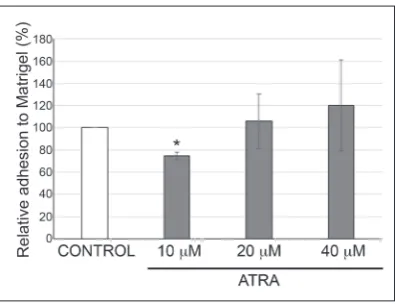
The effect of ATRA on cell-matrix adhesion of U251 glioblastoma cells
Considering that the ability of cancer cells to form me-tastasis is influenced by their capability to adhere to and degrade the extracellular matrix (ECM), our next goal was to analyze if ATRA treatment changes the cell-matrix adhesion capacity of U251 glioblastoma cells. The 5-day treatment of U251 cells with 10 μM ATRA decreased the cell-matrix adhesion ability of U251 cells by approximately 25% compared to control cells (Fig. 4). On the other hand, treatment with 20 and 40 μM ATRA did not affect the cell-matrix adhe-sion ability of U251 cells compared to their DMSO-treated counterparts.
DISCUSSION
Glioblastoma is one of the most common, highly ma-lignant and most therapy-resistant human tumors, with high morbidity and mortality [7]. One of the promising strategies for cancer therapy is treatment with ATRA [22]. The effect of ATRA on glioblasto-mas and heterogeneous responses depending on the
concentration of ATRA, exposure times and types of glioblastoma cells have been noted [9,17-19,23]. Some studies have demonstrated that ATRA inhibits prolif-eration, migration and invasion and induces differen-tiation and apoptosis of glioblastoma cells [9,19,23]. On the other hand, differences in response to ATRA between long-term glioblastoma cell lines and pri-mary cultures isolated from glioblastoma tumors were Fig. 2. ATRA treatment changed the morphology, but did not induce neural
dif-ferentiation of U251 glioblastoma cells. A – Changes in cell morphology were analyzed by fluorescence staining of microtubule cytoskeleton protein α-tubulin. Arrows indicate thin branching processes; scale bar=20 μm. B – Expression of
SOX3 and GFAP was examined by semi-quantitative RT-PCR analysis. Three independent experiments were performed and representative images are shown. The bands were digitalized, quantified with ImageJ software and normalized for GAPDH values. The relative gene expression was calculated as the fold expression compared to control cells which were set as 100%. Data from three independent experiments are presented in a histogram as the means±SD; *p<0.05, compared to control cells.
Fig. 3. ATRA changed the mode of U251 cell migra-tion from collective to single cell motility. A – Migra-tion capability was analyzed by the scratch wound healing assay. Confluent cell monolayers of U251 cells treated with DMSO or different concentrations of ATRA were scratched, and wound recovery was monitored 7 h after the wound was made. Represen-tative phase contrast images of 3 independent wound closure experiments are shown; scale bar=50 μm.
reported [17]. In addition, it was shown that ATRA treatment elevated the transcription of a group of cancer-associated genes in glioblastoma cells [18].
In this study, we demonstrated that ATRA af-fected the viability of U251 cells. Additionally, our results strongly suggest that only high concentrations of ATRA (10, 20, 40 and 60 μM) might significantly inhibit the viability of U251 glioblastoma cells. Con-trary to our results, Lu et al. [23] showed that treat-ment with a lower dose of ATRA (2.5 μM) reduced the growth of U251 cells by approximately 60%. A possible explanation for the observed differences be-tween our results and theirs could be variations in genotype, phenotype and growth characteristics of different subclones of U251 cells, as was observed re-cently by Torsvik et al. [24]. Furthermore, we found that among the pharmacologic doses of retinoic acid (1-10 μM) [25], only 10 μM ATRA affected the vi-ability of U251 cells, which is in accordance with the results of Pijuan-Thompson et al. [25], who showed that treatment with 10 μM ATRA decreased the pro-liferation of U251 cells.
Similar to the results obtained on U87 glioblas-toma cells [26], we observed that ATRA affected U251 cell viability in a dose- and time-dependent manner. Interestingly, we noted that after 3 days of treatment,
1 μM ATRA slightly increased the total number of live cells (by about 10%). This is in accordance with the re-sults obtained on GL-15 glioblastoma cells where low concentrations of retinoic acid (0.1-1 μM) increased the proliferation of cells, while higher concentrations (5-10 μM) reduced the cell proliferation rate [27].
Induction of differentiation in cancer cells in or-der to eliminate tumor phenotypes is the main goal of differentiation therapy of cancer [22]. It was shown that ATRA could induce astrocytic differentiation of T98G and U87MG glioblastoma cells [7]. Our re-sults showed that U251 cells undergo morphological changes after ATRA treatment. Although association between morphological changes and the induction of neural differentiation has been demonstrated in sev-eral glioma cell lines [7,28-30], we did not detect that U251 cells undergo neural differentiation. A possible explanation is that neural differentiation of U251 cells could be induced only with concentrations of ATRA that are smaller than 10 μM, as previously described for glioma stem/progenitor cells [31].
Metastasis is a complex multistep process that includes the ability of malignant cells to migrate and adhere [32,33]. Data about the effect of ATRA on the migration ability of glioblastoma cells are sparse. We observed that ATRA changed the mode of U251 cell migration from collective to single cell motility. Since it has been shown that glioma cells migrate into dis-tant parts of normal brain tissue as single cells [34], we postulated that ATRA treatment might enhance the infiltration of tumor cells into normal brain tissue.
Adhesion of cancer cells to the ECM represents another important step in the metastasis process and is critical for enabling metastatic spread [35,36]. It has been demonstrated that impaired cell-cell adhesion re-sults in an increased metastatic potential of cancer cells (reviewed in [37,38]). However, data regarding the im-pact of cell-matrix adhesion on the metastatic potential of cancer cells are different. Some authors have indi-cated that impaired cell-matrix adhesion decreases the metastatic potential of cancer cells [38,39]. On the other side, it was suggested that a reduction in cell-matrix adhesion is accompanied by an increased metastatic potential of cells [37,40,41]. Our results revealed that only the pharmacologically relevant concentration of ATRA (10 µM) after a 5-day treatment significantly Fig. 4. Pharmacologically relevant concentration of
decreased the cell-matrix adhesion capability of U251 cells. The anti-adhesive influence of ATRA was also observed in cervical SiHa cells [39] and A375 human melanoma cells [42]. Further investigations, including the determination of the strength of cell-matrix adhe-sion, the ability of matrix-remodelling, the invasion potential and the rate of cell spreading, should clarify whether the impaired cell-matrix adhesion capability of ATRA-treated U251 cells is accompanied by a de-creased or an inde-creased metastatic potential.
In summary, here we studied the effects of ATRA treatment on features of human glioblastoma U251 cells. Our results demonstrate that treatment with ATRA might reduce the viability and cell-matrix adhesion capability of U251 glioblastoma cells and increase the number of single cells in the wounded area. Moreover, despite visible changes in cell mor-phology, the expression of neural-specific markers was not changed. Based on these results, we conclude that further studies are warranted before ATRA could be considered as a therapy for glioblastoma.
Acknowledgments: This work was supported by the Ministry of Education, Science and Technological Development, Republic of Serbia, Grant No: 173051.
Conflict of interest disclosure: The authors declare that there is no conflict of interest.
REFERENCES
1. Adamski V, Schmitt AD, Fluh C, Synowitz M, Hattermann K, Held-Feindt J. Isolation and characterization of fast migrating human glioma cells in the progression of malignant gliomas. Oncol Res. 2017;25(3):341-53.
2. Rao SS, Lannutti JJ, Viapiano MS, Sarkar A, Winter JO.
Toward 3D biomimetic models to understand the behav-ior of glioblastoma multiforme cells. Tissue Eng Part B Rev. 2014;20:314-27.
3. Sottoriva A, Spiteri I, Piccirillo SG, Touloumis A, Collins VP, Marioni JC, Curtis C, Watts C, Tavare S. Intratumor hetero-geneity in human glioblastoma reflects cancer evolutionary dynamics. Proc Natl Acad Sci U S A. 2013;110:4009-14. 4. Stupp R, Mason WP, van den Bent MJ, Weller M, Fisher B,
Taphoorn MJ, Belanger K, Brandes AA, Marosi C, Bogdahn U, Curschmann J, Janzer RC, Ludwin SK, Gorlia T, Allgeier A, Lacombe D, Cairncross JG, Eisenhauer E, Mirimanoff RO, European Organisation for R, Treatment of Cancer Brain T, Radiotherapy G, National Cancer Institute of Canada Clinical Trials G. Radiotherapy plus concomitant and adjuvant temo-zolomide for glioblastoma. N Engl J Med. 2005;352:987-96.
5. Castellino RC, Durden DL. Mechanisms of disease: the PI3K-Akt-PTEN signaling node--an intercept point for the con-trol of angiogenesis in brain tumors. Nat Clin Pract Neurol. 2007;3:682-93.
6. Fathima Hurmath K, Ramaswamy P, Nandakumar DN.
IL-1beta microenvironment promotes proliferation, migra-tion, and invasion of human glioma cells. Cell Biol Int. 2014;38:1415-22.
7. Haque A, Das A, Hajiaghamohseni LM, Younger A, Banik NL, Ray SK. Induction of apoptosis and immune response by all-trans retinoic acid plus interferon-gamma in human malignant glioblastoma T98G and U87MG cells. Cancer Immunol Immunother. 2007;56:615-25.
8. Kagechika H, Shudo K. Synthetic retinoids: recent develop-ments concerning structure and clinical utility. J Med Chem. 2005;48:5875-83.
9. Liang C, Yang L, Guo S. All-trans retinoic acid inhibits migra-tion, invasion and proliferamigra-tion, and promotes apoptosis in glioma cells in vitro. Oncol Lett. 2015;9:2833-8.
10. Xia SL, Wu ML, Li H, Wang JH, Chen NN, Chen XY, Kong QY, Sun Z, Liu J. CRABP-II- and FABP5-independent respon-siveness of human glioblastoma cells to all-trans retinoic acid. Oncotarget. 2015;6:5889-902.
11. Fenaux P. The role of all-trans-retinoic acid in the treat-ment of acute promyelocytic leukemia. Acta Haematol. 1993;89(Suppl 1):22-7.
12. Simeone AM, Tari AM. How retinoids regulate breast cancer cell proliferation and apoptosis. Cell Mol Life Sci. 2004;61:1475-84.
13. Pasquali D, Chieffi P, Deery WJ, Nicoletti G, Bellastella A, Sinisi AA. Differential effects of all-trans-retinoic acid (RA) on Erk1/2 phosphorylation and cAMP accumulation in nor-mal and nor-malignant human prostate epithelial cells: Erk1/2 inhibition restores RA-induced decrease of cell growth in malignant prostate cells. Eur J Endocrinol. 2005;152:663-9. 14. Zheng Y, Kramer PM, Lubet RA, Steele VE, Kelloff GJ, Pereira
MA. Effect of retinoids on AOM-induced colon cancer in rats: modulation of cell proliferation, apoptosis and aberrant crypt foci. Carcinogenesis. 1999;20:255-60.
15. Herreros-Villanueva M, Er TK, Bujanda L. Retinoic Acid Reduces Stem Cell-Like Features in Pancreatic Cancer Cells. Pancreas. 2015;44:918-24.
16. Schug TT, Berry DC, Shaw NS, Travis SN, Noy N. Oppos-ing effects of retinoic acid on cell growth result from alter-nate activation of two different nuclear receptors. Cell. 2007;129:723-33.
17. Bouterfa H, Picht T, Kess D, Herbold C, Noll E, Black PM, Roosen K, Tonn JC. Retinoids inhibit human glioma cell pro-liferation and migration in primary cell cultures but not in established cell lines. Neurosurgery. 2000;46:419-30. 18. Schug TT, Berry DC, Toshkov IA, Cheng L, Nikitin AY, Noy
N. Overcoming retinoic acid-resistance of mammary carcino-mas by diverting retinoic acid from PPARbeta/delta to RAR. Proc Natl Acad Sci U S A. 2008;105:7546-51.
20. Lee HJ, Park IH, Kim HJ, Kim SU. Human neural stem cells overexpressing glial cell line-derived neurotrophic factor in experimental cerebral hemorrhage. Gene Ther. 2009;16:1066-76. 21. Drakulic D, Krstic A, Stevanovic M. Establishment and initial
characterization of SOX2-overexpressing NT2/D1 cell clones. Genet Mol Res. 2012;11:1385-400.
22. Yan M, Liu Q. Differentiation therapy: a promising strategy for cancer treatment. Chin J Cancer. 2016;35:3.
23. Lu J, Zhang F, Zhao D, Hong L, Min J, Zhang L, Li F, Yan Y, Li H, Ma Y, Li Q. ATRA-inhibited proliferation in glioma cells is associated with subcellular redistribution of beta-catenin via up-regulation of Axin. J Neurooncol. 2008;87:271-7. 24. Torsvik A, Stieber D, Enger PO, Golebiewska A, Molven A,
Svendsen A, Westermark B, Niclou SP, Olsen TK, Chekenya Enger M, Bjerkvig R. U-251 revisited: genetic drift and phe-notypic consequences of long-term cultures of glioblastoma cells. Cancer Med. 2014;3:812-24.
25. Pijuan-Thompson V, Grammer JR, Stewart J, Silverstein RL, Pearce SF, Tuszynski GP, Murphy-Ullrich JE, Gladson CL.
Retinoic acid alters the mechanism of attachment of malig-nant astrocytoma and neuroblastoma cells to thrombospon-din-1. Exp Cell Res. 1999;249:86-101.
26. Papi A, Bartolini G, Ammar K, Guerra F, Ferreri AM, Rocchi P, Orlandi M. Inhibitory effects of retinoic acid and IIF on growth, migration and invasiveness in the U87MG human glioblastoma cell line. Oncol Rep. 2007;18:1015-21.
27. Paillaud E, Costa S, Fages C, Plassat JL, Rochette-Egly C, Monville C, Tardy M. Retinoic acid increases proliferation rate of GL-15 glioma cells, involving activation of STAT-3 transcription factor. J Neurosci Res. 2002;67:670-9.
28. Das A, Banik NL, Ray SK. Retinoids induced astrocytic dif-ferentiation with down regulation of telomerase activity and enhanced sensitivity to taxol for apoptosis in human glioblas-toma T98G and U87MG cells. J Neurooncol. 2008;87:9-22. 29. Ying M, Wang S, Sang Y, Sun P, Lal B, Goodwin CR,
Guer-rero-Cazares H, Quinones-Hinojosa A, Laterra J, Xia S. Reg-ulation of glioblastoma stem cells by retinoic acid: role for Notch pathway inhibition. Oncogene. 2011;30:3454-67. 30. Zeng Y, Yang Z, Xu JG, Yang MS, Zeng ZX, You C.
Differen-tially expressed genes from the glioblastoma cell line SHG-44 treated with all-trans retinoic acid in vitro. J Clin Neurosci. 2009;16:285-94.
31. Shi Z, Lou M, Zhao Y, Zhang Q, Cui D, Wang K. Effect of all-trans retinoic acid on the differentiation of U87 glioma stem/progenitor cells. Cell Mol Neurobiol. 2013;33:943-51.
32. Liotta LA. Tumor invasion and metastases--role of the extra-cellular matrix: Rhoads Memorial Award lecture. Cancer Res. 1986;46:1-7.
33. Sternlicht MD, Werb Z. How matrix metalloproteinases regu-late cell behavior. Annu Rev Cell Dev Biol. 2001;17:463-516. 34. Brosicke N, Faissner A. Role of tenascins in the ECM of
glio-mas. Cell Adh Migr. 2015;9:131-40.
35. Toda D, Ota T, Tsukuda K, Watanabe K, Fujiyama T, Murakami M, Naito M, Shimizu N. Gefitinib decreases the synthesis of matrix metalloproteinase and the adhesion to extracellular matrix proteins of colon cancer cells. Anticancer Res. 2006;26:129-34.
36. Todd JR, Ryall KA, Vyse S, Wong JP, Natrajan RC, Yuan Y, Tan AC, Huang PH. Systematic analysis of tumour cell-extracellu-lar matrix adhesion identifies independent prognostic factors in breast cancer. Oncotarget. 2016;7:62939-53.
37. Cavallaro U, Christofori G. Cell adhesion in tumor invasion and metastasis: loss of the glue is not enough. Biochim Bio-phys Acta. 2001;1552:39-45.
38. Jiang WG, Sanders AJ, Katoh M, Ungefroren H, Gieseler F, Prince M, Thompson SK, Zollo M, Spano D, Dhawan P, Sliva D, Subbarayan PR, Sarkar M, Honoki K, Fujii H, Georgakilas AG, Amedei A, Niccolai E, Amin A, Ashraf SS, Ye L, Helferich WG, Yang X, Boosani CS, Guha G, Ciriolo MR, Aquilano K, Chen S, Azmi AS, Keith WN, Bilsland A, Bhakta D, Halicka D, Nowsheen S, Pantano F, Santini D. Tissue invasion and metastasis: Molecular, biological and clinical perspectives. Semin Cancer Biol. 2015;35(Suppl):S244-S275.
39. Chattopadhyay N, Ray S, Biswas N, Chatterjee A. Effect of all-trans-retinoic acid on integrin receptors of human cervical cancer (SiHa) cells. Gynecol Oncol. 1999;75:215-21. 40. Li Y, Francia G, Zhang JY. p62/IMP2 stimulates cell
migra-tion and reduces cell adhesion in breast cancer. Oncotarget. 2015;6(32):32656-68.
41. Park JE, Tan HS, Datta A, Lai RC, Zhang H, Meng W, Lim SK, Sze SK. Hypoxic tumor cell modulates its microenvironment to enhance angiogenic and metastatic potential by secretion of proteins and exosomes. Mol Cell Proteomics. 2010;9:1085-99.