© 2019 by the Serbian Biological Society How to cite this article: Li X, Yu T, Li M, Wang Y, Meng B, Mu Y. NANOG improves 63 type I collagen expression in human fetal scleral fibroblasts. Arch Biol Sci.
2019;71(1):63-70.
NANOG
improves type I collagen expression in human fetal scleral fibroblasts
Xuyan Li1,2, Tianfei Yu1,2, Ming Li2, Youqi Wang2, Bo Meng2 and Yanshuang Mu1,*
1Key Laboratory of Animal Cellular and Genetic Engineering of Heilongjiang Province, Northeast Agricultural University, Harbin, China 150030
2College of Life Science and Agriculture Forestry, Qiqihar University, Qiqihar, China 161006
*Corresponding author: [email protected]
Received: July 11, 2018; Revised: September 15, 2018; Accepted: October 11, 2018; Published online: October 23, 2018
Abstract: Human fetal scleral fibroblasts (HFSFs) are components of the sclera and play important roles in its structure and function. In myopia, scleral remodeling reduces collagen fibers and the sclera begins to thin. NANOG is a key transcrip-tion factor essential for pluripotent and self-renewing phenotypes of undifferentiated embryonic stem cells. To determine whether NANOG improves human fetal scleral fibroblast quality and the underlying mechanisms in these cells, we estab-lished stable NANOG-overexpressing HFSFs. We studied type I collagen (COL1A 1) and Rho-associated coiled-coil protein kinase 1 (ROCK1) expression in transfected cells. We also investigated POU5F1, SOX2, KLF4, MYC and SALL4 expression in NANOG stably-overexpressed fibroblasts. Our data show that NANOG expression increased proliferation rates in fibro-blasts. When compared to controls, expression of COL1A 1 in transfected fibroblasts was increased and the expression of ROCK1 was decreased. Similarly, the expression of POU5F1, SOX2 and KLF4 was downregulated, the expression of MYC was upregulated and there was no significant change in the expression of SALL4 in transfected fibroblasts. Our results suggest that in fibroblasts, NANOG regulates ROCK1 expression and improves COL1A 1 expression to delay scleral remodeling.
Keywords: NANOG, human fetal scleral fibroblasts, myopia, collagen
INTRODUCTION
Myopia is the most common eye disease in humans [1]. The human sclera, which is formed by fibroblasts and the collagen fiber sheath, is intimately involved in eye growth regulation and the scleral remodeling of myopia formation. In human sclera, the content of type I collagen fibrils is 50-70% of the total collagen fibers, which are mostly distributed along the equator and posterior pole regions of the eyeball [2]. During scleral remodeling in myopia, the posterior sclera becomes thinner and the diameter of collagen fibrils smaller compared to the sclera in normal eyes [3,4]. Scleral fibroblasts, which synthesize and secrete collagen fibrils and matrices, monitor changes in the surrounding extracellular matrix to maintain a balance of synthesis and degradation [5,6]. If scleral fibroblasts decrease proteoglycan synthesis, the biomechanics of the sclera are affected and elongation of the eye is induced [7]. Scleral remodeling and axial length extension can induce refractive errors, retinal degeneration and/or detachment [8,9].
NANOG is essential for transforming dedifferenti-ated intermediate cells to ground state pluripotency cells in reprogramming processes [10]. Nanog over-expression in embryonic stem cells (ESCs) enhances the transfer of pluripotency in fusion experiments and converts epiblast stem cells to ground state plu-ripotency [11]. Nanog is a novel pluripotent gene that plays crucial roles in maintaining the undifferentiated state of mouse embryonic stem cells (mESCs) [12,13]. In generating induced pluripotent stem cells (iPSCs),
NANOG participates indirectly in cell reprogramming
by interacting with the four pluripotent-associated genes, POU5F1, SOX2, KLF4 and MYC [14]. It has been shown that NANOG directly participates in re-programming human somatic cells by combining with
Lin28, POU5F1 and SOX2 [15-17]. The activation of
In this study, we cloned the full length of the human
NANOG coding region, constructed the eukaryotic
expression vector of NANOG and transfected it into HFSFs, where we systematically investigated biologi-cal cellular characteristics (proliferation) and assessed
COL1A 1 and ROCK1 expression in stably-transfected
cells. The expression of POU5F1, SOX2, NANOG,
KLF4, MYC and SALL4 was also assessed. Our objective
was to detect the effects of NANOG expression on the sclera by evaluating its influence on cell proliferation
and COL1A 1 expression in HFSFs. By determining
the underlying mechanisms of NANOG expression in these cells, we hoped to clarify the mechanism of myopia formation and provide theoretical support for myopia treatment.
MATERIALS AND METHODS
Gene cloning and vector construction
The coding sequence of human NANOG was PCR-amplified from pPyCAG:hNANOG [12] using the fol-lowing sense 5’-ATGAGTGTGGATCCAGCTTGTC-3’ and antisense primers 5’-TCACATATCTTCAGGCT-GTATG-3’. The PCR amplification was carried out as follows: one cycle at a denaturing temperature of 94°C for 4 min, and 35 subsequent cycles; denaturation at 95°C for 30 s, annealing at 56.5°C for 30 s, extension at 72°C for 30 s, and a final extension at 72°C for 10 min. To construct a NANOG mammalian expression plasmid, the NANOG coding sequence was inserted into pcDNA3.1 (+) (Invitrogen, Shanghai, China) between the Hind III and Xho I restriction enzyme sites to generate pcDNA3.1(t)/NANOG. The NANOG
cDNA was also inserted into pEGFP-C1 (Invitrogen, Shanghai, China) betweenthe Bgl II and Xho I restric-tion enzyme sites to generate pEGFP-C1/NANOG, which expresses a GFP-NANOG fusion protein.
Cell culture and transfection
This study was approved by the Ethics Committee of Harbin Medical University in China. HFSFs were obtained from Beijing Institute of Ophthalmology (Beijing, China) [22,[23]. The fibroblasts were cultured in Dulbecco’s modified Eagle’s medium (DMEM) (Invitrogen, Shanghai, China) with 1%
antibiotic/anti-mycotic penicillin-streptomycin (Invitrogen, Shanghai, China), 10% fetal bovine serum (FBS), (Invitrogen, Shanghai, China) and incubated at 37°Cin a humidi-fied incubator with 5% CO2. The growth medium was changed every two days. When the cultures reached 90% confluence, the cells were trypsinized for 1 min at 37°C in 0.25% trypsin/EDTA and subcultured at a ratio of 1:3 (cells to media) in 25 mm2 plastic cell
culture bottles (Invitrogen, Shanghai, China). The cells were randomized into 4 groups: control-untreated HFSFs group (control), the empty pcDNA3.1 vector-transfected HFSFs group (mock), NANOG-transfected HFSFs group (NANOG-trans) and GFP-NANOG-transfected HFSFs group (GFP-NANOG-transfected). For transfection experiments, the cells were plated in 3.5-cm plates (Invitrogen, Shanghai, China) in HFSFs culture medium without penicillin/ streptomycin to achieve 70-80% confluence over 24 h. Cells were transfected with lipofectamine 2000 (Invitrogen, Shanghai, China) according to the manu-facturer’s instructions at a ratio of 3:1 (transfection reagent (mL):DNA (mg)). Cells were passaged 24 h after transfection. The flasks were then supplemented with DMEM selection medium (high glucose supple-mented with 10% FBS and 400 ng/mL G418), after cell adhesion. Cells for stable selection were rendered by G418 selection for 14 days until all untransfected cells died. For the pcDNA3.1 (t)/NANOG plasmid and pEGFP-C1/NANOG plasmid transfections, all growth and selection conditions were identical.
Cell growth curve and cell viability
For the cell growth curve, cells were seeded at 1x10⁴
RNA isolation, reverse transcription and real time PCR
HFSFs cells were seeded in 25-mm2 plastic bottles
at 5×10⁵ cells/mL. After culturing for 24 h, the cells
were harvested for total RNA extraction using Trizol Reagent (Invitrogen, Shanghai, China). Complemen-tary DNA (cDNA) was synthesized according to the manufacturer’s instructions (Fermentas, Shanghai, China), generating about 4 μg of total RNA. Based on sequences from the GenBank database (Supplementary Table S1), COL1A1, ROCK1, NANOG, POU5F1, SOX2,
KLF4, MYC, SALL4, ACTB and GAPDH primers were
designed using Primer-Premier 5 (Premier Biosoft Interpairs, CA, USA).
End-point PCR was performed on a Biometra PCR System (Biometra, Munchen, DE). A typical reaction was performed in 25 μL; it consisted of 2 μL cDNA, 12.5 μL 2×ExTaq PCR Master Mix buffer (TaKaRa, Dalian, China) and primer pairs (10 pmol each). An initial PCR temperature cycle was performed for 4 min at 95°C, followed by 28 cycles of primer annealing for 30 s at 59°C, and extension for 30 s at 72°C. Negative controls for end-point PCR contained no template. The PCR products were examined by 1% agarose gel electrophoresis (TaKaRa, Dalian, China) in TAE buffer at 11 V/cm for 30 min.
Quantitative real-time PCR (qPCR) was performed on an ABI7500 Fast Real-Time PCR System (Ap-plied Biosystems, CA, USA). A typical reaction was performed in 25 μL; it consisted of 2 μL cDNA, 12.5 μL 2×SYBR Green PCR buffer and primer pairs (10 pmol each). An initial PCR temperature cycle was performed for 2 min at 95°C, followed by 40 cycles of primer annealing for 30 s at 60°C, and extension for 30 s at 72°C. Negative controls for the qPCR contained no template. The change in threshold cycle (ΔCt) was calculated by subtracting the average Ct of GAPDH mRNA from the average Ct of target genes. NANOG-trans and mock samples were normalized to the con-trol, and the t-test was performed on NANOG-trans versus mock groups. All experiments were performed in triplicate. Comparative quantification values were obtained from the Ct number, where an increase in signal was associated with an exponential increase in PCR products.
Hoechst 33342 staining and fluorescence microscopy
After transfection with the plasmid encoding EGFP-NANOG, the HFSFs were stained using Hoechst 33342 (Sigma, Shenyang, China). Cells were washed three times in Dulbecco’s phosphate-buffered saline (DPBS) and fixed in 4% (w/v) paraformaldehyde/4% (w/v) sucrose in DPBS for 40 min at room temperature. The HFSFs were then stained with 5 mg/mL Hoechst 33342 for 8 min and washed twice in DPBS. Cells were placed on a glass slide, and Antifade mounting medium (Abcam, Beijing, China) was added and sealed with nail polish. Samples were assessed by fluorescence microscopy at 530 nm and 480 nm excitation wave-lengths (Olympus BX51, Japan).
Western blotting
intensity was quantified by densitometry using ImageJ software (version 1.38). The relative level of protein expression was expressed as the density ratio of the protein compared to ACTB levels in the same sample.
Statistical analysis
Statistical analyses were performed using SPSS 16.0 Statistical Software (SPSS, Inc., Chicago, IL, USA). All data were expressed as the mean±SD of at least three separate repeated experiments. The differences between exposed and untreated cells were analyzed by t-test. Values of p < 0.05 were considered statisti-cally significant.
RESULTS
Expression of human NANOG in HFSFs
Two eukaryotic expression vectors were constructed to express human NANOG and GFP-NANOG fu-sion proteins, respectively (Fig.1A). RT-qPCR results demonstrated that NANOG mRNA was successfully expressed in NANOG stably-transfected HFSFs (Fig. 1B). Western blot analysis also showed thatNANOG was expressed in HFSFs, which were stably transfected by pcDNA3.1 (t)/NANOG (Fig. 1C).
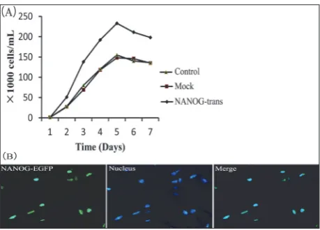
Ectopically expressed NANOG localizes to the nucleus and promotes HFSFs cell growth
We measured the effects of NANOG expression on HFSFs cell growth. NANOG transfected HFSFs showed an increased proliferation rate compared to those of the mock HFSFs (P<0.05), while the rate of cell growth was similar between the mock and control HFSFs (P>0.05) (Fig. 2A).
In ESCs, NANOG contains a homeodomain, sug-gesting it acts as a transcriptional regulator and should localize to the nucleus [11,24]. To verify the subcellular localization of ectopically-expressed NANOG, we generated a construct comprising NANOG fused to Enhanced Green Fluorescent Protein (EGFP-NANOG). The results showed that EGFP-NANOG was localized to the nucleus in HFSFs (Fig. 2D). This observation suggests that NANOG encodes a nuclear localization signal and regulates gene transcription in nucleus [25].
NANOG affects COL1A1 and ROCK1 expression
in HFSFs
NANOG expression significantly increased COL1A1
mRNA expression in NANOG-trans HFSFs compared to the mock HFSFs (P<0.05), while for expression
of COLA1 mRNA, there were no significant
differ-ences between mock and control HFSFs (P>0.05)
Fig. 1. Expression of human NANOG in HFSFs. A – NANOG plas-mid (GFP and pcDNA3.1) construction. B – NANOG expression in HFSFs was detected by end-point PCR. ACTB acted as a loading control. Control – the control-untreated HFSFs. Mock – the empty pcDNA3.1 vector-transfected HFSFs. NANOG-trans – NANOG -transfected HFSFs. C – NANOG protein expression levels were determined by Western blotting. Equal protein concentrations were loaded in each lane. ACTB acted as a loading control. Control – the control-untreated HFSFs. Mock – the empty pcDNA3.1 vector-transfected HFSFs. NANOG-trans – NANOG-transfected HFSFs.
(Fig.3A). NANOG expression significantly increased the COL1A1 protein level in NANOG-trans HFSFs compared to mock HFSFs (P<0.05) (Fig. 3B and C). There were no significant differences in protein levels between the mock and control HFSFs (P>0.05) (Fig. 3B and C). ROCK1 mRNA levels were decreased in NANOG-trans HFSFs as compared to mock HFSFs (P<0.05) (Fig.3A). Reduced ROCK1 protein levels also correlated with decreased mRNA levels in
NANOG-trans HFSFs compared to the mock HFSFs (P<0.05) (Fig. 3B and C). These data suggest that COL1A1 and
ROCK1 expression in HFSFs was significantly affected
by NANOG expression.
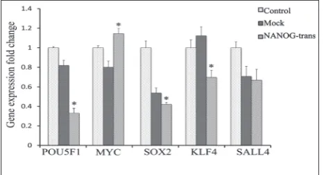
Activation of pluripotent genes by NANOG
overexpression in HFSFs
To clarify whether NANOG interacted with other key pluripotent genes thereby increasing COL1A1
expression in HFSFs, we investigated the expression levels of pluripotent genes including POU5F1, SOX2,
KLF4, MYC and SALL4 in NANOG stably-transfected HFSFs, using qPCR (Fig. 4). Compared with the mock HFSFs, the expression of POU5F1, SOX2 and KLF4
significantly declined in NANOG-trans HFSFs (p < 0.05). The expression of MYC significantly increased in NANOG-trans HFSFs compared to the mock HFSFs (p < 0.05). There was no significant change in SALL4
in NANOG-trans HFSFs compared to the mock HFSFs (p > 0.05).
DISCUSSION
The development of myopia in humans is associated with marked thinning of the sclera, the tough outer coat of the eye that facilitates change in eye size [4]. This altered scleral morphology is associated with local changes in collagen fibril ultrastructure and
Fig. 3.NANOG affects COL1A1 and ROCK1 expression in HFSFs. A – NANOG affects COL1A1 and ROCK1 mRNA expression. Fold difference was calculated with respect to the control-untreated HFSFs and the HFSFs transfected with pcDNA3.1 empty vector. Control – the control-untreated HFSFs. Mock – the empty pcDNA3.1 vector-transfected HFSFs. NANOG-trans – NANOG-transfected HFSFs. Bars represent the mean±standard errors of three independent experiments. NANOG-trans and mock were normalized to the control and statistical analysis was performed on NANOG-trans vs mock. B – COL1A1 and ROCK1 protein expression levels were determined by Western blotting. Control – the control-untreated HFSFs. Mock – the empty pcDNA3.1 vector-transfected HFSFs. NANOG-trans –
NANOG-transfected HFSFs. C – Corresponding densitometric analyses of the protein bands of COL1A1 and ROCK1 protein. Control – the control-untreated HFSFs. Mock – the empty pcDNA3.1 vector-transfected HFSFs. NANOG-trans – NANOG-transfected HFSFs. NANOG-trans and mock were normalized to the control, and statistical analysis was performed on NANOG-trans versus mock. *P<0.05 indicates significant differences.
increased numbers of small-diameter collagen fibrils [26]. In addition, there is a more lamellar organization of posterior scleral collagen fibril bundles [27]. Scleral fibroblasts, which synthesis and secrete the collagen fibrils, are involved in scleral remodeling and play important roles in the control of eye size and myopia development [28,29]. In a previous study, the overex-pression of NANOG in mouse fibroblasts promoted cell entry into the S phase and cell proliferation [20]. Our results show that constitutive overexpression of
NANOG significantly increased HFSFs proliferation.
NANOG, through CDK6 and CDC25A binding, allows human ESCs to enter the S phase from the G1 phase [30]. Compared with normal ESCs, the ESCs with a higher expression level of CDK6 and CDC25A in the S phase had a shorter cycle time to enter the next S phase [30]. In murine NIH3T3 cells, cell proliferation was accelerated when Nanog was highly expressed [20,31], therefore reducing NANOG expression could reduce breast cancer cell proliferation [32]. From these observations, it appears that NANOG plays an impor-tant role in regulating HFSFs proliferation.
Collagen accounts for 90% of scleral dry weight, the majority of this being type I collagen [33]. As the main component of the sclera, type I collagen is in-volved in connective tissue growth and extracellular matrix reorganization [2]. The COL1A1 gene produces the pro-alpha1 chain, which combines with another pro-alpha1 chain and also with a pro-alpha2 chain (produced by the COL1A2 gene) to create a molecule of type I procollagen [34]. It has been reported that
a COL1A1 polymorphism was statistically associated
with simple myopia phenotypes [35,36]. Similarly,
COL1A1 reductions appear to trigger scleral
remodel-ing and induce myopia [3]. Our results demonstrated that NANOG upregulated COL1A1 mRNA and protein expression in HFSFs, indicating that NANOG increases collagen synthesis in the sclera and may delay sclera remodeling, which is a potential risk factor for eye elongation.
The small molecule Rho GTP enzyme family has regulatory roles in fibroblast cytoskeleton rearrange-ment, cell proliferation and gene transcription [37].
ROCK1 is an extremely important downstream protein
in the Rho signaling pathway [37]. When extracellular stimuli activate G-protein-coupled receptors, Rho pro-teins are activated and combine with ROCK to induce
downstream biological effects, such as cytoskeletal reorganization and fiber synthesis of fibroblasts [38]. Activation of the Rho A/ROCK1 signaling pathway upregulates COL1A1 expression at the extracellular matrix (ECM); however, this reaction can be altered by using the inhibitor Y-27632, which inhibits ROCK1
expression, blocking the RhoA/ROCK1 signaling path-way and thereby downregulating COL1A1 expression at the ECM [39]. By contrast, in this study, ROCK1
expression decreased after NANOG overexpression in HFSFs, suggesting that NANOG may improve COL1A1
synthesis by activating other signaling pathways. In ESCs, NANOG, POU5F1, SOX2 and KLF4 were the core pluripotency transcription factors maintaining the pluripotent state of these cells [40]. POU5F1, SOX2
and NANOG function together to form a regulatory
loop to maintain ES cell pluripotency and self-renewal [24]. KLF4 is upstream of NANOG and cooperates with PBX1 directly to regulate NANOG expression in human ESCs [41]. To determine whether NANOG
regulates the cell cycle, COL1A1 and ROCK1 expression through other pluripotency-related genes in HFSFs, we examined the expression of POU5F1, SOX2 and KLF4. Our results demonstrated that although NANOG was maintained at the 20-fold higher level than normal HFSFs, the expression of POU5F1, SOX2 and KLF4
was downregulated. These results showed that only
NANOG overexpression cannot reactivate the
expres-sion of pluripotency-related genes in HFSFs. Sall4 is an important component of the transcription regulatory networks in ESCs and cooperates with NANOG [42]. In this study, SALL4 expression remained unchanged after NANOG overexpression. This is because SALL4
activation by NANOG requires POU5F1 and SOX2
participation. MYC is an important cell-cycle regulator and promotes cell proliferation [43]. In a previous study, it was shown that NANOG binds on the MYC promoter region to promote expression [32]. In the present study, MYC expression was significantly increased after NANOG overexpression; therefore, MYC may be the main factor in promoting HFSF proliferation.
CONCLUSION
The present study shows that the expression of NANOG
increased HFSF proliferation and improved COL1A1
mecha-nisms underlying NANOG gene function in HFSFs is not yet clear. Additional studies will be required to clarify the precise mechanism by which NANOG
participates in regulating collagen synthesis in HFSFs.
Acknowledgment: This work was supported by the Open Projects of Key Laboratory of Animal Cellular and Genetic Engineering of Heilongjiang Province (KF201709).
Author contributions: Yanshuang Mu, designed the study, con-ducted the experimental work, analyzed the results and wrote the first draft of the manuscript. Xuyan Li, Tianfei Yu, Ming Li and Youqi Wang participated in designing the study, the experimen-tal work, and in obtaining and analyzing the results. Bo Meng participated in drafting the article and critically revising it. All authors contributed to and have approved the final manuscript.
Conflict of interest disclosure: The authors have no financial interests or potential conflicts of interests to declare.
REFERENCES
1. Holden B, Sankaridurg P, Smith E, Aller T, Jong M, He M. Myopia, an underrated global challenge to vision: where the current data takes us on myopia control. Eye (Lond).
2014;28:142-6.
2. Watson PG, Young RD. Scleral structure, organisation and disease. A review. Exp Eye Res. 2004;78:609-23.
3. McBrien NA, Cornell LM, Gentle A. Structural and ultra-structural changes to the sclera in a mammalian model of high myopia. Invest Ophthalmol Vis Sci. 2001;42:2179-87. 4. Gentle A, Liu Y, Martin JE, Conti GL, McBrien NA. Collagen
gene expression and the altered accumulation of scleral col-lagen during the development of high myopia. J Biol Chem.
2003;278:16587-94.
5. McBrien NA, Gentle A. Role of the sclera in the development and pathological complications of myopia. Prog Retin Eye Res. 2003;22:307-38.
6. Hu S, Cui D, Yang X, Hu J, Wan W, Zeng J. The crucial role of collagen-binding integrins in maintaining the mechani-cal properties of human scleral fibroblasts-seeded collagen matrix. Mol Vis. 2011;17:1334-42.
7. Rada JA, Nickla DL, Troilo D. Decreased proteoglycan syn-thesis associated with form deprivation myopia in mature primate eyes. Invest Ophthalmol Vis Sci. 2000;41:2050-8. 8. Rada JA, Johnson JM, Achen VR, Rada KG. Inhibition of
scleral proteoglycan synthesis blocks deprivation-induced axial elongation in chicks. Exp Eye Res. 2002;74:205-15. 9. Cui W, Bryant MR, Sweet PM, McDonnell PJ. Changes in
gene expression in response to mechanical strain in human scleral fibroblasts. Exp Eye Res. 2004;78:275-84.
10. Silva J, Chambers I, Pollard S, Smith A. Nanog pro-motes transfer of pluripotency after cell fusion. Nature.
2006;441:997-1001.
11. Silva J, Nichols J, Theunissen TW, Guo G, van Oosten AL, Barrandon O, Wray J, Yamanaka S, Chambers I, Smith A.
Nanog is the gateway to the pluripotent ground state. Cell.
2009;138: 722-37.
12. Chambers I, Colby D, Robertson M, Nichols J, Lee S, Tweedie S, Smith A. Functional expression cloning of Nanog, a plu-ripotency sustaining factor in embryonic stem cells. Cell.
2003;113:643-55.
13. Mitsui K, Tokuzawa Y, Itoh H, Segawa K, Murakami M, Taka-hashi K, Maruyama M, Maeda M, Yamanaka S. The homeo-protein Nanog is required for maintenance of pluripotency in mouse epiblast and ES cells. Cell. 2003;113:631-42.
14. Miyanari Y, Torres-Padilla ME. Control of ground-state pluri-potency by allelic regulation of Nanog. Nature. 2012;483:470-3. 15. Yu J, Vodyanik MA, Smuga-Otto K, Antosiewicz-Bourget J,
Frane JL, Tian S, Nie J, Jonsdottir GA, Ruotti V, Stewart R, Slukvin II, Thomson JA. Induced pluripotent stem cell lines derived from human somatic cells. Science. 2007;318:1917-20. 16. Choi KD, Yu J, Smuga-Otto K, Salvagiotto G, Rehrauer W,
Vodyanik M, Thomson J, Slukvin I. Hematopoietic and endo-thelial differentiation of human induced pluripotent stem cells. Stem Cells. 2009;27:559-67.
17. Hanna J, Saha K, Pando B, van Zon J, Lengner CJ, Creyghton MP, van Oudenaarden A, Jaenisch R. Direct cell reprogram-ming is a stochastic process amenable to acceleration. Nature.
2009;462:595-601.
18. Takahashi K , Yamanaka S. Induction of pluripotent stem cells from mouse embryonic and adult fibroblast cultures by defined factors. Cell. 2006;126:663-76.
19. Okita K, Ichisaka T, Yamanaka S. Generation of germ-line-competent induced pluripotent stem cells. Nature.
2007;448:313-7.
20. Zhang J, Wang X, Chen B, Suo G, Zhao Y, Duan Z, Dai J. Expression of Nanog gene promotes NIH3T3 cell prolifera-tion. Biochem Biophys Res Commun. 2005;338:1098-102. 21. Piestun D, Kochupurakkal BS, Jacob-Hirsch J, Zeligson
S, Koudritsky M, Domany E, Amariglio N, Rechavi G, Givol D. Nanog transforms NIH3T3 cells and targets cell-type restricted genes. Biochem Biophys Res Commun.
2006;343:279-85.
22. Huo L, Cui D, Yang X, Gao Z, Trier K, Zeng J. All-trans reti-noic acid modulates mitogen-activated protein kinase path-way activation in human scleral fibroblasts through retinoic acid receptor beta. Mol Vis. 2013;19:1795-803.
23. Cui D, Trier K, Chen X, Zeng J, Yang X, Hu J, Ge J. Distribu-tion of adenosine receptors in human sclera fibroblasts. Mol Vis. 2008;14:523-9.
24. Boyer LA, Lee TI, Cole MF, Johnstone SE, Levine SS, Zucker JP, Guenther MG, Kumar RM, Murray HL, Jenner RG, Gif-ford DK, Melton DA, Jaenisch R, Young RA. Core transcrip-tional regulatory circuitry in human embryonic stem cells. Cell. 2005;122:947-56.
25. Do HJ, Lim HY, Kim JH, Song H, Chung HM, Kim JH. An intact homeobox domain is required for complete nuclear localization of human Nanog. Biochem Biophys Res Com-mun. 2007;353:770-5.
27. Curtin BJ, Iwamoto T, Renaldo DP. Normal and staphyloma-tous sclera of high myopia. An electron microscopic study. Arch Ophthalmol. 1979;97:912-5.
28. Seyhan N, Canseven AG. In vivo effects of ELF MFs on col-lagen synthesis, free radical processes, natural antioxidant system, respiratory burst system, immune system activities, and electrolytes in the skin, plasma, spleen, lung, kidney, and brain tissues. Electromagn Biol Med. 2006;25:291-305. 29. Nakajima H, Kishi T, Tsuchiya Y, Yamada H, Tajima S.
Expo-sure of fibroblasts derived from keloid patients to low-energy electromagnetic fields: preferential inhibition of cell prolif-eration, collagen synthesis, and transforming growth factor beta expression in keloid fibroblasts in vitro. Ann Plast Surg.
1997;39:536-41.
30. Zhang X, Neganova I, Przyborski S, Yang C, Cooke M, Atkin-son SP, Anyfantis G, Fenyk S, Keith WN, Hoare SF, Hughes O, Strachan T, Stojkovic M, Hinds PW, Armstrong L, Lako M. A role for NANOG in G1 to S transition in human embryonic stem cells through direct binding of CDK6 and CDC25A. J Cell Biol. 2009;184:67-82.
31. Jeter CR, Liu B, Liu X, Chen X, Liu C, Calhoun-Davis T, Repass J, Zaehres H, Shen JJ, Tang DG. NANOG promotes cancer stem cell characteristics and prostate cancer resistance to androgen deprivation. Oncogene. 2011;30:3833-45. 32. Han J, Zhang F, Yu M, Zhao P, Ji W, Zhang H, Wu B, Wang
Y, Niu R. RNA interference-mediated silencing of NANOG reduces cell proliferation and induces G0/G1 cell cycle arrest in breast cancer cells. Cancer Lett. 2012;321:80-8.
33. Jobling AI, Gentle A, Metlapally R, McGowan BJ, McBrien NA. Regulation of scleral cell contraction by transforming growth factor-beta and stress: competing roles in myopic eye growth. J Biol Chem. 2009;284:2072-9.
34. Wessel H, Anderson S, Fite D, Halvas E, Hempel J, Sundar-Raj N. Type XII collagen contributes to diversities in human corneal and limbal extracellular matrices. Invest Ophthalmol Vis Sci. 1997;38:2408-22.
35. Metlapally R, Li YJ, Tran-Viet KN, Abbott D, Czaja GR, Male-caze F, Calvas P, Mackey D, Rosenberg T, Paget S, Zayats T, Owen MJ, Guggenheim JA, Young TL. COL1A1 and COL2A1
genes and myopia susceptibility: evidence of association and suggestive linkage to the COL2A1 locus. Invest Ophthalmol Vis Sci. 2009;50:4080-6.
36. Nakanishi H, Yamada R, Gotoh N, Hayashi H, Yamashiro K, Shimada N, Ohno-Matsui K, Mochizuki M, Saito M, Iida T, Matsuo K, Tajima K, Yoshimura N, Matsuda F. A genome-wide association analysis identified a novel suscep-tible locus for pathological myopia at 11q24.1. PLoS Genet.
2009;5:e1000660.
37. Nakayamada S, Kurose H, Saito K, Mogami A, Tanaka Y. Small GTP-binding protein Rho-mediated signaling pro-motes proliferation of rheumatoid synovial fibroblasts. Arthritis Res Ther. 2005;7:R476-84.
38. Ridley AJ. Rho family proteins: coordinating cell responses. Trends Cell Biol. 2001;11:471-7.
39. Zhu J, Nguyen D, Ouyang H, Zhang XH, Chen XM, Zhang K. Inhibition of RhoA/Rho-kinase pathway suppresses the expression of extracellular matrix induced by CTGF or TGF-beta in ARPE-19. Int J Ophthalmol. 2013;6:8-14.
40. Huang CE, Hu FW, Yu CH, Tsai LL, Lee TH, Chou MY, Yu CC. Concurrent expression of Oct4 and Nanog maintains mesenchymal stem-like property of human dental pulp cells. Int J Mol Sci. 2014;15:18623-39.
41. Chan KK, Zhang J, Chia NY, Chan YS, Sim HS, Tan KS, Oh SK, Ng HH, Choo AB. KLF4 and PBX1 directly regulate NANOG expression in human embryonic stem cells. Stem Cells. 2009;27:2114-25.
42. Wu Q, Chen X, Zhang J, Loh YH, Low TY, Zhang W, Zhang W, Sze SK, Lim B, Ng HH. Sall4 interacts with Nanog and co-occupies Nanog genomic sites in embryonic stem cells. J Biol Chem. 2006;281:24090-4.
43. Bretones G, Delgado MD, Leon J. Myc and cell cycle control. Biochim Biophys Acta. 2015;1849:506-16.
Supplementary Data
Supplementary Table S1.
![OllyDbg 1 [Beist Security Study Group] pdf](data:image/gif;base64,R0lGODlhAQABAIAAAP///wAAACH5BAEAAAAALAAAAAABAAEAAAICRAEAOw==)