UCSF
UC San Francisco Electronic Theses and Dissertations
TitleDevelopment and applications of CRISPR-based functional genomics platforms in human iPSC-derived neurons Permalink https://escholarship.org/uc/item/83c7n47k Author Tian, Ruilin Publication Date 2020 Peer reviewed|Thesis/dissertation
eScholarship.org Powered by the California Digital Library
by
Submitted in partial satisfaction of the requirements for degree of in
in the
GRADUATE DIVISION of the
UNIVERSITY OF CALIFORNIA, SAN FRANCISCO
Approved: ______________________________________________________________________________ Chair ______________________________________________________________________________ ______________________________________________________________________________ ______________________________________________________________________________ ______________________________________________________________________________ Committee Members
Copyright 2020 by
Ruilin Tian
ACKNOWLEDGEMENTS
I would like to thank my advisor Martin Kampmann for his guidance and support throughout my PhD, for providing me space and opportunities to explore and develop my scientific interests, and for always being encouraging. He is not only a great scientist but also a model mentor. I am very fortunate to work with and learn from him in the past five years.
I would also like to thank all the members past and present in the Kampmann lab for making the lab an amazing place to work in everyday. In particular, I would like to thank Connor Ludwig, Jason Hong and Anthony Abarientos for their tremendous
contributions to this project, and Poornima Ramkumar, Xiaoyan Guo, Kun Leng, Emmy Li and Stephanie See for giving me advice and ideas, discussing questions and sharing reagents with me and encouraging me when I was depressed. I would also like to thank Jaime Leong for taking care of my reagents orders, receipts for reimbursement and other logistics so that I can focus on my research.
I would also like to thank my thesis committee members, Li Gan and Bruce Conklin for their invaluable advice and ideas throughout my PhD, and their generous support during my academic job application.
Lastly but most importantly, this dissertation would not have been possible without my family. My parents, Jing Wang and Jinyuan Tian, have always been
supportive and encouraging. They came to the US from China to help me take care of my baby so that I could focus on my study. My wife, Ling Zhu, has always been my
biggest cheerleader and a constant source of love. And to my son, David T. Tian, who brings me endless joy and from his eyes I see light.
Chapter 2 is reprinted as it appears in:
Tian, R., Gachechiladze, M. A., Ludwig, C. H., Laurie, M. T., Hong, J. Y., Nathaniel, D., Prabhu, A. V., Fernandopulle, M. S., Patel, R., Abshari, M., Ward, M. E., & Kampmann, M. (2019). CRISPR Interference-Based Platform for Multimodal Genetic Screens in Human iPSC-Derived Neurons. Neuron 104, 239-255.e12.
Author contributions:
R.T., M.A.G., C.H.L., M.E.W., and M.K. contributed to the study’s overall
conception, design, and interpretation and wrote the manuscript and created the figures with input from the other authors. R.T. designed and conducted the validation screens and CROP-seq screens and designed and conducted computational analyses for all screens (including survival screens, CROP- seq, and longitudinal imaging screens), with guidance from M.K. M.A.G. designed and conducted imaging screens with guidance from M.E.W. and M.K. C.H.L. established CRISPRi in iPSC-derived neurons and designed and conducted the primary screens with guidance from R.T. and M.K. C.H.L. also conducted and analyzed Quant-seq experiments and created the iNeuronRNASeq web application. M.A.G., M.T.L., M.S.F., and R.P. designed, generated, and
characterized constructs and cell lines, with guidance from M.E.W. and M.K. J.Y.H. and D.N. generated and characterized con- structs and cell lines, with guidance from R.T. and M.K. A.V.P. contributed to validation experiments and M.A. optimized FACS for iPSC line construction.
Chapter 3 is reprinted as a manuscript in preparation:
Tian, R., Abarientos, A., Hong, J., Yan, R., Xu, K., Kampmann, M. (2020).
Genome-wide CRISPRi/a screens in human neurons link lysosomal failure to ferroptosis
Author contributions:
R.T and M.K. designed the project; R.T., A.A., J.H. and R.Y. performed experiments; R.T. analyzed data; R.T. and M.K. wrote the manuscript
Development and applications of CRISPR-based functional genomics platforms in human iPSC-derived neurons
By Ruilin Tian
ABSTRACT
CRISPR/Cas9-based functional genomics have transformed our ability to elucidate mammalian cell biology. However, most previous CRISPR-based screens were conducted in cancer cell lines rather than healthy, differentiated cells such as neurons. Neurons possess unique cell biological properties that enable them to exert highly specialized physiological functions. To enable systematic characterization of neuronal cell biology, we established CRISPR interference (CRISPRi)- and CRISPR activation (CRISPRa)-based platforms in human neurons derived from induced
pluripotent stem cells (iPSCs) that allow robust repression or activation of endogenous genes and large-scale genetic screens in human neurons. Using this toolkit, we
performed multiple genome-wide screens to identity genes controlling neuronal survival under unstressed and oxidative stress conditions and genes regulating neuronal redox homeostasis. These screens provide numerous novel biological insights. We also demonstrate that our platforms can be coupled with single-cell RNA sequencing and longitudinal high-content imaging to reveal consequences of genetic perturbations on gene expression and neuronal morphology respectively. Our results highlight the power of unbiased genetic screens in iPSC-derived differentiated cell types and provide a platform for systematic interrogation of normal and disease states of neurons.
TABLE OF CONTENTS CHAPTER ONE 1 Introduction Introduction 2 References 7 CHAPTER TWO 12
CRISPR interference-based platform for multimodal genetic screens in human iPSC-derived neurons Introduction 13 Results 16 Discussion 34 Figures 38 Tables 63
Materials and Methods 72
References 106
CHAPTER THREE 117
Genome-wide CRISPRi/a screens in human neuronslink lysosomal failure to ferroptosis
Introduction 118
Results 122
Discussion 143
Figures 148
Materials and Methods 166
LIST OF FIGURES
Fig.2.1.Durable gene knockdown by CRISPR interference in human
iPSC-derived neurons 38
Fig.2.2. Normal karyotype, neuronal differentiation and activity and CRISPRi
activity of the CRISPRi- i3N iPSC monoclonal line 40 Fig.2.3. Massively parallel screen for essential genes in iPSCs and
iPSC-derived neurons 42
Fig.2.4. Characterization of results from massively parallel screens for
essential genes in iPSCs and neurons 44 Fig.2.5. Functional analysis of hit genes from primary survival screens in
iPSCs and iPSC-derived neurons 46 Fig.2.6. Pooled validation of hit genes from the primary screen 48 Fig.2.7. CROP-Seq reveals transcriptome changes in iPSCs and iPSC-derived
neurons induced by knockdown of survival-relevant genes 50 Fig.2.8. Pooled validation of hit genes from the primary screen 52 Fig.2.9. Characterization of CROP-Seq screen results 55 Fig.2.10. Cell-type specific responses to gene knockdown on the
transcriptomic level 57
Fig.2.11. CROP-Seq reveals neuron-specific transcriptomic consequences
of MAT2A knockdown 59
Fig.2.12 .Longitudinal imaging to track the effect of selected hit gene
knockdown on iPSC growth, neuronal survival and neurite morphology 60
Fig.2.13. Reproducibility of longitudinal imaging results 62 Fig.3.1. Genome-wide CRISPRi & CRISPRa screens in human iPSC-derived
neurons identify regulators of neuronal survival 148 Fig.3.2. Karyotyping of the monoclonal CRISPRa-iPSC line and comparison
of CRISPRi and CRISPRaa screen results for neuronal survival with
other published survival screens for different cell types 150 Fig.3.3. Genome-wide CRISPRi & a screens in human iPSC-derived neurons
identify regulators of oxidative stress response and redox homeostasis 151 Fig.3.4. Comparing CRISPRa survival screens in +AO and -AO conditions and
common hits in ROS and lipid peroxidation screens 153 Fig.3.5. CROP-seq reveals transcriptomic responses to perturbations of
neurodegenerative disease-associated hit genes
in human iPSC-derived neurons 154 Fig.3.6. Shared signatures of transcriptomic responses to the knockdown
of VPS54, PAXIP1 and PON2 in human iPSC-derived neurons 156 Fig.3.7. Overexpression of NQO1 induces unexpected transcriptome changes
in human iPSC-derived neurons 157 Fig.3.8. Depletion of PSAP induces ROS and lipid peroxidation in neurons
and causes neuronal ferroptosis in the absence of antioxidants 158 Fig.3.9. Depletion of PSAP in neurons disrupts glycosphingolipid degradation
and causes lipofuscin formation in the lysosome,
Fig.3.10. Characterization of PSAP KO neurons 163 Fig.3.11. A working model for PSAP depletion inducing neuronal ferroptosis 165
LIST OF TABLES
CHAPTER ONE
Introduction
Introduction
The human body comprises hundreds of different cell types. Even though their genomes are nearly identical, cell types are characterized by vastly different cell biologies, enabling them to fulfill diverse physiological functions. Transcriptomic
profiling, fueled by recent advances in single-cell- and single-nucleus-RNA sequencing technologies, has revealed cell-type specific gene expression signatures (Gao et al., 2018; Han et al., 2020; Lake et al., 2018; Muraro et al., 2016). In addition to gene expression, gene function can also be cell type-specific, as evidenced by the fact that mutations in broadly expressed or housekeeping genes can lead to strongly cell-type specific defects and disease states. Striking examples are familial mutations causing neurodegenerative diseases, which are often characterized by the selective vulnerability of specific neuronal subtypes, even if the mutated gene is expressed throughout the brain or even throughout the body. Cell-type specific gene function is also supported by our recent finding that knockdown of certain genes can have remarkably different impacts on cell survival and gene expression in different isogenic human cell types, including stem cells and neurons (Tian et al., 2019). Therefore, understanding the function of human genes in different cell types is the next step toward elucidating tissue-specific cell biology and uncovering disease mechanisms.
A powerful approach to functionally annotate the human genome is genetic screening in cultured cells. The robustness of such screens has improved substantially through the recent introduction of CRISPR/Cas9-based approaches. Cas9 nuclease can be targeted by single guide RNAs (sgRNAs) to introduce DNA breaks in coding regions
of genes, which are subsequently repaired by non-homologous end-joining pathways. This process frequently causes short deletions or insertions that disrupt gene function. This CRISPR nuclease (CRISPRn) strategy has enabled genetic screens through the use of pooled sgRNA libraries targeting large numbers of genes (Koike-Yusa et al., 2014; Shalem et al., 2014; Wang et al., 2014; Zhou et al., 2014). We previously
developed an alternative platform for loss-of-function screens in mammalian cells based on CRISPR interference (CRISPRi) (Gilbert et al., 2014). In CRISPRi screens, sgRNAs target catalytically dead Cas9 (dCas9) fused to a KRAB transcriptional repression domain to transcription start sites in the genome, thereby inhibiting gene transcription. CRISPRn and CRISPRi screening platforms each have their advantages for specific applications (Kampmann, 2018); (Rosenbluh et al., 2017), but generally yield similar results (Horlbeck et al., 2016). Most previous CRISPR-based screens were
implemented in cancer cell lines or stem cells rather than healthy differentiated human cells such as neurons, thereby limiting potential insights into cell type-specific roles of human genes.
To address this need, we developed CRISPRi- and CRISPRa-based platforms for genetic screens in human induced pluripotent stem cell (iPSC)-derived neurons. To our knowledge, this is the first description of a large-scale CRISPR-based screening platform in any differentiated, human iPSC-derived cell type. We focused on neurons as our first application, since functional genomic screens in human neurons have the potential to reveal mechanisms of selective vulnerability in neurodegenerative diseases (Kampmann, 2017) and convergent mechanisms of neuropsychiatric disorders (Willsey
et al., 2018), thus addressing urgent public health issues. iPSC technology is
particularly relevant to the study of human neurons, since primary neurons are difficult to obtain from human donors, and non-expandable due to their post-mitotic nature.
We integrated CRISPRi and CRISPRa technologies with our previously described i3Neuron platform (Fernandopulle et al., 2018, Wang et al., 2017), which yields large quantities of highly homogeneous neurons, a prerequisite for robust population-based screens. We decided to use CRISPRi rather than CRISPRn, since CRISPRn-associated DNA damage is highly toxic to iPSCs and untransformed cells (Haapaniemi et al., 2018, Schiroli et al., 2019, Ihry et al., 2018). Furthermore, CRISPRi perturbs gene function by partial knockdown, rather than knockout, thereby enabling the investigation of the biology of essential genes. While large-scale genetic screens in mouse primary neurons have previously been implemented using RNA interference (RNAi) technology (Nieland et al., 2014; Sharma et al., 2013), CRISPRi represents an important advance over RNAi, since it lacks the pervasive off-target effects (Gilbert et al., 2014) inherent to RNAi-based screening approaches (Adamson et al., 2012; Jackson et al., 2003; Kaelin, 2012).
We demonstrate the versatility of our approach in three complementary genetic screens, based on neuronal survival, single-cell RNA sequencing (scRNA-Seq), and neuronal morphology. These screens revealed striking examples of cell-type specific gene functions and identified new genetic modifiers of neuronal biology.
Neurons, as one of the longest-living cell types in the human body, are
neurons do not have the ability to ‘self-renew’ by cell division. Therefore, robust stress response mechanisms are required for neurons to maintain long-term health. One of the predominant stresses in aging and neurodegenerative diseases is oxidative stress (Barnham et al., 2004; Finkel and Holbrook, 2000), which is induced by excessive accumulation of reactive oxygen species (ROS) in the cell. ROS are highly reactive oxygen-derived molecules that are generated as by-products of normal oxygen metabolism. At low levels, ROS have physiological functions in cellular signaling and activate pro-survival pathways such as MAPK pathways (Kim et al., 2015).
Various antioxidant systems have evolved to control ROS levels and maintain redox homeostasis, including non-enzymatic antioxidants such as vitamin E, vitamin C and glutathione, and enzymatic antioxidants such as superoxide dismutase (SOD), glutathione peroxidases (GPX), peroxiredoxins (PRX) and catalase (Kim et al., 2015). There are also dedicated cellular pathways that sense and respond to ROS levels such as the Keap1-Nrf2 pathway (Sies et al., 2017). An imbalance of ROS production and antioxidant defenses leads to excessive accumulation of ROS, which can cause oxidative damage to proteins, lipids and DNA and ultimately lead to cell death (Kim et al., 2015; Sies et al., 2017). In particular, peroxidation of lipids containing
polyunsaturated fatty acids (PUFAs) can cause a non-apoptotic cell death termed ferroptosis, which is iron-dependent (Li et al., 2020).
The brain is highly susceptible to ROS and ferroptosis, due to its high levels of oxygen consumption, abundant redox-active metals such as iron and copper, limited antioxidants and high levels of PUFAs (Patel, 2016). A large body of evidence has
indicated the implications of oxidative stress, iron accumulation and ferroptosis in many neurodegenerative diseases, including Alzheimer’s disease (AD), Parkinson’s disease (PD) and Amyotrophic Lateral Sclerosis (ALS) (Lin and Beal, 2006; Niedzielska et al., 2016; Rouault, 2013), yet a comprehensive understanding of how neurons regulate redox homeostasis and maintain survival under oxidative stress is lacking.
We applied our functional genomics platforms to systematically identify genetic modifiers of ROS levels, lipid peroxidation, and neuronal survival under oxidative stress. These screens uncovered an unexpected role for prosaposin (PSAP), knockdown of which strongly induced ROS and lipid peroxidation levels in neurons and led to neuronal ferroptosis under oxidative stress. We elucidated the underlying mechanism: depletion of PSAP and resulting defects in glycosphingolipids (GSLs) degradation lead to the formation of lipofuscin in the lysosome, which is a hallmark of aged neurons,, driving the accumulation of iron and generation of ROS that oxidize lipids. Intriguingly, the strong phenotypes of PSAP depletion are only presented in neurons, but not iPSCs or HEK293s. These results demonstrate the power of our platforms in uncovering novel cell-type specific human cell biology.
REFERENCES
Adamson, B., Smogorzewska, A., Sigoillot, F.D., King, R.W., and Elledge, S.J. (2012). A genome-wide homologous recombination screen identifies the RNA-binding protein RBMX as a component of the DNA-damage response. Nat. Cell Biol. 14, 318–328.
Barnham, K.J., Masters, C.L., and Bush, A.I. (2004). Neurodegenerative diseases and oxidative stress. Nat. Rev. Drug Discov. 3, 205–214.
Fernandopulle, M.S., Prestil, R., Grunseich, C., Wang, C., Gan, L., and Ward, M.E. (2018). Transcription Factor-Mediated Differentiation of Human iPSCs into Neurons. Curr. Protoc. Cell Biol. 79, e51.
Finkel, T., and Holbrook, N.J. (2000). Oxidants, oxidative stress and the biology of ageing. Nature 408, 239–247.
Gao, S., Yan, L., Wang, R., Li, J., Yong, J., Zhou, X., Wei, Y., Wu, X., Wang, X., Fan, X., et al. (2018). Tracing the temporal-spatial transcriptome landscapes of the human fetal digestive tract using single-cell RNA-sequencing. Nat. Cell Biol. 20, 721–734.
Gilbert, L.A., Horlbeck, M.A., Adamson, B., Villalta, J.E., Chen, Y., Whitehead, E.H., Guimaraes, C., Panning, B., Ploegh, H.L., Bassik, M.C., et al. (2014).
Genome-scale CRISPR-mediated control of gene repression and activation. Cell 159, 647–661.
Haapaniemi, E., Botla, S., Persson, J., Schmierer, B., and Taipale, J. (2018).
CRISPR-Cas9 genome editing induces a p53-mediated DNA damage response. Nat. Med. 24, 927–930.
Han, X., Zhou, Z., Fei, L., Sun, H., Wang, R., Chen, Y., Chen, H., Wang, J., Tang, H., Ge, W., et al. (2020). Construction of a human cell landscape at single-cell level. Nature.
Horlbeck, M.A., Gilbert, L.A., Villalta, J.E., Adamson, B., Pak, R.A., Chen, Y., Fields, A.P., Park, C.Y., Corn, J.E., Kampmann, M., et al. (2016). Compact and highly active next-generation libraries for CRISPR-mediated gene repression and activation. Elife 5.
Ihry, R.J., Worringer, K.A., Salick, M.R., Frias, E., Ho, D., Theriault, K., Kommineni, S., Chen, J., Sondey, M., Ye, C., et al. (2018). p53 inhibits CRISPR-Cas9
engineering in human pluripotent stem cells. Nat. Med. 24, 939–946.
Jackson, A.L., Bartz, S.R., Schelter, J., Kobayashi, S.V., Burchard, J., Mao, M., Li, B., Cavet, G., and Linsley, P.S. (2003). Expression profiling reveals off-target gene regulation by RNAi. Nat. Biotechnol. 21, 635–637.
Kaelin, W.G. (2012). Molecular biology. Use and abuse of RNAi to study mammalian gene function. Science 337, 421–422.
Kampmann, M. (2017). A CRISPR approach to neurodegenerative diseases. Trends Mol. Med. 23, 483–485.
Kampmann, M. (2018). Crispri and crispra screens in mammalian cells for precision biology and medicine. ACS Chem. Biol. 13, 406–416.
Kim, G.H., Kim, J.E., Rhie, S.J., and Yoon, S. (2015). The role of oxidative stress in neurodegenerative diseases. Exp. Neurobiol. 24, 325–340.
Koike-Yusa, H., Li, Y., Tan, E.-P., Velasco-Herrera, M.D.C., and Yusa, K. (2014). Genome-wide recessive genetic screening in mammalian cells with a lentiviral CRISPR-guide RNA library. Nat. Biotechnol. 32, 267–273.
Lake, B.B., Chen, S., Sos, B.C., Fan, J., Kaeser, G.E., Yung, Y.C., Duong, T.E., Gao, D., Chun, J., Kharchenko, P.V., et al. (2018). Integrative single-cell analysis of transcriptional and epigenetic states in the human adult brain. Nat. Biotechnol. 36, 70–80.
Lin, M.T., and Beal, M.F. (2006). Mitochondrial dysfunction and oxidative stress in neurodegenerative diseases. Nature 443, 787–795.
Li, J., Cao, F., Yin, H.-L., Huang, Z.-J., Lin, Z.-T., Mao, N., Sun, B., and Wang, G. (2020). Ferroptosis: past, present and future. Cell Death Dis. 11, 88.
Muraro, M.J., Dharmadhikari, G., Grün, D., Groen, N., Dielen, T., Jansen, E., van Gurp, L., Engelse, M.A., Carlotti, F., de Koning, E.J.P., et al. (2016). A Single-Cell Transcriptome Atlas of the Human Pancreas. Cell Syst. 3, 385-394.e3.
Niedzielska, E., Smaga, I., Gawlik, M., Moniczewski, A., Stankowicz, P., Pera, J., and Filip, M. (2016). Oxidative stress in neurodegenerative diseases. Mol. Neurobiol. 53, 4094–4125.
Nieland, T.J.F., Logan, D.J., Saulnier, J., Lam, D., Johnson, C., Root, D.E., Carpenter, A.E., and Sabatini, B.L. (2014). High content image analysis identifies novel
regulators of synaptogenesis in a high-throughput RNAi screen of primary neurons. PLoS ONE 9, e91744.
Patel, M. (2016). Targeting oxidative stress in central nervous system disorders. Trends Pharmacol. Sci. 37, 768–778.
Rosenbluh, J., Xu, H., Harrington, W., Gill, S., Wang, X., Vazquez, F., Root, D.E., Tsherniak, A., and Hahn, W.C. (2017). Complementary information derived from CRISPR Cas9 mediated gene deletion and suppression. Nat. Commun. 8, 15403.
Rouault, T.A. (2013). Iron metabolism in the CNS: implications for neurodegenerative diseases. Nat. Rev. Neurosci. 14, 551–564.
Schiroli, G., Conti, A., Ferrari, S., Della Volpe, L., Jacob, A., Albano, L., Beretta, S., Calabria, A., Vavassori, V., Gasparini, P., et al. (2019). Precise Gene Editing Preserves Hematopoietic Stem Cell Function following Transient p53-Mediated DNA Damage Response. Cell Stem Cell 24, 551-565.e8.
Shalem, O., Sanjana, N.E., Hartenian, E., Shi, X., Scott, D.A., Mikkelson, T., Heckl, D., Ebert, B.L., Root, D.E., Doench, J.G., et al. (2014). Genome-scale CRISPR-Cas9 knockout screening in human cells. Science 343, 84–87.
Sharma, K., Choi, S.-Y., Zhang, Y., Nieland, T.J.F., Long, S., Li, M., and Huganir, R.L. (2013). High-throughput genetic screen for synaptogenic factors: identification of LRP6 as critical for excitatory synapse development. Cell Rep. 5, 1330–1341.
715–748.
Tian, R., Gachechiladze, M.A., Ludwig, C.H., Laurie, M.T., Hong, J.Y., Nathaniel, D., Prabhu, A.V., Fernandopulle, M.S., Patel, R., Abshari, M., et al. (2019). CRISPR Interference-Based Platform for Multimodal Genetic Screens in Human
iPSC-Derived Neurons. Neuron 104, 239-255.e12.
Wang, C., Ward, M.E., Chen, R., Liu, K., Tracy, T.E., Chen, X., Xie, M., Sohn, P.D., Ludwig, C., Meyer-Franke, A., et al. (2017). Scalable Production of iPSC-Derived Human Neurons to Identify Tau-Lowering Compounds by High-Content
Screening. Stem Cell Reports 9, 1221–1233.
Wang, T., Wei, J.J., Sabatini, D.M., and Lander, E.S. (2014). Genetic screens in human cells using the CRISPR-Cas9 system. Science 343, 80–84.
Willsey, A.J., Morris, M.T., Wang, S., Willsey, H.R., Sun, N., Teerikorpi, N., Baum, T.B., Cagney, G., Bender, K.J., Desai, T.A., et al. (2018). The psychiatric cell map initiative: A convergent systems biological approach to illuminating key molecular pathways in neuropsychiatric disorders. Cell 174, 505–520.
Zhou, Y., Zhu, S., Cai, C., Yuan, P., Li, C., Huang, Y., and Wei, W. (2014).
High-throughput screening of a CRISPR/Cas9 library for functional genomics in human cells. Nature 509, 487–491.
CHAPTER TWO
CRISPR interference-based platform for multimodal genetic screens in human iPSC-derived neurons
INTRODUCTION
While DNA sequencing has provided us with an inventory of human genes, and RNA sequencing is revealing when and where these genes are expressed, the next challenge is to systematically understand the function of human genes in different cell types. A powerful approach to functionally annotate the human genome is genetic screening in cultured cells. The robustness of such screens has improved substantially through the recent introduction of CRISPR/Cas9-based approaches. Cas9 nuclease can be targeted by single guide RNAs (sgRNAs) to introduce DNA breaks in coding regions of genes, which are subsequently repaired by non-homologous end-joining pathways. This process frequently causes short deletions or insertions that disrupt gene function. This CRISPR nuclease (CRISPRn) strategy has enabled genetic screens through the use of pooled sgRNA libraries targeting large numbers of genes (Koike-Yusa et al., 2014; Shalem et al., 2014; Wang et al., 2014; Zhou et al., 2014). We previously
developed an alternative platform for loss-of-function screens in mammalian cells based on CRISPR interference (CRISPRi) (Gilbert et al., 2014). In CRISPRi screens, sgRNAs target catalytically dead Cas9 (dCas9) fused to a KRAB transcriptional repression domain to transcription start sites in the genome, thereby inhibiting gene transcription. CRISPRn and CRISPRi screening platforms each have their advantages for specific applications (Kampmann, 2018; Rosenbluh et al., 2017), but generally yield similar results (Horlbeck et al., 2016). Most previous CRISPR-based screens were
implemented in cancer cell lines or stem cells rather than healthy differentiated human cells, thereby limiting potential insights into cell type-specific roles of human genes.
Here, we present a CRISPRi-based platform for genetic screens in human induced pluripotent stem cell (iPSC)-derived neurons. To our knowledge, it is the first description of a large-scale CRISPR-based screening platform in any differentiated, human iPSC-derived cell type. We focused on neurons as our first application, since functional genomic screens in human neurons have the potential to reveal mechanisms of selective vulnerability in neurodegenerative diseases (Kampmann, 2017) and
convergent mechanisms of neuropsychiatric disorders (Willsey et al., 2018), thus addressing urgent public health issues. iPSC technology is particularly relevant to the study of human neurons, since primary neurons are difficult to obtain from human donors, and non-expandable due to their post-mitotic nature.
We integrated CRISPRi technology with our previously described i3Neuron platform (Fernandopulle et al., 2018; Wang et al., 2017), which yields large quantities of highly homogeneous neurons, a prerequisite for robust population-based screens. We decided to use CRISPRi rather than CRISPRn, since CRISPRn-associated DNA
damage is highly toxic to iPSCs and untransformed cells (Haapaniemi et al., 2018; Ihry et al., 2018; Schiroli et al., 2019). Furthermore, CRISPRi perturbs gene function by partial knockdown, rather than knockout, thereby enabling the investigation of the
biology of essential genes. While large-scale genetic screens in mouse primary neurons have previously been implemented using RNA interference (RNAi) technology (Nieland et al., 2014; Sharma et al., 2013), CRISPRi represents an important advance over RNAi, since it lacks the pervasive off-target effects (Gilbert et al., 2014) inherent to
RNAi-based screening approaches (Adamson et al., 2012; Jackson et al., 2003; Kaelin, 2012).
We demonstrate the versatility of our approach in three complementary genetic screens, based on neuronal survival, single-cell RNA sequencing (scRNA-Seq), and neuronal morphology. These screens revealed striking examples of cell-type specific gene functions and identified new genetic modifiers of neuronal biology. Our results provide a strategy for systematic dissection of normal and disease states of neurons, and highlight the potential of interrogating human cell biology and gene function in iPSC-derived differentiated cell types.
RESULTS
Robust CRISPR interference in human iPSC-derived neurons
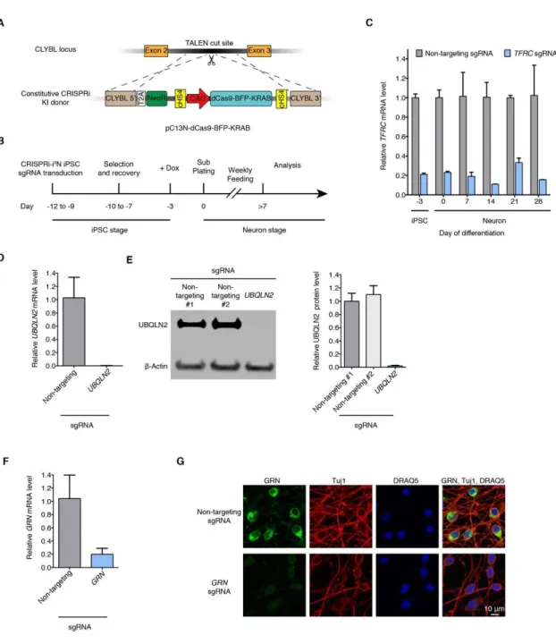
As a first step towards a high-throughput screening platform in neurons, we developed a scalable CRISPRi-based strategy for robust knockdown of endogenous genes in homogeneous populations of human iPSC-derived neurons. We built on our previously described i3Neuron (i3N) platform, which enables large-scale production of iPSC-derived glutamatergic neurons. Central to this platform is an iPSC line with an inducible Neurogenin 2 (Ngn2) expression cassette (Zhang et al., 2013) in the AAVS1 safe-harbor locus (Fernandopulle et al., 2018; Wang et al., 2017). To enable stable CRISPRi in iPSC-derived neurons, we generated a plasmid (pC13N-dCas9-BFP-KRAB) to insert an expression cassette for CAG promoter-driven dCas9-BFP-KRAB into the CLYBL safe harbor locus, which enables robust transgene expression throughout
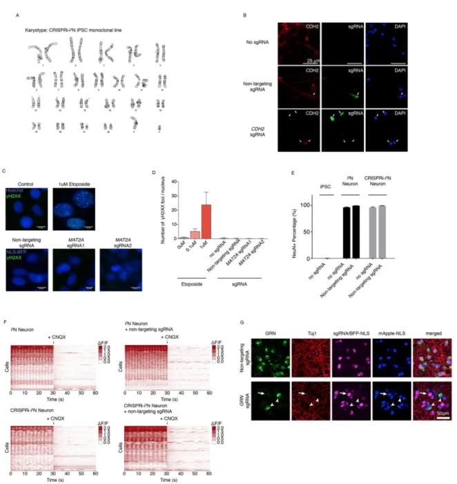
neuronal differentiation at higher levels than the AAVS1 locus (Cerbini et al., 2015) (Fig. 2.1A). We then integrated this cassette into our i3N iPSC line, and called the resulting monoclonal line CRISPRi-i3N iPSCs. A normal karyotype was confirmed for
CRISPRi-i3N iPSCs (Fig. 2.2A).
To validate CRISPRi activity, we transduced these iPSCs with a lentiviral construct expressing an sgRNA targeting the transferrin receptor gene (TFRC). Knockdown of TFRC mRNA was robust in iPSCs and in i3Neurons for several weeks after differentiation (Fig. 2.1B,C). We also validated knockdown of three additional genes, UBQLN2 (Fig. 2.1D,E), GRN (Fig. 2.1F,G) and CDH2 (Fig. 2.2B) by qRT-PCR,
Western blot, and/or immunofluorescence. Our platform thus enables potent CRISPRi knockdown of endogenous genes in iPSC-derived neurons.
Since CRISPRn-associated DNA damage has been found to be highly toxic to iPSCs (Ihry et al., 2018), we evaluated whether the CRISPRi machinery caused DNA damage in iPSCs or otherwise interfered with neuronal differentiation or activity. We found that expression of CRISPRi machinery and/or sgRNAs did not cause detectable DNA damage (Fig. 2.2C,D), as expected based on the abrogation of nuclease activity in dCas9, and did not affect neuronal differentiation (Fig. 2.2E) or activity as evaluated by calcium imaging (Fig. 2.2F).
We established the CRISPRi-i3N system used throughout this study in the background of the well-characterized WTC11 iPSC line (Miyaoka et al., 2014). In addition, we also generated an equivalent line in the NCRM5 iPSC line (Luo et al., 2014) and validated its CRISPRi activity (Fig. 2.2G).
A pooled CRISPRi screen reveals neuron-essential genes
We then used this platform to identify cell type-specific genetic modifiers of survival in pooled genetic screens in iPSCs and iPSC-derived neurons (Fig. 2.3A). We first transduced CRISPRi-i3N iPSCs with our lentiviral sgRNA library H1 (Horlbeck et al., 2016). The H1 library targets 2,325 genes encoding kinases and other proteins
representing the “druggable genome” with at least five independent sgRNAs per gene, plus 500 non-targeting control sgRNAs, for a total of 13,025 sgRNAs. Transduced iPSCs were either passaged for 10 days, or differentiated into neurons by
doxycycline-induced Ngn2 expression. Neurons were collected 14, 21 and 28 days post-induction. Frequencies of cells expressing each sgRNA at each time point were determined by next-generation sequencing of the sgRNA-encoding locus. We observed highly correlated sgRNA frequencies between independently cultured experimental replicates (Fig. 2.4A), supporting the robustness of these measurements.
To analyze the screen results, we developed a new bioinformatics pipeline, MAGeCK-iNC (MAGeCK including Negative Controls, available at
kampmannlab.ucsf.edu/mageck-inc). This pipeline integrates a published method, MAGeCK (Li et al., 2014) with aspects of our previous bioinformatics pipeline (Kampmann et al., 2013, 2014) to take full advantage of the non-targeting control
sgRNAs in our library when computing P values (see Methods for details). Based on the depletion or enrichment of sgRNAs targeting specific genes at different time points compared to day 0, we identified hit genes for which knockdown was toxic or beneficial to either iPSCs or neurons at different time points (Fig. 2.3B, Fig. 2.4B). We then
calculated a knockdown phenotype score and significance P value for each gene (Table S1). The large number of non-targeting sgRNAs in our library enabled us to generate “quasi-genes” from random groupings of non-targeting sgRNAs to empirically estimate a false-discovery rate (FDR) for a given cutoff of hit strength (defined as the product of phenotype score and –log10(P value)), see Methods for details. We defined genes passing an FDR < 0.05 as hit genes. For the majority of hit genes, two or three sgRNAs in the library resulted in strong phenotypes (Fig. 2.4C,D), justifying the use of five
Knockdown phenotypes of hit genes were strongly correlated between neurons at different time points, but distinctly less correlated between neurons and iPSCs (Fig. 2.3C). Next, we compared genes that were essential in iPSCs and/or neurons in our screens with “gold-standard” essential genes that were previously identified through genetic screens in cancer cell lines (Hart et al., 2017). This analysis revealed a shared core set of essential genes, as expected, and additional iPSC-specific and
neuron-specific essential genes (Fig. 2.3D).
Using Gene Set Enrichment Analysis (GSEA) (Mootha et al., 2003; Subramanian et al., 2005), we found enrichment of distinct groups of survival-related genes in
neurons compared to iPSCs, such as genes associated with sterol metabolism (Fig. 2.5A). We validated the strong neuronal dependence on the cholesterol biogenesis pathway pharmacologically using the HMG-CoA reductase inhibitor mevastatin (Fig. 2.3E) and found that CRISPRi knockdown of HMG-CoA reductase (HMGCR) can be partially rescued by supplementing its product mevalonate (Fig. 2.3F).
We determined expression levels of genes at different time points during
neuronal differentiation by Quant-Seq (Data deposited in GEO, GSE124703; the results can be visualized at kampmannlab.ucsf.edu/ineuron-rna-seq). As a group,
neuron-essential genes were expressed at significantly higher levels than non-essential genes in iPSC-derived neurons (one-sided Mann-Whitney U test, Fig. 2.5B). The vast majority of neuron-essential genes were detectable at the transcript level, further supporting the specificity of our screen results.
Intriguingly, we identified several genes that specifically enhanced neuronal survival when knocked down, including MAP3K12 (encoding dual leucine zipper kinase DLK), MAPK8 (encoding Jun kinase JNK1), CDKN1C (encoding the cyclin-dependent kinase inhibitor p57) and EIF2AK3 (encoding the eIF2alpha kinase PERK) (Table S1). A pathway involving DLK, JNK and PERK has previously been implicated in neuronal death (Ghosh et al., 2011; Huntwork-Rodriguez et al., 2013; Larhammar et al., 2017; Miller et al., 2009; Pozniak et al., 2013; Watkins et al., 2013; Welsbie et al., 2013), validating our approach.
In summary, our large-scale CRISPRi screen in human iPSC-derived neurons uncovered genes that control the survival of neurons, but not cancer cells or iPSCs, demonstrating the potential of our platform to characterize the biology of differentiated cell types.
Pooled validation of hit genes
To validate and further characterize hit genes from the primary large-scale screen, we performed a series of secondary screens. For this purpose, we generated a new lentiviral sgRNA plasmid (pMK1334) that enables screens with single-cell RNA-Seq (scRNA-Seq) readouts (based on the CROP-Seq format (Datlinger et al., 2017)), and high-content imaging readouts (expressing a bright, nuclear-targeted BFP) (Fig. 2.6A). We individually cloned 192 sgRNAs into this plasmid (184 sgRNAs targeting 92 different hit genes with two sgRNAs per gene and eight non-targeting control sgRNAs). Then, to confirm essential genes identified in our primary screen, we pooled these plasmids and
conducted a survival-based validation screen (Fig. 2.6A). Because the library size was small compared to the primary screen, we obtained a high representation of each sgRNA in the validation screen. As in the primary screen, CRISPRi-i3N iPSCs
transduced with the plasmid pool were either passaged as iPSCs or differentiated into glutamatergic neurons, and then harvested at different time points for next-generation sequencing and calculation of survival phenotypes for each sgRNA (Table S2). We observed a high correlation of raw sgRNA counts between two independently
differentiated biological replicates (R2 > 0.9, Fig. 2.6B), supporting the robustness of phenotypes measured in the pooled validation screen. We then compared the results from the validation screen with those from the primary screen. In both iPSCs and neurons, all positive hits and most of the negative hits from the primary screen were confirmed in the validation screen (Fig. 2.6C). These findings indicate that hits identified in the primary screen are highly reproducible.
In the brain, many neuronal functions are supported by glial cells, particularly astrocytes. To rule out the possibility that hits from the primary screen were artifacts of an astrocyte-free culture environment, we included an additional condition in the validation screen, in which neurons were co-cultured with primary mouse astrocytes. Neuronal phenotypes in the presence or absence of astrocytes were highly correlated (Fig. 2.6D,E and Fig. 2.8A), indicating that the vast majority of the neuron-essential genes we identified are required even in the presence of astrocytes. However, we identified a small number of genes, including PPCDC, UROD and MAT2A, for which knockdown was less toxic in the presence of astrocytes (Fig. 2.6F). This suggests that
astrocytes may compensate for the loss of function for these genes in neurons. We also identified a small number of other genes, including MMAB, UBA1 and PPP2R2A, for which knockdown was more toxic in the presence of astrocytes (Fig. 2.6F). These genes may function in pathways affected by crosstalk between neurons and astrocytes.
Inducible CRISPRi distinguishes neuronal differentiation and survival phenotypes
A caveat of our primary screen is that we introduced the sgRNA library into cells constitutively expressing CRISPRi machinery at the iPSC stage. Therefore, some hit genes detected in the primary screen may play a role in neuronal differentiation rather than neuronal survival. To explore this possibility, we developed a system to allow independent control of neuronal differentiation and CRISPRi activity. We generated inducible CRISPRi constructs by tagging the CRISPRi machinery (dCas9-BFP-KRAB) with dihydrofolate reductase (DHFR) degrons. In the absence of the small molecule trimethoprim (TMP), these DHFR degrons cause proteasomal degradation of fused proteins. Addition of TMP counteracts degradation (Iwamoto et al., 2010). Our initial construct contained a single N-terminal DHFR degron (Fig. 2.8B), which was insufficient to fully suppress CRISPRi activity in the absence of TMP (Fig. 2.8C). Therefore, we generated another plasmid (pRT029) with DHFR degrons on both the N- and C-termini of dCas9-BFP-KRAB (Fig. 2.8G). This dual-degron CRISPRi construct was then
integrated into the CLYBL locus of i3N-iPSCs. In the absence of TMP, the
double-degron construct had no CRISPRi activity in iPSCs or neurons (Fig. 2.8D). TMP addition starting at the iPSC stage resulted in robust CRISPRi activity in iPSCs and
neurons (Fig. 2.8D), and TMP addition starting at the neuronal stage resulted in moderate CRISPRi activity (Fig. 2.8E). While future optimization of the inducible CRISPRi construct will be necessary, these results indicate that temporal regulation of CRISPRi activity can be achieved in iPSCs and differentiated neurons.
We used the inducible CRISPRi platform to determine if hit genes from our
primary screen were related to neuronal survival or differentiation. iPSCs expressing the dual-degron construct were transduced with the pooled validation sgRNA library. Cells were then cultured under three different conditions, including no TMP, TMP added starting at the iPSC stage, and TMP added at the neuronal stage (Fig. 2.6H). In the population cultured without TMP, none of the sgRNAs showed strong phenotypes compared to cells to which TMP was added at the iPSC stage (Fig. 2.6I), confirming the tight control of the inducible system. To determine if any of the neuron-essential genes identified in our primary screen were in actuality required for differentiation, we
compared neurons in which knockdown was induced either at the iPSC stage or later at the neuronal stage of the protocol. Phenotypes observed in these two conditions were highly correlated (r = 0.98, Fig. 2.6J), indicating that the vast majority of hits identified from the original screen are indeed essential for neuronal survival, rather than
differentiation (Fig. 2.6H).
Interestingly, there was one exception: sgRNAs targeting PPP1R12C were strongly enriched when TMP was added at the iPSC stage, but this phenotype was substantially weaker when TMP was added at the neuron stage. Based on this finding, we hypothesized that these sgRNAs may interfere with neuronal differentiation. Indeed,
we observed that two independent sgRNAs targeting PPP1R12C each caused continued proliferation instead of neuronal differentiation in a subset of iPSCs (Fig. 2.8F,G), providing an explanation for the enrichment of cells expressing
PPP1R12C-targeted sgRNAs in the primary screen. Thus, our inducible CRISPRi approach successfully uncovered a false-positive hit from the primary screen, which affected differentiation as opposed to neuronal survival. Interestingly, the AAVS1 locus, into which the inducible Ngn2 transgene was integrated, resides within the PPP1R12C gene. An open question remains as to whether PPP1R12C plays a role in neuronal differentiation, or whether sgRNAs directed against PPP1R12C interfered with doxycycline-mediated induction of Ngn2.
Taken together, these pooled validation screens confirmed that hits from the primary screen were highly reproducible and that we were able to identify genes specifically essential for neuronal survival.
CROP-Seq generates mechanistic hypotheses for genes controlling neuronal survival
Recently developed strategies to couple CRISPR screening to scRNA-Seq readouts yield rich, high-dimensional phenotypes from pooled screens (Adamson et al., 2016; Datlinger et al., 2017; Dixit et al., 2016). As a first step towards understanding the mechanisms by which hit genes affect the survival of iPSCs and neurons, we
investigated how gene knockdown altered transcriptomes of single cells (Fig. 2.7A). We selected 27 genes that exemplified different categories of hits based on their pattern of
survival phenotypes in iPSCs and neurons (Fig. 2.6E). A pool of 58 sgRNAs (two sgRNAs targeting each selected gene and four non-targeting control sgRNAs) in the secondary screening plasmid pMK1334 (Fig. 2.6A) was transduced into CRISPRi-i3N iPSCs. We used the 10x Genomics platform to perform scRNA-Seq of ~ 20,000 iPSCs and 20,000 Day 7 neurons. We chose to monitor transcriptomic effects of hit gene knockdown at the early Day 7 time point to capture earlier, gene-specific effects of knockdown, as opposed to later nonspecific effects reflecting toxicity. Transcripts containing sgRNA sequences were further amplified to facilitate sgRNA identity assignment, adapting a previously published strategy (Hill et al., 2018). Following
sequencing, transcriptomes and sgRNA identities were mapped to individual cells (Data deposited in GEO, GSE124703). High data quality was evident from the mean reads per cell (~84,000 for iPSCs, ~91,000 for neurons), the median number of genes detected per cell (~5,000 for iPSCs, ~4,600 for neurons) and the number of cells to which a unique sgRNA could be assigned after quality control (~15,000 iPSCs, ~8,400 neurons). Based on the expression of canonical marker genes, we excluded the
possibility that gene knockdown interfered with differentiation to glutamatergic neurons (Fig. 2.9A).
Next, we examined the transcriptomes of groups of cells expressing a given sgRNA (which we refer to as “sgRNA groups”). In both iPSCs and neurons, the two sgRNA groups expressing sgRNAs targeting the same gene tended to form clusters in t-Distributed stochastic neighbor embedding (tSNE) plots (Fig. 2.9B), confirming that independent sgRNAs targeting the same gene had highly similar phenotypic
consequences. The extent of gene knockdown varied across cells within an sgRNA group and between the two sgRNAs targeting the gene. Given that many genes
selected for the CROP-Seq screen are essential, it is likely that cells with lower levels of knockdown had a survival advantage and are enriched in the sequenced population. To characterize phenotypes in cells with the most stringent gene knockdown, we took advantage of the single-cell resolution of the CROP-Seq data to select the top 50% of cells with the best on-target knockdown for each gene for further analysis. We refer to this group of cells as the “gene knockdown group”. Compared to cells with non-targeting sgRNAs, the expression levels of the targeted genes in a gene knockdown group were greatly repressed (Fig. 2.7B). For most genes (24/27 in iPSCs, 18/27 in neurons) knockdown levels of greater than 80% were achieved. Together, these findings further support the robustness of CRISPRi knockdown and of the transcriptomic phenotypes determined by our modified CROP-Seq platform.
To characterize how gene knockdown altered transcriptomes of iPSCs and neurons, we performed differential expression analysis between gene knockdown groups and the negative control group (Table S3). While knockdown of some genes induced the expression of cell-death related genes (including PDCD2, AEN, GADD45A and ATF3), no generic signature of dying cells dominated the differentially expressed genes. Rather, knockdown of different genes resulted in gene-specific transcriptomic signatures (Fig. 2.7C). By clustering gene knockdown groups based on the signature of differential gene expression, we found transcriptomic signatures associated with
resulted in upregulation of functionally related genes. For example, knockdown of genes involved in cholesterol and fatty acid biosynthesis, including HMGCS1, HMGCR, PMVK, MVK, MMAB, and HACD2, caused induction of other genes in the same pathway (Fig. 2.7C, Table S3). Thus, pooled CROP-Seq screens can identify and group functionally related genes in human neurons.
The CROP-Seq screen also generated mechanistic hypotheses. For example, knockdown of MAP3K12 specifically improved neuronal survival. Signaling by the MAP3K12-encoded kinase DLK was previously implicated in neuronal death and
neurodegeneration (Ghosh et al., 2011; Huntwork-Rodriguez et al., 2013; Larhammar et al., 2017; Miller et al., 2009; Pozniak et al., 2013; Watkins et al., 2013; Welsbie et al., 2013). In our screen, knockdown of MAP3K12 resulted in coherent changes in neuronal gene expression (Fig. 2.10A and Table S3). Ribosomal genes and the anti-apoptotic transcription factor Brn3a (encoded by POU4F1) were upregulated.
Conversely, we observed downregulation of the pro-apoptotic BCL-2 protein Harakiri/DP5 (encoded by HRK), the neurodegeneration-associated amyloid precursor protein (APP), and the pro-apoptotic transcription factor JUN, which is also a
downstream signaling target of DLK (Welsbie et al., 2013). Furthermore, MAP3K12 knockdown caused downregulation of a vast array of proteins involved in cytoskeletal organization, and upregulation of specific synaptotagmins, which act as calcium sensors in synaptic vesicles. These changes in gene expression may relate to the function of DLK in synaptic terminals and its reported role as a neuronal sensor of cytoskeletal damage (Valakh et al., 2015). Lastly, MAP3K12 knockdown induced expression of
neuritin (NRN1), a neurotrophic factor associated with synaptic plasticity and
neuritogenesis (Cantallops et al., 2000; Javaherian and Cline, 2005; Naeve et al., 1997; Yao et al., 2016). Intriguingly, neuritin levels are decreased in Alzheimer’s Disease patient brains, and overexpression of neuritin was found to be protective in a mouse model of Alzheimer’s Disease (Choi et al., 2014). Thus, CROP-Seq provides a wealth of testable hypotheses for neuroprotective mechanisms and specific effectors downstream of DLK/MAP3K12 inhibition.
CROP-Seq reveals neuron-specific transcriptomic consequences of gene knockdown
The results from our parallel CROP-Seq screens in iPSCs and neurons enabled us to compare transcriptomic consequences of gene knockdown across both cell types (Fig. 2.9C). Interestingly, only a few genes, including SQLE, MMAB, MVK, UQCRQ, and ATP5B, showed high similarity (similarity score > 0.15) in the transcriptomic changes they induced in iPSCs versus neurons. Knockdown of most genes induced distinct transcriptomic responses in the two cell types. This suggests that either gene
knockdown caused different stress states in the two cell types or that gene regulatory networks are wired differently in iPSCs and iPSC-derived neurons.
To further dissect these cell type-specific phenotypes, we ranked genes by the similarity of their knockdown phenotypes in iPSCs and neurons with respect to survival and transcriptomic response (Fig. 2.9D). For some genes, both survival and
category of genes is UQCRQ, which encodes a component of the mitochondrial complex III in the electron transport chain. UQCRQ is essential in both cell types (Fig. 2.10B), and knockdown of UQCRQ had similar transcriptomic consequences in both iPSCs and neurons – upregulation of mitochondrially encoded electron transport chain components and of ribosomal proteins (Fig. 2.10C, Table S3). Similarly, knockdown of cholesterol and fatty acid biosynthesis genes induced expression of other cholesterol and fatty acid biosynthesis genes in both iPSCs and neurons (Fig. 2.7C, Table S3).
Interestingly, we also found examples of genes that were essential in both neurons and iPSCs, yet caused substantially different transcriptomic phenotypes when knocked down (Fig. 2.9D). For example, knockdown of the essential E1 ubiquitin activating enzyme, UBA1 (Fig. 2.10B) caused neuron-specific induction of a large number of genes (Fig. 2.10D, Table S3), including those encoding heat shock proteins (cytosolic chaperones HSPA8 and HSPB1 and endoplasmic reticulum chaperones HSPA5 and HSP90B1). This suggests that compromised UBA1 function triggered a broad proteotoxic stress response in neurons, but not iPSCs, consistent with the role of UBA1 in several neurodegenerative diseases (Groen and Gillingwater, 2015). Thus, even ubiquitously expressed housekeeping genes can play distinct roles in different cell types.
Lastly, we discovered that some genes differed with respect to both survival and transcriptomic phenotypes in neurons and iPSCs (Fig. 2.9D). This was expected for genes predominantly expressed in neurons, such as MAP3K12 (Fig. 2.10A). However, we also found examples of genes in which knockdown had strikingly different
transcriptomic consequences in neurons and iPSCs despite high expression in both cell types. Such a gene is MAT2A, encoding methionine adenosyl transferase 2a, which catalyzes the production of the methyl donor S-adenosylmethionine (SAM) from methionine and ATP (Fig. 2.11A). MAT2A is essential in neurons, but not iPSCs (Fig. 2.11B). Knockdown of MAT2A in iPSCs did not substantially affect the expression of any gene other than MAT2A itself (Fig. 2.11C). In neurons, however, knockdown of MAT2A caused differential expression of thousands of genes (Fig. 2.11D, Table S3). Genes downregulated in neurons in response to MAT2A knockdown were enriched for neuron-specific functions (Fig. 2.11E), providing a possible explanation for the
neuron-selective toxicity of MAT2A knockdown.
In summary, results from CROP-Seq screens in iPSCs and iPSC-derived
neurons further highlight differences in gene function across the two cell types, provide rich insights into consequences of gene knockdown, and generate mechanistic
hypotheses. They further support the idea that it is critically important to study gene function in relevant cell types, even for widely expressed genes.
An arrayed CRISPRi platform for rich phenotyping by longitudinal imaging
While pooled genetic screens are extremely powerful due to their scalability, many cellular phenotypes cannot be evaluated using a pooled approach. Such phenotypes include morphology, temporal dynamics, electrophysiological properties, and non-cell-autonomous phenotypes. To expand the utility of our screening platform, we therefore optimized an arrayed CRISPRi platform for iPSC-derived neurons.
As a proof-of-concept arrayed screen, we established a longitudinal imaging platform to track the effect of knocking down selected hit genes from our primary screen on neuronal survival and morphology over time. First, we stably expressed cytosolic mScarlet (for neurite tracing) and nuclear-localized mNeon-Green (for survival analysis) in CRISPRi-i3N iPSCs. Then, we infected these iPSCs in multi-well plates with lentiviral preparations encoding 48 individual sgRNAs (23 genes selected from the gene set from the CROP-Seq screen targeted by two sgRNAs each, and two non-targeting sgRNAs), followed by puromycin selection and longitudinal imaging of iPSCs, or neuronal
differentiation. After three days, we re-plated pre-differentiated neurons on 96-well plates alongside similarly prepared cells that did not express sgRNAs or the cytosolic mScarlet marker at a 1:20 ratio to allow more accurate tracing of mScarlet-expressing neurons. These plates were then longitudinally imaged every few days using an automated microscope with a large area of each well imaged at each time point,
allowing us to re-image the same populations of neurons over time (Fig. 2.12A, Movies S3, S4).
We developed an automated image analysis pipeline to segment neuronal cell bodies and neurites (Fig. 2.12B). By tracking cell numbers over time, we could measure neuronal survival and iPSC proliferation (Fig. 2.12C,D). Quantification of survival based on longitudinal imaging was robust across independent experiments (Fig. 2.13A). Three individual sgRNAs were so toxic that they prevented longitudinal imaging, and were removed from further analysis. As anticipated, the vast majority of sgRNAs that altered survival in pooled screens also altered survival in our arrayed longitudinal survival
analysis (Fig. 2.12D). However, longitudinal imaging provided additional information on the timeline of toxicity caused by knockdown of different genes and revealed
gene-specific temporal patterns (Fig. 2.12D,E).
We then analyzed the effect of gene knockdown on neurite morphology. Our neurite segmentation algorithm extracted multiple morphology metrics, including neurite length, number of neurite trunks and neurite branching (Fig. 2.12B,C). Our longitudinal imaging approach also enabled us to evaluate adverse effects of the expression of CRISPRi machinery and/or non-targeting sgRNA using highly sensitive readouts. We found that neither CRISPRi machinery nor non-targeting sgRNAs affected neuronal survival (Fig. 2.13B) or neurite growth (Fig. 2.13C).
Surprisingly, knockdown of genes that we selected based on their impact on neuronal survival also had distinct effects on neuronal morphology (Fig. 2.12C,F). Knockdown of the geranylgeranyltransferase PGGT1B promoted both neurite growth and branching, consistent with previous findings that protein prenylation inhibits axon growth (Li et al., 2016). Neurite length and the number of neurite trunks were under independent genetic control (Fig. 2.12G).
Taken together, the profile of features extracted from our imaging platform was so information-rich and gene-specific that hierarchical clustering of individual sgRNAs based on these features led to co-clustering of the two sgRNAs targeting a given gene for the majority of genes (Fig. 2.12E). Conceptually, knockdown phenotypes of specific genes occupy distinct regions in a high-dimensional neuronal morphology space (Fig. 2.12E,G).
In combination with survival-based and CROP-Seq screens, our arrayed
high-content CRISPRi platform will enable the deep characterization of gene function in a plethora of human cell types.
DISCUSSION
Here, we describe a platform for large-scale, multimodal CRISPRi-based genetic screens in human iPSC-derived neurons. While CRISPR screens in cancer cells and stem cells have revealed numerous biological insights, we reasoned that screens in differentiated, non-cancerous cell types could elucidate novel, cell-type specific gene functions. Indeed, our survival screens uncovered genes that were essential for neurons, but not iPSCs or cancer cells. We also found that knockdown of some broadly-expressed housekeeping genes, such as UBA1, caused strikingly distinct transcriptomic phenotypes in neurons compared to iPSCs, consistent with the idea that gene functions can vary across distinct cell types. Lastly, our arrayed screening platform uncovered gene-specific effects on longitudinal survival and neuronal morphology. These proof-of-concept screens have generated a wealth of phenotypic data, which will provide a rich resource for further analysis and the generation of mechanistic
hypotheses.
The combination of CRISPRi functional genomics and iPSC-derived neuron technology leverages the strengths of both approaches. Neurons are a highly
specialized and disease-relevant cell type, and thus it is crucial to study certain human gene functions in these cells. However, primary human neurons cannot readily be
obtained in the quantities and homogeneity needed for large-scale screens. By contrast, human iPSCs have several fundamental qualities ideally suited for screens. They can be made from readily available cells, such as skin fibroblasts or peripheral mononuclear
blood cells; they can be genetically engineered and subsequently expanded to generate large numbers of isogenic cells; and they can then be differentiated into a variety of cell types, including specific neuronal subtypes. Differentiation protocols based on induced expression of transcription factors are particularly useful for screens, as they are rapid and yield large numbers of homogeneous neurons. In addition to the Ngn2-driven generation of glutamatergic neurons (Fernandopulle et al., 2018; Wang et al., 2017; Zhang et al., 2013) used here, induced expression of different transcription factors yield other types of neurons, such as motor neurons (Hester et al., 2011; Shi et al., 2018) and inhibitory neurons (Yang et al., 2017). Systematic screens are beginning to uncover additional combinations of transcription factors driving specific neuronal fates (Liu et al., 2018; Tsunemoto et al., 2018). Thus, iPSC technology could be used to generate different neuron types from an isogenic parental cell line, which would facilitate parallel CRISPR screens to dissect neuronal subtype specific gene function. Such screens will address fundamental questions in neuroscience, such as why specific neuronal
subtypes are selectively vulnerable in neurodegenerative diseases (Kampmann, 2017). Furthermore, genetic modifier screens in neurons derived from patient iPSCs and isogenic controls have the potential to uncover new disease mechanisms. These discoveries may, in turn, yield new therapeutic strategies to correct cellular defects linked to disease genes. Despite their usefulness, iPSC-derived neurons have
limitations – in particular, they do not fully recapitulate all features of mature (or aging) neurons in the human brain. We anticipate that functional genomics approaches, such
as ours, may hold the key to improving protocols that lead to ever more faithful models of mature human neurons.
CRISPRi is particularly well suited as a method to study gene function in
iPSC-derived neurons, for several reasons. First, it does not cause DNA damage (Fig. 2.2C,D), and thus lacks the non-specific p53-mediated toxicity observed with CRISPRn approaches in iPSCs and untransformed cells (Haapaniemi et al., 2018; Ihry et al., 2018). Second, it is inducible and reversible (Gilbert et al., 2014), enabling the time-resolved dissection of human gene function. Third, it perturbs gene function via partial knockdown, as opposed to knockout, thereby enabling functional characterization of essential genes, as demonstrated in this study.
There are several areas for further development of our platform. Further
optimization of inducible CRISPRi will result in more potent gene repression in mature neurons, leading to increased sensitivity. The standard use of inducible CRISPRi would be preferable in order to initiate gene perturbation in the differentiated cell state, thereby avoiding false-positive phenotypes due to interference with the differentiation process. Also, establishment of our CRISPR activation (CRISPRa) approach in iPSC-derived neurons will enable gain-of-function genetic screens, which yield complementary insights to CRISPRi loss-of-function screens (Gilbert et al., 2014). Finally, using
synthetic sgRNAs instead of lentivirus in arrayed CRISPRi screens would substantially increase scalability.
We anticipate that the technology described here can be broadly applied to include additional neuron-relevant readouts, such as multi-electrode arrays (to measure
electrophysiological properties) and brain organoids (to assay interactions of neurons with other cell types). However, our technology is not limited to neurons, and should provide a paradigm for investigating the specific biology of numerous other types of differentiated cells. Parallel genetic screens across the full gamut of human cell types cells may systematically uncover context-specific roles of human genes, leading to a deeper mechanistic understanding of how they control human biology and disease.
FIGURES
Fig. 2.1. Durable gene knockdown by CRISPR interference in human iPSC-derived neurons
(A) Construct pC13N-dCas9-BFP-KRAB for the expression of CRISPRi machinery from the CLYBL safe-harbor locus: catalytically dead Cas9 (dCas9) fused to blue fluorescent protein (BFP) and the KRAB domain, under the control of the constitutive CAG
(B) Timeline for sgRNA transduction, selection and recovery, doxycycline-induced neuronal differentiation and functional analysis of CRISPRi-i3N iPSCs.
(C) Knockdown of the transferrin receptor (TFRC) in CRISPRi-i3N iPSCs and neurons. CRISPRi-i3N iPSCs were lentivirally infected with an sgRNA targeting TFRC or a non-targeting negative control sgRNA. Neuronal differentiation was induced by addition of doxycycline on Day −3 of the differentiation protocol and plating cells in neuronal medium on Day 0. Cells were harvested at different days for qPCR. After normalizing by GAPDH mRNA levels, ratios of TFRC mRNA were calculated for cells expressing the TFRC-targeting sgRNA versus the non-targeting sgRNA; mean ± SD (two biological replicates).
(D, E) Knockdown of ubiquilin 2 (UBQLN2) in CRISPRi-i3N neurons. CRISPRi-i3N neurons infected with UBQLN2 sgRNA or non-targeting control sgRNA were harvested on Day 11 for qPCR (D) or Western blot (E) to quantify UBQLN2 knockdown at the mRNA level or protein level, respectively. (D) Relative UBQLN2 mRNA level was determined by normalizing UBQLN2 mRNA level by GAPDH. Relative UBQLN2 mRNA was calculated for cells expressing the UBQLN2-targeting sgRNA versus the
non-targeting sgRNA; mean ± SD (three biological replicates). (E) Left, representative Western blot (Loading control β-Actin). Right, quantification of UBQLN2 protein levels normalized by β-Actin for cells with non-targeting sgRNAs or UBQLN2 sgRNA; mean ± SD (two independent Western blots).
(F,G) Knockdown of progranulin (GRN) in CRISPRi-i3N neurons. CRISPRi-i3N neurons infected with GRN sgRNA or non-targeting control sgRNA were harvested on Day 11 for qPCR (F) or monitored by immunofluorescence (IF) microscopy on Day 5. (G) Relative GRN mRNA level normalized by GAPDH mRNA. Ratio of relative GRN mRNA for cells expressing the GRN-targeting sgRNA versus the non-targeting sgRNA; mean ± SD (three biological replicates). (G) Top row, non-targeting negative control sgRNA. Bottom row, sgRNA targeting progranulin. Progranulin signal (IF, green), neuronal marker Tuj1 (IF, red) and nuclear counterstain DRAQ5 (blue) are shown.
Figure 2.2. Normal karyotype, neuronal differentiation and activity and CRISPRi activity of the CRISPRi- i3N iPSC monoclonal line
(A) Karyotyping of the monoclonal CRISPRi- i3N iPSC line confirmed a normal male karyotype.
(B) Knockdown of N-cadherin (CDH2) in iPSC-derived neurons on Day 18 monitored on the protein level by immunofluorescence (IF) microscopy. White arrows mark cells infected with a lentiviral plasmid expressing an sgRNA and GFP (green). Top row, uninfected cells. Middle