N A N O E X P R E S S
Open Access
Synthesis, Study, and Discrete Dipole
Approximation Simulation of Ag-Au
Bimetallic Nanostructures
Yang Hu
1, An-Qi Zhang
2, Hui-Jun Li
1, Dong-Jin Qian
1and Meng Chen
1*Abstract
Water-soluble Ag-Au bimetallic nanostructures were prepared via co-reduction and seed-mediated growth routes employing poly-(4-styrenesulfonic acid-co-maleic acid) (PSSMA) as both a reductant and a stabilizer. Ag-Au alloy nanoparticles were obtained by the co-reduction of AgNO3and HAuCl4, while Ag-Au core-shell nanostructures were prepared through seed-mediated growth using PSSMA-Au nanoparticle seeds in a heated AgNO3solution. The optical properties of the Ag-Au alloy and core-shell nanostructures were studied, and the growth mechanism of the bimetallic nanoparticles was investigated. Plasmon resonance bands in the range 422 to 517 nm were observed for Ag-Au alloy nanoparticles, while two plasmon resonances were found in the Ag-Au core-shell nanostructures. Furthermore, discrete dipole approximation theoretical simulation was used to assess the optical property differences between the Ag-Au alloy and core-shell nanostructures. Composition and morphology studies confirmed that the synthesized materials were Ag-Au bimetallic nanostructures.
Keywords:Alloy, Bimetallic nanostructures, Core-shell, Discrete dipole approximation
Background
Noble metal nanoparticles (NPs) have recently attracted great attention due to their special electronic, optical, magnetic, and catalytic properties, which vary consider-ably from that of their bulk materials. Moreover, bimetallic NPs such as Ag-Au alloys and core-shell structures exhibit characteristic properties attributed to the synergic effect between the two metals and not observed in monometallic Au and Ag NPs [1]. The Au:Ag molar ratios and their geometrical arrangement have a significant influence on the resulting optical properties of bimetallic nanostruc-tures [2]. Studies have shown that the surface plasmon resonance of bimetallic Ag-Au NPs could be adjusted within the range 400–520 nm, while for spherical Ag or Au NPs, these can be restricted to approximately 400 or 520 nm, respectively [3]. In addition, bimetallic NPs have excellent properties for surface-enhanced Raman spec-troscopy, which can be exploited in potential bioanalytical and biomedical applications [4]. Ag-Au alloy NPs are also
more catalytically active than their counterparts in cata-lytic reactions such as CO oxidation [5]. Thus, the synthe-sis of bimetallic Ag-Au NPs with controlled structures and properties is relevant for various applications.
Over the past decade, there has been substantial interest in the preparation of Ag-Au core-shell and alloy nanostructures through methods such as the sim-ultaneous co-reduction of Au and Ag salts in solution or seed-mediated growth through the deposition of metal nanostructures on the surface of metallic seeds. Chemical-reducing agents such as sodium borohydride [6–8], hydroxylamine hydrochloride [4, 9, 10] ascorbic acid [1, 11], formaldehyde [12], and citrate [13] have been commonly used in the co-reduction of Au and Ag. Wilson et al. [2] used sodium borohydride as a re-ductant to synthesize dendrimer-encapsulated bimetal-lic Ag-Au alloy and core-shell NPs (1–3 nm in size). Cheng et al. [14] utilized ascorbic acid in the synthesis of star-shaped Ag-Au bimetallic NPs. Additionally, radi-ation methods, including γ-ray, ultraviolet light, and microwave, have also been frequently considered. Hodak et al. [15] reported on the laser-assisted synthesis of Ag-Au core-shell structures through seed-mediated growth, * Correspondence:[email protected]
1Department of Chemistry, Shanghai Key Laboratory of Molecular Catalysis
and Innovative Materials, Fudan University, Shanghai 200433, P. R. China Full list of author information is available at the end of the article
whereas Gonzalez et al. [16] utilized an ultraviolet initiator to produce Ag-Au alloy and core-shell bimetallic NPs.
In the present study, Ag-Au alloy and core-shell nano-structures with a plasmon resonance absorption within the range 400–520 nm were successfully synthesized through reduction and seed-mediated growth methods. The co-reduction method was mainly employed to prepare Ag-Au alloy nanostructures by direct mixing of the metal salts. In the seed-mediated growth method, the cores were initially prepared, and the shells were then deposited on the surface of the core seeds using poly-(4-styrenesulfonic acid-co-maleic acid) (PSSMA) as the reductant and stabilizer. Com-parison of the two synthetic methods allowed assessment of the growth mechanism of Ag-Au alloy and core-shell nanostructures. In addition, the plasmon resonance absorp-tions were examined through theoretical extinction spectra simulated by the discrete dipole approximation (DDA) model. The surface plasmon resonances of Ag-Au core-shell and alloy NPs were found to be similar to those of monometallic Ag or Au NPs [17].
Methods
Materials and Synthesis
PSSMA sodium salt, with a styrenesulfonic acid to maleic acid ratio of 3:1 and average molecular weight of 20,000 gmol−1, was purchased from Sigma-Aldrich. AgNO3, HAuCl4, and NH3H2O (25 %) were obtained
from the Shanghai Chemicals Co. All reagents were used as received without further purification. The solvent was deionized with water purified by a Millipore system.
Ag-Au alloy and core-shell NPs were prepared by the seed-mediated growth method. In detail, PSSMA-stabilized Ag and Au NPs (PSSMA-Ag NPs and PSSMA-Au NPs, re-spectively) were initially prepared through a hydrothermal method detailed below. HAuCl4 and AgNO3 salts were
then separately added into the Ag NP or PSSMA-Au NP seed solutions, respectively, to obtain Ag-PSSMA-Au alloy and core-shell NPs.
Synthesis of PSSMA-Stabilized Ag-Au Alloy NPs
In a typical synthesis of PSSMA-stabilized Ag-Au alloy NPs, 20 mL of aqueous solution of PSSMA (40 mM, calculated in terms of the repeating unit) was added to 20 mL of AgNO3 solution (2.5 mM). The pH value of
the resulting solution was adjusted to 10 by addition of NH3H2O. The final concentrations of AgNO3 and the
PSSMA repeating unit were 1.25 and 20 mM, respectively. The mixture was then loaded into an autoclave and heated at 120 °C for 12 h.
After the reaction, 4.4 mL of the synthesized PSSMA-Ag NPs was diluted to 40 mL in a three-neck flask to yield a 0.14-mM PSSMA-Ag NPs suspension. An appropriate amount of HAuCl4 salts (4 wt%) was injected into the
suspension. The mixture was then heated in an oil bath at
90 °C for 19 h. The Ag+:AuCl4−molar ratio was adjusted to
1:2, 1:1, 1:0.5, 1:0.333, 1:0.2, and 1:0.125. During the reac-tion, 2 mL aliquots of the NP suspension was retrieved at 10, 20, 40, 60, 90, 120 min, 4, and 19 h and cooled in an ice bath for UV–Vis absorption characterization.
Synthesis of PSSMA-Stabilized Ag-Au Core-Shell NPs In a typical synthesis of PSSMA-stabilized Ag-Au core-shell NPs, 39.8 mL of deionized water was added into a beaker with 0.294 g of PSSMA. Following dissolution of the PSSMA, 0.2 mL of HAuCl4(4 wt%) was injected into
the beaker. The pH value of the resulting solution was approximately 6.5. The final concentrations of HAuCl4
and the PSSMA repeating unit were 0.5 and 9.45 mM, respectively. The mixture was heated in the three-neck flask at 90 °C for 3 h.
After the reaction, 20 mL of the synthesized PSSMA-Au NP suspension was added into another three-neck flask. A different amount of AgNO3salts was then injected into
the suspension. The mixture was heated in an oil bath at 90 °C for 25 h. The Ag+:AuCl4−molar ratio was adjusted to
1:0.17, 1:0.5, 1:1, 1:2, and 1:4. In order to monitor the formation process of the bimetallic NPs, 2 mL aliquots of the NP dispersion was retrieved at 1, 2, 4, 6, 9, 12, and 25 h and cooled in an ice bath for UV–Vis absorption characterization.
Characterization and Measurements
UV–Vis absorption spectra were recorded on a UV-2550 spectrophotometer (Shimadzu, Japan) at room temperature using a glass cuvette with a 1-cm optical path, the wave-length of which varied between 200 and 800 nm. X-ray photoelectron spectra (XPS) measurements were per-formed on a VGESCALAB MKII X-ray photoelectron spectrometer, using non-monochromatized Mg-Kα X-rays as the excitation source. The binding energies obtained in the XPS analysis were corrected by refer-encing the C1speak to 284.60 eV. X-ray diffraction was
carried out using a Bruker D8 advance X-ray diffract-ometer with Cu-Kα radiation (λ= 1.54056 Å). Samples for measurement were prepared by placing bimetallic colloid solution droplets on quartz plates and allowing them to dry at 50 °C and repeating it for three times.
from which the average size and standard deviation of metal NPs could be calculated.
Results and Discussion
Optical Properties and DDA Simulation of Ag-Au Alloy NPs
Highly stable Ag-Au alloy NPs with varied molar ra-tios were synthesized through the thermal reaction of PSSMA-Ag NPs and HAuCl4 solutions at 90 °C. The
color of the Ag NP colloids changed from deep brown to pale yellow upon addition of HAuCl4and subsequently
[image:3.595.60.538.394.671.2]to light red 10 min after. In addition, the reddish color gradually deepened as the reaction proceeded or with an increase in the Au:Ag molar ratio.
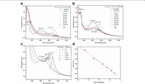
Figure 1 shows the time-dependent UV–Vis absorption spectra of the Ag-Au alloy nanostructures obtained with the Ag+:AuCl4− molar ratios of 1:2, 1:1, 1:0.5, 1:0.33, 1:0.2,
and 1:0.125. The single and symmetric peaks centered at 405 nm were attributed to the primary dipolar excitation of PSSMA-Ag NPs, which disappeared upon the addition of AuCl4− solution. This change is attributed to the galvanic
replacement reaction, namely, Ag(NP) + AuCl4−→Au(NP) +
Ag+[18, 19].
When the mixture was heated at 90 °C for 10 min, a new absorption peak began to evolve at a longer wave-length, nearer to the characteristic absorption peak of Au NPs. Continued heating lead to a slow increase in the absorption intensity and a successive blueshift of the surface plasmon resonance bands, indicating that the synthesized NPs were mostly composed of elemental Au. In addition, with an increasing Ag:Au molar ratio, the position of the final absorption band was closer to the absorption peak of Ag NPs (compare Fig. 1a and Fig.1b). After heating for 19 h, the final absorption bands were located at 517, 481, 460, 445, 432, and 422 nm, respect-ively (Fig. 1c). Further, a linear relationship between the resonance locations and the Ag mole fraction could be observed (the linear correlation coefficient was 0.99; Fig. 1d), indicating the successful formation of compos-ites. The plasmon resonance absorptions of bimetallic nanocrystals vary considerably from those of their monometallic NP counterparts since their surface plas-mon polaritons are determined by two different dielectric functions [20]. As reported in previous studies [13, 16], the formation of Ag-Au alloy structures can be confirmed by the presence of one absorption band, which would blueshift with an increase in the molar ratio of Ag [21–23]
Fig. 1Time-dependent UV–Vis spectra of Ag-Au alloy nanoparticles (NPs) prepared from PSSMA-Ag NPs:HAuCl4at molar ratiosa1:2 andb
1:0.125.cUV–Vis absorption spectra of Ag-Au alloy NPs at different Ag mole fractions.dPlot of the wavelength corresponding to the maximum absorbance against the Ag mole fraction for Ag-Au alloy NPs obtained by varying the mole fractions of HAuCl4while keeping the concentration
contrary to what would be observed in the formation of core-shell nanostructures.
Alloy NPs were also prepared through co-reduction of Ag and Au salt solutions at various pHs in an autoclave heated at 90 °C. In our previous work [24], it was estab-lished that a pH value of 10 is suitable for the synthesis of Ag NPs. UV–Vis absorption spectra of the samples (Fig. 2) clearly indicate that the plasmon resonance bands changed when the pH was adjusted to 10. How-ever, the linear correlation coefficient for the reaction without pH adjustment was 0.91, while that for the reac-tion with pH adjustment was only 0.84, implying that such a pH was not favorable for the synthesis of Ag-Au alloy NPs.
To verify these results, computer simulations were performed to assess whether the real UV–Vis spectra corresponded with theoretical calculations. The simu-lated UV–Vis spectra showed an extinction efficiency of the synthesized product. The relation of extinction, scat-tering, and absorption efficiency factors are as follows:
Qext¼QscaþQabs ð1Þ
The simulation methods of calculating the absorption and scattering efficiency usually belong to two categories: exact and approximated solutions [25]. For precise, spher-ically symmetric targets, such as homogenous spheres and multilayered concentric spheres, the Mie theory [26], the very first exact solution of Maxwell equations, can be applied. To date, several Mie theory computer codes have been developed [27]. Herein, the MieLab code [28], a free software specially designed for computing optical proper-ties of multilayered spheres, was used. The theoretical basis of MieLab has been illustrated by Yang [29]. Users should provide initial parameters, including the number of layers, size distribution of each layer, complex refractive
index tables for each layer, and refractive index of ambient medium, among others.
For homogeneous Ag-Au alloy spheres, the dielectric constants can be calculated as follows:
εAlloy χAg;ω
¼χAgεAgð Þ þω 1−χAg
εAuð Þω ; ð2Þ
whereχAgis the Ag fraction in the Ag-Au alloy,ωis the frequency of incident light, and εAg and εAu are the dielectric constants for Ag and Au, respectively. The relationship between dielectric constant and refractive index is described in Eq. 3, where n is the complex re-fractive index.
ε ωð Þ ¼nð Þω 2 ð
3Þ
By simply calculating the dielectric constant of the Ag-Au alloy, we obtained its refractive index table as the input file. Initial parameters were set as number of layers = 1, ra-dius = 10 nm, and refractive index of ambient medium = 1.4466. The simulation results are shown in Fig. 3. As the Ag fraction increased from 0 to 100 %, there was a signifi-cant increase in intensity, along with a blueshifting of the extinction maximum. Each curve shows only one peak, shifting from 537 (pure gold) to 415 nm (pure silver).
We compared UV–Vis absorption spectra of the pre-pared samples with simulated data for different Ag fractions of the Ag-Au alloy. The simulated spectrum indicated that Ag-Au alloy NPs only presented one plasmon resonance band and UV–Vis absorption wavelength shifts to shorter wavelengths with an increase in Ag fraction from 0 to 100 %; the linear correlation coefficient for the simulated spectrum was 0.97. Thus, experimental data was consistent with simulated data. Nevertheless, contrary to experimental data, the intensity of the UV–Vis absorption for the Ag fraction from 0 to 100 % in the simulated data gradually increased.
Fig. 2UV–Vis absorption spectra of Ag-Au alloy samples prepared at 90 °C for 19 h with different molar ratios of Ag+:AuCl 4
−in the solutionsa
[image:4.595.58.539.547.694.2]The existence of a single absorption band and a good linear relationship between the plasmon resonance bands with the increasing molar ratio of Ag confirmed the formation of Ag-Au alloy NPs. The mechanism of alloy formation with varying Ag:Au molar ratios was evaluated through analysis of the UV–Vis absorption spectra (Fig. 1). When HAuCl4 was added, the 405-nm
plasmon resonance band quickly disappeared for the materials synthesized with Ag+:AuCl4− ratios from 1:2 to
1:0.2, while a 387-nm new absorption peak appeared for the sample with a Ag+:AuCl4− ratio of 1:0.125. The
PSSMA-Ag NP solution was a yellowish brown color, indicative of the presence of Ag NPs, which rapidly changed to light yellow color following the addition of HAuCl4in agreement with the results of previous
stud-ies [7, 8, 10, 11, 30]. This color change was attributed to an oxidation-reduction reaction of AuCl4−, given that the
reduction potential of AuCl4− to Au (1.498 V) is higher
than that of Ag+to Ag (0.799 V); this also led to the dis-appearance of the 405-nm peak. The stoichiometric equivalent of Ag(0) to AuCl4− is 3 following a complete
reduction reaction. Thus, when the Ag+:AuCl4− ratio is
above 1:3, a reduction from Au3+to Au2+, Au+, and sub-sequently Au easily occurs. Nevertheless, the reduction of Au3+ to Au2+ and Au+ yields no absorptions in the UV–Vis region [16], thus explaining why the UV absorp-tion peak of AgNPs disappeared without the appearance of any Au NP peaks upon addition of HAuCl4.
For the reaction at a Ag+:AuCl4− ratio of 1:0.125, the
solution turned from yellowish brown to a pale brown color following the addition of HAuCl4to the
PSSMA-Ag NPs solution. In addition, the emergence of an absorption peak at 387 nm could be attributed to a de-crease in Ag NP size, since the Ag:AuCl4− molar ratio
was less than 1:3, and therefore Ag NPs could not be
completely oxidized to Ag+ given the low AuCl4−
concentrations.
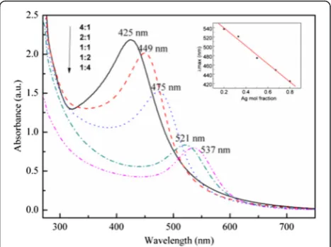
A set of seed-mediated growth experiments was con-ducted at a fixed total concentration of PSSMA-Ag NPs and HAuCl4of 0.5 mM (Fig. 4). A linear correlation
co-efficient of 0.99, comparing the maximum absorption wavelength to Ag content, was obtained. The absorption intensity of samples with Ag+:AuCl4− ratios of 4:1, 2:1,
1:1, 1:2, and 1:4 gradually increased with an increasing Ag concentration. The intensity increase was consistent with the simulated Ag-Au alloy results, thus confirming the formation of an Ag-Au alloy.
Optical Properties and DDA Simulation of Ag-Au Core-Shell NPs
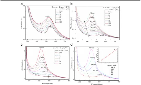
PSSMA-stabilized Ag-Au core-shell NPs were prepared by heating the mixtures of PSSMA-Au NPs, and AgNO3
at 90 °Cas high temperature in synthesis of Ag-Au core-shell nanoparticles leads to nonuniform the sizes of NPs. UV–Vis absorption spectra for the prepared samples with Au/Ag molar ratios of 1:0.17, 1:0.5, 1:1, 1:2, and 1:4 are shown in Fig. 5. The Ag-Au bimetallic nanostruc-tures with an Au:Ag molar ratio of 1:0.17 had only one absorption band in the 522–547-nm absorption range during the heating process. Samples with an Au:Ag molar ratio of 1:1 and 1:4 showed two plasmon resonance bands during the early stage of the reaction (Fig. 5b, c). After heating for several hours, only one peak was observed and attributed to the increasing thickness of the Ag shell.
After heating for the same amount of time, UV–Vis ab-sorption bands of the five samples were located at 522, 467, 446, 434, and 417 nm and were blueshifted with the increasing Au:Ag ratio. The linear correlation coeffi-cient for the maximum absorption wavelength versus Ag concentration was 0.91 and much lower than that
Fig. 3Simulated UV–Vis spectrum of Ag-Au alloy with different Ag contents by discrete dipole approximation.Inset: linear relationship between maximum absorption wavelength and Ag mole fraction
[image:5.595.57.291.87.261.2] [image:5.595.305.539.519.693.2]of the Ag-Au alloy NPs (Fig. 5d). Thus, the possibility of alloy NP formation is excluded. On the other hand, the appearance of two plasmon resonance bands and the consistent blueshifted absorptions which eventu-ally merged into one peak strongly prove the forma-tion of core-shell Ag-Au NPs [6].
To further analyze the synthesized core-shell NPs, UV–Vis absorption spectra of the prepared samples were compared with the simulated data (Fig. 6) for different Ag concentrations in Ag-Au core-shell NP formation.
For the simulation results of citric acid-coated Ag@Au spheres, all the particles were immersed in cyclohexane, leading to a three-layered sphere model. According to TEM images of the materials, the initial parameters were set at an Au core size of 532 nm. In the simulated model, as the Au:Ag+ ratio progressed from 1:0 to 1:2.36, the spectrum showed two characteristic peaks. With increas-ing particle size, the plasmon resonance bands gradually blueshifted and the absorption intensity increased with the increasing Ag ratio until only one peak could be ob-served, consistent with the experimental results obtained (Fig. 5). Changes in the peak positions and absorption in-tensity in both experimental and simulated spectra were influenced by an increase in Ag shell thickness around Au NPs following an increase in the Ag ratio. When the
Au:Ag molar ratio was less than 1:0.33, only one absorp-tion peak was observed. With a relative increase in Ag, two plasmon resonance bands appeared, representing the typical plasmon resonance absorption of Ag-Au core-shell NPs as described by Murphy et al. [6]. Finally, with high Ag concentrations, only the characteristic peak of Ag
Fig. 5Time-dependent UV–Vis spectra of the prepared samples at different CPSSMA-Au NPs:CAgNO3ratiosa1:0.17,b1:1, andc1:4 at a PSSMA-Au
nanoparticle concentration of 0.25 mM.dWavelength corresponding to the maximum absorbance for different CPSSMA-Au NPs:CAgNO3ratios
[image:6.595.57.539.87.378.2] [image:6.595.306.538.523.703.2]could be observed. The reducing ability of Au NPs is weaker than that of Ag NPs, and therefore Au NPs could not be oxidized by Ag+to form Ag NPs when AgNO3was
added to the Au seed suspension. In fact, Ag NPs were formed by PSSMA reduction of AgNO3on the surface of
the Au seed through electrostatic adsorption.
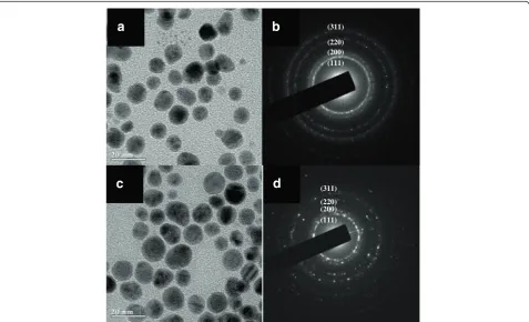
Morphological Study of Ag-Au Bimetallic Nanostructures Representative TEM images of the PSSMA-stabilized metal NPs indicating a particle size of approximately 20 nm are shown in Fig. 7. The sample morphology varied according to the Ag content. The mechanism of formation of the alloy involves etching of the Ag NP sur-face by HAuCl4before Au NP deposition on the Ag NP
surface, followed by PSSMA reduction of Ag ions. The alloy process is incomplete and only prominent at the interface between the Ag core and Au shell [31]. Several lattice planes could be observed in their corresponding selected area electron diffraction patterns (Fig. 7b, d). Since Ag and Au have very similar lattice parameters and are miscible over the entire composition range [13, 32], the two series of lattice planes were practically identical.
Ag-Au alloy NPs were also synthesized in an auto-clave with variation in the Ag:AuCl4− ratios. The seed
Ag NPs were not well-distributed in size and had varied
morphologies ranging from spherical to triangular or polygonal. The Ag seed diameters were approximately 9.7 nm (Additional file 1: Figure S1a). Following addition of HAuCl4(Ag:AuCl4–ratios were 1:0.25 and 1:1), the
ob-tained alloy NPs were mainly spherical with the existence of a few triangular NPs. The NP diameters were approxi-mately 10.6 and 11.2 nm, respectively (Additional file 1: Figure S1b and S1c). This increase in size was attributed to the formation of Ag-Au alloy NPs. A further increase in HAuCl4concentration lead to the formation of
irregular-shaped NPs with a decrease in mean diameter to 10.6 nm, probably due to the formation of small Au NPs, which did not form an alloy with Ag seeds.
Ag-Au core-shell nanostructures were observed under TEM. Nearly spherical core-shell NPs of approximately 15 nm were observed to coexist with moderate amounts of NPs smaller than 2 nm (Additional file 1: Figure S2). It is hypothesized that the smaller particles were pure Ag due to the reduction of Ag+by PSSMA. Reduction of Ag+ on the Au NPs resulted in the formation of core-shell nanostructures, while nucleation and growth of Ag without the substrate may have led to the formation of the smaller Ag NPs. In addition, the Ag shell thickened with an increasing Ag+to Au3+ratio, consistent with the UV–Vis spectra results.
a
c
b
(111) (200) (220) (311)
d
(111) (200) (220) (311)
[image:7.595.60.538.413.703.2]Characterization of Structure and Composition of Ag-Au Bimetallic Nanostructures
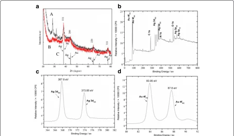
[image:8.595.56.539.421.702.2]X-ray diffraction patterns corresponding to the synthe-sized bimetallic NPs are shown in Fig. 8. Peaks at 38.2°, 44.4°, 64.5°, and 77.5° (Fig. 8a, b) can be indexed as the (111), (200), (220), and (311) lattice planes of the cubic Ag or Au nanostructures within experimental error [8, 32–34]. Since Ag and Au have very similar lattice parameters and complete miscibility for the composition range [13, 32, 33], these lattice planes are also observed in cubic Ag-Au alloy or core-shell structures. Further, the lat-tice plane of (111) is the most obvious, as it has a higher free energy and faster grow rate [35].
Additional evidence of the composition of Ag-Au alloy NPs was obtained by XPS. Figure 8b shows the XPS spec-tra of the Ag-Au alloy sample with an Ag:AuCl4− molar
ratio of 1:2. Peaks of C 1s, N 1s, and O 1s confirm the existence of a PSSMA-stabilized structure. Further, prom-inent peaks of Ag 3d and Au 4f regions, with a 6.0-eV difference between Ag 3d5/2and 3d3/2, and 3.7-eV
differ-ence between Au 4f7/2and 4f5/2, were recorded and
attrib-uted to the presence of Ag and Au atoms (Fig. 8c, d). According to the XPS handbook, peaks at 368.3 and 374.3 eV corresponding to Ag 3d5/2 and Ag 3d3/2 were
assigned to pure Ag and those at 367.5 and 373.5 eV to Ag2O. The binding energies of Ag 3d5/2and Ag 3d3/2of
the alloy nanostructures with a Ag:AuCl4− ratio of 1:2
observed herein were similar to those stated for Ag and Ag2O [34]. The 0.5-eV energy shift (Ag:AuCl4−= 1:2) for
the Ag peaks was attributed to the interaction between the carboxyl oxygen of PSSMA and the Ag core. For samples with Ag:AuCl4− ratios of 1:0.5 and 1:0.33, a slight binding
energy difference of 0.2 eV deviation from the standard values was observed, which suggests the formation of Ag-Au alloy NPs as reported in the literature [36].
Conclusions
Water-soluble Ag-Au bimetallic NPs were synthesized by using polymer PSSMA as both a reducing agent and a stabilizer. By adjusting the Ag:Au molar ratio, different optical properties were observed during the formation of Ag-Au alloy and core-shell NPs. One plasmon resonance band of the Ag-Au alloy NPs could be adjusted within the range 422–517 nm by varying the HAuCl4
concentra-tion in the aqueous PSSMA-stabilized Ag NP soluconcentra-tions. Due to the co-reduction of Ag and Au salts in PSSMA aqueous solution, UV–Vis absorption bands were observed to redshift with an increase in Au content. However, two plasmon resonance bands were observed during the forma-tion of the Ag-Au core-shell NPs, with a blueshift of the UV–Vis absorption bands due to the reduction of Ag salts on the surface of Au seeds. Furthermore, changes in the Ag:Au ratio had an effect on the composition and morph-ology of the Ag-Au alloy and core-shell nanostructures.
b
c
d
a
A
B
C
Additional File
Additional file 1: Figure S1.Transmission electron microscopy images and corresponding particle sizes. Histograms of the synthesized Ag NPs (a and b) and Ag-Au alloy nanostructures with CPSSMA-Ag NPs:CHAuCl4of 1:0.25
(c and d) and 1:1 (e and f). The concentration of PSSMA-Ag nanoparticles was 0.14 mM. The size distribution of the particles was calculated from the TEM images of the prepared samples.Figure S2.Transmission electron microscopy images of different magnification factor of the as-prepared nanoparticles with CPSSMA-Au NPs:CAgNO3of 1:0.5 (a and b) and 1:2 (c and d).
The concentration of PSSMA-Au nanoparticles was 0.25 mM. (DOC 1549 kb)
Abbreviations
DDA:discrete dipole approximation; NPs: nanoparticles; PSSMA: poly(4-styrenesulfonic acid-co-maleic acid); PSSMA-Ag NPs: PSSMA-stabilized Ag nanoparticles; PSSMA-Au NPs: PSSMA-stabilized Au nanoparticles; TEM: transmission electron microscopy; XPS: X-ray photoelectron spectra.
Competing Interests
The authors declare that they have no competing interests.
Authors’contributions
YH carried out the synthesis and characterizations of the products, analysed data, and drafted the manuscript. AQZ carried out the theoretical simulations of UV-vis spectra. HJL performed the synthetic experiments of Au-Ag alloy nanostructures. DJQ and MC supervised the project, contributed in the de-sign and discussion of this work, and in the revision of the manuscript. All authors read and approved the finalmanuscript.
Acknowledgements
The financial support from the National Science Foundation of China (51073039, 21471036, 11179015, and 51173108) and Innovation Program of Shanghai Municipal Education Commission (12ZZ143) are gratefully acknowledged.
Author details
1Department of Chemistry, Shanghai Key Laboratory of Molecular Catalysis
and Innovative Materials, Fudan University, Shanghai 200433, P. R. China.
2Department of Materials Science, Fudan University, Shanghai 200433, P. R.
China.
Received: 7 November 2015 Accepted: 14 April 2016
References
1. Hong S, Choi Y, Park S (2011) Shape control of Ag shell growth on Au nanodisks. Chem Mat 23:5375–5378
2. Wilson OM, Scott RWJ, Garcia-Martinez JC, Crooks RM (2005) Synthesis, characterization, and structure-selective extraction of 1-3-nm diameter AuAg dendrimer-encapsulated bimetallic nanoparticles. J Am Chem Soc 127:1015–1024 3. Wang C, Peng S, Chan R, Sun SH (2009) Synthesis of AuAg alloy
nanoparticles from core/shell-structured Ag/Au. Small 5:567–570 4. Kumar GVP, Shruthi S, Vibha B, Reddy BAA, Kundu TK, Narayana C (2007)
Hot spots in Ag core-Au shell nanoparticles potent for surface-enhanced Raman scattering studies of biomolecules. J Phys Chem C 111:4388–4392 5. Liu JH, Wang AQ, Chi YS, Lin HP, Mou CY (2005) Synergistic effect in an
Au-Ag alloy nanocatalyst: CO oxidation. J Phys Chem B 109:40–43 6. Mallin MP, Murphy CJ (2002) Solution-phase synthesis of sub-10 nm Au-Ag
alloy nanoparticles. Nano Lett 2:1235–1237
7. Yang J, Lee JY, Too HP (2005) Core-shell Ag-Au nanoparticles from replacement reaction in organic medium. J Phys Chem B 109:19208–19212 8. Shin Y, Bae IT, Arey BW, Exarhos GJ (2008) Facile stabilization of gold-silver
alloy nanoparticles on cellulose nanocrystal. J Phys Chem C 112:4844–4848 9. Ah CS, Do Hong S, Jang DJ (2001) Preparation of AucoreAgshell nanorods
and characterization of their surface plasmon resonances. J Phys Chem B 105:7871–7873
10. Jin YD, Dong SJ (2003) One-pot synthesis and characterization of novel silver-gold bimetallic nanostructures with hollow interiors and bearing nanospikes. J Phys Chem B 107:12902–12905
11. Zhang X, Tsuji M, Lim S, Miyamae N, Nishio M, Hikino S, Umezu M (2007) Synthesis and growth mechanism of pentagonal bipyramid-shaped gold-rich Au/Ag alloy nanoparticles. Langmuir 23:6372–6376
12. Gheorghe DE, Cui LL, Karmonik C, Brazdeikis A, Penaloza JM, Young JK, Drezek RA, Bikram M (2011) Gold-silver alloy nanoshells: a new candidate for nanotherapeutics and diagnostics. Nanoscale Res Lett 6:554
13. Link S, Wang ZL, El-Sayed MA (1999) Alloy formation of gold-silver nanoparticles and the dependence of the plasmon absorption on their composition. J Phys Chem B 103:3529–3533
14. Cheng LC, Huang JH, Chen HM, Lai TC, Yang KY, Liu RS, Hsiao M, Chen CH, Her LJ, Tsai DP (2012) Seedless, silver-induced synthesis of star-shaped gold/ silver bimetallic nanoparticles as high efficiency photothermal therapy reagent. J Mater Chem 22:2244–2253
15. Hodak JH, Henglein A, Giersig M, Hartland GV (2000) Laser-induced inter-diffusion in AuAg core-shell nanoparticles. J Phys Chem B 104:11708–11718
16. Gonzalez CM, Liu Y, Scaiano JC (2009) Photochemical strategies for the facile synthesis of gold-silver alloy and core-shell bimetallic nanoparticles. J Phys Chem C 113:11861–11867
17. Hubenthal F, Ziegler T, Hendrich C, Alschinger M, Trager F (2005) Tuning the surface plasmon resonance by preparation of gold-core/silver-shell and alloy nanoparticles. Eur Phys J D 34:165–168
18. Chen JY, Wiley B, McLellan J, Xiong YJ, Li ZY, Xia YN (2005) Optical properties of Pd-Ag and Pt-Ag nanoboxes synthesized via galvanic replacement reactions. Nano Lett 5:2058–2062
19. Sun YG, Xia YN (2002) Shape-controlled synthesis of gold and silver nanoparticles. Science 298:2176–2179
20. Sinzig J, Quinten M (1994) Scattering and absorption by spherical multilayer particles. Appl Phys A-Mater Sci Process 58:157–162
21. Udayabhaskararao T, Sun Y, Goswami N, Pal SK, Balasubramanian K, Pradeep T (2012) Ag7Au6: a 13-atom alloy quantum cluster. Angew Chem Int Ed 51:2155–2159
22. Tong L, Cobley CM, Chen JY, Xia YN, Cheng JX (2010) Bright three-photon luminescence from gold/silver alloyed nanostructures for bioimaging with negligible photothermal toxicity. Angew Chem Int Ed 49:3485–3488 23. Song JM, Chen WT, Hsieh KH, Kao TH, Chen IG, Chiu SJ, Lee HY (2014) An in
situ study on the coalescence of monolayer-protected Au-Ag nanoparticle deposits upon heating. Nanoscale Res Lett 9:438
24. Cai LJ, Wang M, Hu Y, Qian DJ, Chen M (2011) Synthesis and mechanistic study of stable water-soluble noble metal nanostructures. Nanotechnology 22:285601
25. Zhao J, Pinchuk AO, McMahon JM, Li SZ, Ausman LK, Atkinson AL, Schatz GC (2008) Methods for describing the electromagnetic properties of silver and gold nanoparticles. Acc Chem Res 41:1710–1720
26. Mie G (1908) Beiträge zur Optik trüber Medien, speziell kolloidaler Metallösungen. Ann Phys 330:377–445
27. Du H (2004) Mie-scattering calculation. Appl Opt 43:1951–1956 28. Ovidio P, Pablo P, Umapada P (2011) MieLab: a software tool to perform
calculations on the scattering of electromagnetic waves by multilayered spheres. Int J Spectrosc 2011:583743
29. Yang W (2003) Improved recursive algorithm for light scattering by a multilayered sphere. Appl Opt 42:1710–1720
30. Sun L, Luan WL, Shan YJ (2012) A composition and size controllable approach for Au-Ag alloy nanoparticles. Nanoscale Res Lett 7:225 31. Pedersen DB, Wang SL, Duncan EJS, Liang SH (2007) Adsorbate-induced
diffusion of Ag and Au atoms out of the cores of Ag@Au, AIJ@Ag, and Ag@AgI core-shell nanoparticles. J Phys Chem C 111:13665–13672 32. Devarajan S, Bera P, Sampath S (2005) Bimetallic nanoparticles: a single step
synthesis, stabilization, and characterization of Au-Ag, Au-Pd, and Au-Pt in sol-gel derived silicates. J Colloid Interface Sci 290:117–129
33. Pal A, Shah S, Devi S (2008) Preparation of silver-gold alloy nanoparticles at higher concentration using sodium dodecyl sulfate. Aust J Chem 61:66–71 34. Deng ZW, Chen M, Wu LM (2007) Novel method to fabricate SiO(2)/Ag
composite spheres and their catalytic, surface-enhanced Raman scattering properties. J Phys Chem C 111:11692–11698
35. Zhang JT, Li XL, Sun XM, Li YD (2005) Surface enhanced Raman scattering effects of silver colloids with different shapes. J Phys Chem B 109:12544–12548 36. Han SW, Kim Y, Kim K (1998) Dodecanethiol-derivatized Au/Ag bimetallic