JOURNAL OFCLINICALMICROBIOLOGY, Dec. 1995, p. 3138–3145 Vol. 33, No. 12 0095-1137/95/$04.0010
Copyrightq1995, American Society for Microbiology
Comparison of Three Staining Methods for Detecting
Microsporidia in Fluids
ELIZABETH S. DIDIER,1* JAN M. ORENSTEIN,2ABDALLA ALDRAS,1† DONNA BERTUCCI,1
LINDA B. ROGERS,1
ANDF. AILLEEN JANNEY3
Department of Microbiology, Tulane Regional Primate Research Center, Covington, Louisiana 704331;
Department of Pathology, George Washington University Medical Center, Washington, D.C. 200372;
and Department of Pathology, Louisiana State University Medical Center,
New Orleans, Louisiana 701123
Received 26 May 1995/Returned for modification 20 July 1995/Accepted 6 September 1995
Calcofluor white 2MR, modified trichrome blue, and indirect immunofluorescent antibody (IFA) staining
methods were evaluated and compared for detecting microsporidia in stool. Serial 10-fold dilutions of
Enceph-alitozoon(Septata)intestinaliswere prepared in three formalinized stool specimens or in Tris-buffered saline. Ten-microliter aliquots were smeared onto glass slides, fixed with methanol, stained, and read by at least three individuals. The results indicated that the calcofluor stain was the most sensitive method, required approxi-mately 15 min to perform, but did generate some false-positive results due to similarly staining small yeast cells. The modified trichrome blue stain was nearly as sensitive as the calcofluor stain and allowed for easier distinction between microsporidia and yeast cells. This stain, however, required approximately 60 min to
perform. The IFA stain with polyclonal murine antiserum againstE. intestinaliswas the least sensitive of the
methods and required approximately 130 min to perform. The lower limit of detection with the calcofluor and
modified trichrome stains was a concentration of about 500 organisms in 10ml of stool to detect one
micro-sporidian after viewing 50 fields at a final magnification of31,000. Reliability was also addressed by use of 74
stool, urine, and intestinal fluid specimens, 50 of which were confirmed for the presence of microsporidia by transmission electron microscopy (TEM). All TEM-positive specimens were detected by calcofluor and mod-ified trichrome blue staining. Ten specimens were not detected by the IFA stain. An additional seven TEM-negative specimens were read positive for microsporidia with the calcofluor stain, and of these, five also were read positive with the modified trichrome blue stain. The resulting diagnostic paradigm was to screen speci-mens with the calcofluor stain and to confirm the results with the modified trichrome stain. IFA, which was less sensitive, may become useful for microsporidian species identification as specific antibodies become available.
Microsporidia are small protozoan parasites which infect a wide range of hosts, including insects, fish, birds, and mammals (4). These organisms are reported increasingly as causing op-portunistic infections in persons with AIDS, and clinical signs of disease include chronic diarrhea, cystitis, nephritis, cholan-gitis, hepatitis, peritonitis, myositis, keratitis, sinusitis, and pneumonia (18, 23, 31). Diagnosis of microsporidiosis has been difficult because of the small spore sizes of the microsporidian species infecting man, which range from 1.0 by 1.5 to 2.0 by 4.0
mm. Definitive biopsy diagnosis often has depended upon transmission electron microscopy (TEM) for observing the po-lar tubule which is the unique structure found only in micro-sporidia. TEM, however, is time-consuming and costly, re-quires a great deal of expertise, and is believed to be less sensitive than desired. As a result, attention has been directed toward development of reliable, sensitive, and efficient meth-ods for detecting microsporidia in patient fluid specimens. Detection of specific antibodies against microsporidia in serum is variable in AIDS patients as a result of progressing immu-nodeficiency, resulting in unreliable diagnoses (7), although improvements for defining specific antibodies by Western blot-ting (immunoblotblot-ting) have been reported more recently (17).
Diagnosis of microsporidiosis, however, still appears to depend upon direct detection of the parasite.
Several (immuno)histochemical staining methods for detect-ing microsporidia in patient specimens have been developed. One histochemical method is the modified trichrome stain which was first applied to microsporidia by Weber et al. (30) and later modified by Ryan et al. (20), who used aniline blue instead of fast green as the counterstain. A further modifica-tion was applied by Kokoskin et al. (13), who used the modified trichrome blue stain at higher temperatures to increase the intensity of staining and shorten the staining time. Methods employing fluorescent brighteners that stain chitin in the en-dospore layer of microsporidia were described by Vavra and Chalupsky (26) and more recently by van Gool et al. (24). Indirect immunofluorescent antibody (IFA) staining methods using polyclonal and monoclonal antibodies have been used to detect microsporidia as well (1, 32, 36). These three methods have different advantages and disadvantages. The purpose of this study, therefore, was to measure the levels of sensitivity, specificity, and reliability of the modified trichrome blue stain, the calcofluor stain, and IFA staining methods.
MATERIALS AND METHODS
Specimens for sensitivity assays.Encephalitozoon intestinalis, originally named Septata intestinalis (3) and subsequently reclassified (11), was grown in RK-13
cells (6, 8, 24), harvested from tissue culture supernatants, washed once with Tris-buffered saline (TBS) containing 0.3% Tween 20 (TBS-Tween), resus-pended in TBS, and counted on a hemacytometer. Aliquots of microsporidia
containing 10-fold dilutions ranging from 53108
to 53104
/ml were prepared. Three microsporidian-negative stool specimens were mixed with 10% buffered
* Corresponding author. Mailing address: Department of Microbi-ology, Tulane Regional Primate Research Center, 18703 Three Rivers Rd., Covington, LA 70433. Fax: (504) 893-1352. Electronic mail ad-dress: [email protected].
† Present address: Department of Medical and Molecular Parasitol-ogy, New York University Medical Center, New York, NY 10010.
3138
on May 15, 2020 by guest
http://jcm.asm.org/
formalin (1 part stool with 3 parts 10% formalin) and vortexed to resuspend the fecal matter. One milliliter of each stool was mixed with 1 ml of each micros-poridian aliquot to generate three sets of stool samples containing the five
dilutions of microsporidia. The samples were centrifuged at 4003g for 15 min,
the supernatants were removed, and 10% buffered formalin was added to gen-erate a final 1-ml suspension. In this way, each dilution of microsporidia con-tained an equal amount of fecal debris.
Smears were prepared by applying 10ml of each parasite suspension from the
three sets of stools and from the TBS control suspensions onto areas (2 by 3 cm2
) marked on glass slides. The smears were allowed to dry, and the slides were fixed in methanol for 10 min at room temperature. Control slides containing tissue-culture-derived microsporidia were included for all staining methods to verify optimum conditions of the procedures.
Specimens for reliability assays.Seventy-four stool, pelletted urine, and pel-letted duodenal lavage fluid specimens were fixed with 10% buffered formalin and assessed for the presence of microsporidia by TEM as described previously (19). Of these, 37 contained Enterocytozoon bieneusi (10 stool and 27 duodenal lavage fluid specimens), and 12 contained Encephalitozoon intestinalis (4 stool, 3 duodenal lavage fluid, and 5 urine specimens). These specimens were coded and tested blind by the calcofluor, modified trichrome blue, and IFA staining meth-ods.
Calcofluor staining method.The fluorescent brightener calcofluor white M2R (Sigma Chemical Co., St. Louis, Mo.) was prepared as a 0.5% (wt/vol) solution in TBS (pH 7.2) and stored in the dark at room temperature (26, 27). Prior to
use, the calcofluor stain was centrifuged at 15,0003g for 2 min to remove
precipitates. Methanol-fixed smears were allowed to air dry, and a drop or two of the calcofluor solution was added. After 2 or 3 min at room temperature, the slides were rinsed with slow-running water and counterstained with 0.1% Evan’s blue (Sigma; E-2129) in TBS (pH 7.2) for 1 min at room temperature. The slides were rinsed under slow-running water, allowed to air dry, sealed with Cytoseal 60 (Stephen’s Scientific, Riverdale, N.J.), and viewed under a UV microscope at a wavelength of 395 to 415 nm (observation light of 455 nm). Organisms appeared as bluish-white or turquoise oval halos.
Modified trichrome blue staining method.The modified trichrome stain orig-inally used for microsporidia by Weber et al. (30) and which was modified by Ryan et al. (20) and Kokoskin et al. (13) was adapted for this study by changing the incubation time and temperature. The staining solution in this study was prepared by dissolving 6.0 g of chromotrope 2R (Sigma; C-3143) with 0.5 g of aniline blue (Fisher, Fairlawn, N.J.; A-969) and 0.7 g of phosphotungstic acid in 3 ml of glacial acetic acid. This solution stood at room temperature for 30 min, after which 100 ml of distilled water was added and 1.0 M HCl was added to generate a pH 2.5 solution. Methanol-fixed smears were stained in this
chromo-trope 2R solution for 30 min at 378C and then rinsed for 10 s with acid alcohol
(4.5 ml of acetic acid in 995.5 ml of 90% ethyl alcohol). The smears were then dehydrated with a 10-s rinse in 95% ethyl alcohol, two 5-min incubations in 95% ethyl alcohol, a 10-min incubation in 100% ethyl alcohol, and a 5-min incubation in xylene (or xylene substitute). Each slide was then sealed with Cytoseal 60, and
the slides were viewed under a light microscope with a3100 oil-immersion lens
to generate a final31,000 magnification.
IFA staining method.The IFA staining was performed as described by Aldras et al. (1). Briefly, polyclonal antiserum was raised in BALB/c mice which were
given three monthly intraperitoneal inoculations of 107
Percoll-purified
Enceph-alitozoon intestinalis organisms. Sera were collected by bleeding from the tail vein
1 week after the third inoculation, pooled, absorbed with pelleted formalin-fixed
fecal debris to remove cross-reacting antibodies, and stored at2408C until used.
Methanol-fixed smears were incubated with antiserum diluted 1:100 with TBS for
60 min at 378C, rinsed with TBS, and stained with fluorescein
isothiocyanate-conjugated goat anti-mouse immunoglobulin G (diluted 1:100; Sigma) for 60 min
at 378C. After rinsing with TBS, the slides were sealed with 95% glycerol in TBS
and viewed with a UV microscope with a wavelength of 380 to 490 nm
(obser-vation light wavelength of.515 nm).
In one set of experiments, the microsporidia were smeared directly onto glass slides prior to staining. In a second set of experiments, and when using the patient specimens, the suspensions were washed once with TBS-Tween by
cen-trifugation at 15,0003g for 2 min, and the pellets were resuspended in the
original volume of 10% buffered formalin prior to preparing the smears for staining.
Statistics.In the sensitivity studies, the numbers of organisms viewed per field
with a3100 oil-immersion objective (total magnification,31,000) were counted
until at least 200 organisms or 100 fields were viewed. Results from three readings per slide were averaged. In the reliability studies, all slides were read by three individuals. In cases with discrepancies, the results of the two like readings were used. Comparisons between any two groups of samples were measured by
the two-tailed Student t test. For measuring the reliability results, sensitivity5
[(number of true positives)/(number of true positives1number of false
nega-tives)]3100; specificity5[(number of true negatives)/(number of true negatives
1number of false positives)]3100; positive predictive value5[(number of true
positives)/(number of true positives1number of false positives)]3100; and
negative predictive value5[(number of true negatives)/(number of true
nega-tives1number of false negatives)]3100.
RESULTS
Staining characteristics of microsporidia.The staining
char-acteristics of Encephalitozoon intestinalis and Candida albicans are shown in Fig. 1. When stained with calcofluor, the
Enceph-alitozoon intestinalis organisms displayed variable fluorescence
intensities in that mature organisms are smaller and stained more intensely, while germinated spores and less mature forms (meronts and sporonts), which are slightly larger, fluoresced less intensely. Depending on the orientation of the microspo-ridia, the anterior end of the spores may appear concavitated. Whereas microsporidia tend to be more oval in shape, small yeast cells are more round, although budding or recently bud-ded yeast cells may appear teardrop shaped. Yeast cells also display a turquoise fluorescent ring but, unlike microsporidia, will counterstain orange in the cytoplasm.
[image:2.612.60.556.72.259.2]Microsporidia stained with modified trichrome blue appear bright pink. Often, a posterior vacuole and central diagonal pink stripe within the spores are visible. Small yeast cells, such as C. albicans cells, also stain bright pink but appear darker
FIG. 1. Staining characteristics of microsporidia and yeast cells. Encephalitozoon intestinalis in stool specimens were stained with calcofluor (A), modified trichrome
blue (B), or IFA (C). Microsporidia (arrows) and yeast cells (arrowheads) are shown in each panel. Final magnification,31,000.
on May 15, 2020 by guest
http://jcm.asm.org/
and no internal structures are visible, making the yeast cells appear more homogeneous in their staining qualities. Bacteria and most fecal debris appear light blue because of the coun-terstain.
By IFA staining with the murine hyperimmune antiserum raised against Encephalitozoon intestinalis and fluorescein iso-thiocyanate-conjugated goat anti-mouse immunoglobulin G, microsporidia stain as bright apple-green oval halos. Back-ground staining appeared to be more orange to brownish-yellow, which was reduced by absorbing the primary hyperim-mune antiserum with nonspiked fecal debris. C. albicans stained brownish-yellow in the IFA test.
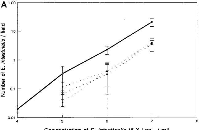
Sensitivity studies.The sensitivity and lower limits of
para-site detection were measured for the calcofluor, modified trichrome blue, and IFA stains. Each value represents the mean of three readings (6standard deviation). Comparisons were made between dilutions of microsporidia suspended in stool and those suspended in TBS. In the calcofluor staining assay (Fig. 2), no statistically significant differences were seen at any dilution when comparing microsporidia added to TBS with those suspended in stool. In the modified trichrome blue staining assay (Fig. 2), statistically significantly lower numbers of microsporidia were viewed per field when the microsporidia were suspended in stool at the higher concentrations (53106 organisms per ml, P,0.05; 53 107organisms per ml, P, 0.05). This was probably due to clumping of the microsporidia such that individual spores were not discerned. At the lower concentrations of 53104and 53105microsporidia per ml of stool, however, there were no statistically significant differ-ences in the number of microsporidia observed per field when compared with the number of microsporidia suspended in TBS.
With the IFA stain, significantly lower numbers of micro-sporidia were detected at all concentrations of organisms sus-pended in stool when compared with the concentrations of organisms suspended in TBS (Fig. 3A). Because microsporidia adsorb proteins onto their surfaces, the IFA staining was re-peated after washing an aliquot of each stool suspension of microsporidia with TBS-Tween (Fig. 3B). At the higher con-centrations of 5 3 106 and 5 3 107 organisms per ml, the average number of organisms detected per field improved if the parasite suspension was washed with TBS-Tween. At the lower concentration of 5 3 105 microsporidia per ml, the number of washed microsporidia detected in stool was still significantly lower (P, 0.05) than the number of microspo-ridia detected in TBS.
The lower limits for detecting microsporidia by the cal-cofluor, modified trichrome blue, and IFA staining methods were measured and compared. Less than one microsporidian per field was observed when microsporidia were suspended at 53104organisms per ml of TBS, and the average numbers of fields viewed to observe one microsporidian were counted. The results in Table 1 show that an average of approximately 50 fields were viewed to detect one microsporidian suspended in TBS by any of the three staining methods. The number of fields viewed to find one microsporidian did not increase significantly if the parasites were suspended in stool and stained with cal-cofluor or modified trichrome blue. However, approximately threefold more fields (160) were viewed to detect one IFA-stained microsporidian in the stool suspensions.
The ability to distinguish between yeast cells and microspo-ridia was also addressed. Equivalent numbers of C. albicans and Encephalitozoon intestinalis were added to the stool spec-imens. Smears were prepared and stained with the calcofluor, modified trichrome blue, and IFA stains, and the number of microsporidia was counted. By calcofluor staining,
approxi-mately 10% more microsporidia were counted at each dilution when compared with the microsporidia suspended in TBS but not spiked with C. albicans (data not shown). No statistically significantly higher numbers of microsporidia were counted in stools containing microsporidia and yeast cells that were stained by the modified trichrome blue or IFA stains.
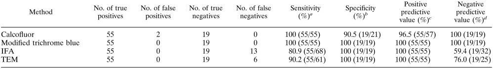
Reliability studies.The calcofluor, modified trichrome blue,
and IFA stains were compared for their levels of reliability in detecting microsporidia in 74 clinical specimens. All 49 TEM-positive specimens were detected by the calcofluor and modi-fied trichrome blue stains, whereas 10 TEM-positive specimens were not detected by the IFA stain (Table 2). An additional eight specimens (five stool and three intestinal fluid samples) were read positive for microsporidia by the calcofluor stain, and six of those also were read positive by the modified trichrome blue stain. At least 200 fields were examined, and only two or three microsporidia were observed in these spec-imens. TEM was repeated on these additional eight specimens, however, and a microsporidian was detected in one stool spec-imen. With TEM as the reference standard, both calcofluor and modified trichrome stains were 100% sensitive but ex-pressed lower levels of specificity (77.4 and 82.8%, respective-ly). Because TEM is believed to be relatively insensitive, and since IFA was even less sensitive than TEM, the reliability calculations were reevaluated to include as true positives those specimens that were read positive by both calcofluor and mod-ified trichrome blue stains (Table 3). This resulted in individ-ually higher levels of specificity and predictive values for both calcofluor and modified trichrome stains.
When comparing time efficiencies for performing these staining methods, the calcofluor stain required the least time of approximately 15 min, followed by the modified trichrome blue stain, which required approximately 60 min, and the IFA stain, which required approximately 130 min. In addition, the cal-cofluor-stained specimens were easiest to read, requiring ap-proximately 2 to 3 min per slide to view 200 fields. Specimens stained with modified trichrome blue or by IFA stain required approximately 5 to 10 min to carefully view 200 fields for detecting microsporidia. The times required to prepare the reagents or to generate the polyclonal antisera were not in-cluded in this comparison.
DISCUSSION
Opportunistic infections are major causes of morbidity and mortality in persons infected with human immunodeficiency virus. Microsporidia are reported increasingly in patients with chronic diarrhea, with prevalence rates reaching 39% (14, 18). Additional cases of keratitis, myositis, sinusitis, nephritis, hep-atitis, peritonitis, and pneumonia have been associated with microsporidiosis in AIDS patients (31). Because all stages of the microsporidian species that infect humans are quite small, it is believed that they are easily overlooked in routine diag-nostic procedures, particularly since they stain variably with hematoxylin-eosin.
Presently, definitive diagnosis of microsporidiosis often de-pends upon TEM, which is time-consuming, requires much expertise, and is relatively insensitive. As such, much effort has been directed toward developing alternative diagnostic meth-ods. In this study, three staining methods, a calcofluor chitin stain, a modified trichrome blue stain, and an IFA stain, were compared. The resulting diagnostic paradigm was to screen all specimens with the calcofluor method because it was the quick-est method of the three, had a high level of sensitivity, and had a 100% negative predictive value. The calcofluor staining method may have generated some false-positive results,
pre-3140 DIDIER ET AL. J. CLIN. MICROBIOL.
on May 15, 2020 by guest
http://jcm.asm.org/
sumably due to similarly staining small yeast cells. However, since TEM was used as the gold standard and its sensitivity for detecting spores in fluid specimens is unknown, the false pos-itives may actually be true pospos-itives. Therefore, the modified trichrome stain could then be used to corroborate the cal-cofluor results because microsporidia can be discerned from yeast cells. In addition, the modified trichrome stain was equally sensitive as the calcofluor stain, although it did require
more time. The IFA stain was the least sensitive of the meth-ods tested here but may become more useful for species iden-tification of microsporidia once species-specific antibodies be-come commercially available.
The calcofluor stain used in this study was based on methods described by Vavra and colleagues (26–28), who used cal-cofluor white M2R, and van Gool et al. (25), who used Uvitex 2B as the fluorescent brightener for detecting microsporidia in
FIG. 2. Sensitivities of the calcofluor (A) and modified trichrome blue (B) staining methods. Serial 10-fold dilutions of microsporidia were prepared in three
formalin-fixed stool specimens (dotted lines) or TBS (solid lines). Values represent the means (6standard deviations) of three readings viewed at a total magnification
of31,000.
on May 15, 2020 by guest
http://jcm.asm.org/
stool and biopsy specimens. Both stains bind to chitin found in the endospore layer of microsporidia. Calcofluor was used in this study because it is readily available from several commer-cial sources, whereas Uvitex 2B is no longer commercommer-cially available from Ciba Geigy. The counterstain, Evan’s blue, was used to help reduce some of the background that has been reported when calcofluor is used. The advantages of the cal-cofluor stain are the short staining time required and the high sensitivity of this stain, both quantitatively and qualitatively. Microsporidia suspended in stool were as readily detected as microsporidia suspended in TBS such that all TEM-positive specimens were detected by the calcofluor stain. Another ad-vantage is that the calcofluor-stained microsporidia did not bleach as quickly as the IFA-stained microsporidia, and if
FIG. 3. Sensitivity of the IFA staining method. Serial 10-fold dilutions of microsporidia were prepared in three formalin-fixed stool specimens (dotted lines) or TBS (solid lines). The parasite suspensions were used directly (A) or washed one time with TBS-Tween prior to preparing the smears (B). Values represent the means
(6standard deviations) of three readings viewed at a total magnification of31,000.
TABLE 1. Sensitivities of methods for detecting microsporidiaa
Method
No. of fields viewed to detect one
microsporidian (mean6SD)b
TBS Stool
Calcofluor 44.67621.45 40.05623.36
Modified trichrome blue 45.33618.45 30.87610.78
IFA 50.0069.38 163.30647.26c
a
Encephalitozoon intestinalis organisms were suspended in TBS or stool at a
concentration of 53104
organisms per ml or 500 organisms in 10ml.
b
Slides were viewed by three individuals at a final magnification of31,000
with oil-immersion objectives.
c
P,0.05 compared with TBS value.
3142 DIDIER ET AL. J. CLIN. MICROBIOL.
on May 15, 2020 by guest
http://jcm.asm.org/
[image:5.612.314.556.616.687.2]stored in the dark, the calcofluor-stained specimens may be viewed several months later with little loss of fluorescence. Furthermore, individual microsporidia can be discerned in ag-gregates, even in thicker areas of the smears.
In addition to the Uvitex 2B staining method described by van Gool et al. (25) and the calcofluor white staining methods described here and by Vavra et al. (26–28), other fluorescent chitin staining methods have been applied to microsporidia. DeGirolami and colleagues (5) used Uvitex 2B and also found that Fungiqual A (Spezialchemikalien fu¨r die medizinische Diagnostik, Kanden, Germany) was equally effective as Uvitex 2B for detecting microsporidia in stool and duodenal aspirates. They also reported that calcofluor white was less satisfactory than these other stains because of high background, but the authors did not mention whether a counterstain was employed. In addition, Luna et al. (15) used a chitin stain named Cellu-fluor (Polysciences, Warrington, Pa.) to reliably detect micro-sporidia in stool. Furthermore, Fungiqual A was favored over Fungifluor (Polysciences) and calcofluor white for detecting microsporidia in fluids and tissues by Orenstein and coworkers (18a). As improvements are made, the chitin staining methods for detecting microsporidia may improve in sensitivity and reliability. Although not described in these other reports, the only difficulty of the calcofluor staining method used in this study was that similarly staining small yeast cells sometimes interfered with detection of microsporidia.
To corroborate the results of the calcofluor stain, the mod-ified trichrome stain can be applied. The use of concentrated chromotrope 2R for detecting microsporidia was first de-scribed by Weber et al. (30); in this method, slides were stained for 90 min at room temperature, and fast green was the coun-terstain. This staining procedure was modified by Ryan et al. (20), who used aniline blue as the counterstain, and by Koko-skin et al. (13), who described the more intense staining of microsporidia by use of a higher incubation temperature of 508C with a shorter incubation time of 10 min. In our study, slides were incubated for 30 min at 378C, which was a more convenient incubation temperature and which prolonged the
life of the staining solution by 1 to 2 weeks. To save the time required for preparing the staining solutions, commercially available modified trichrome kits for staining microsporidia are available from Meridian Diagnostics, Inc. (Cincinnati, Ohio), and Scientific Device Laboratories, Inc. (Des Plaines, Ill.); these kits incorporate different counterstains and have both been used successfully in this lab. As also reported by Garcia et al. (9), the choice of counterstain is a matter of preference since, in our experience, both the fast green and aniline blue counterstain bacteria and fecal debris equally well to provide contrast to the pink-staining microsporidia when the higher staining incubation temperature is used. While the modified trichrome stain required more time than the cal-cofluor stain did (60 versus 15 min), the major advantage of the modified trichrome blue stain was that microsporidia could be distinguished from yeast cells. Although the spores of
Entero-cytozoon bieneusi can be distinguished by differences in their
size and shape from the Encephalitozoon spp. with the modi-fied trichrome blue stain, it is not possible to identify the specific Encephalitozoon species.
IFA staining methods have been employed either for detect-ing antibodies to microsporidia in sera (2, 7, 12, 17) or for detecting microsporidia in tissues and specimens (22, 33, 36). The lower sensitivity of the IFA staining methods using hyper-immune polyclonal antisera was surprising, however. The poly-clonal antiserum raised against Encephalitozoon intestinalis for these experiments also stained Enterocytozoon bieneusi, and this characteristic of cross-reactivity for microsporidia has been documented by others as well (16, 22, 32). The high back-ground often seen with IFA staining was reduced by absorbing the primary antiserum with pelletted fecal debris, but some autofluorescence background was still apparent. Microsporidia tend to adsorb proteins onto their surfaces, so the specimens were washed once with TBS-Tween to reduce this coating effect and thereby increase exposure of the parasite’s antigens to antibodies. This increased the sensitivity when enumerating parasites in stool at the higher concentrations. However, the sensitivity was still significantly lower when counting
IFA-TABLE 2. Performance of methods for detecting microsporidia with TEM as the reference standard
Method No. of true
positivesa No. of false
positives
No. of true negatives
No. of false negatives
Sensitivity
(%)b Specificity
(%)c
Positive predictive
value (%)d
Negative predictive
value (%)e
Calcofluor 50 7 24 0 100 (50/50) 77.4 (24/31) 87.7 (50/57) 100 (24/24)
Modified trichrome blue 50 5 24 0 100 (50/50) 82.8 (24/29) 90.1 (50/55) 100 (24/24)
IFA 50 1 24 10 83.3 (50/60) 96.0 (24/25) 98.0 (50/51) 70.6 (24/34)
TEM 50 0 24 0 100 (50/50) 100 (24/24) 100 (50/50) 100 (24/24)
a
Forty-nine specimens were originally found to be positive for microsporidia by TEM. TEM was repeated on the eight additional calcofluor-positive specimens, and a microsporidian was found in one of the stool specimens.
b
Values in parentheses are (number of true positives)/(number of true positives1number of false negatives).
c
Values in parentheses are (number of true negatives)/(number of true negatives1number of false positives).
d
Values in parentheses are (number of true positives)/(number of true positives1number of false positives).
e
[image:6.612.56.556.86.157.2]Values in parentheses are (number of true negatives)/(number of true negatives1number of false negatives).
TABLE 3. Performance of methods for detecting microsporidia with calcofluor and modified trichrome stains as reference standards
Method No. of true
positives
No. of false positives
No. of true negatives
No. of false negatives
Sensitivity
(%)a Specificity
(%)b
Positive predictive
value (%)c
Negative predictive
value (%)d
Calcofluor 55 2 19 0 100 (55/55) 90.5 (19/21) 96.5 (55/57) 100 (19/19)
Modified trichrome blue 55 0 19 0 100 (55/55) 100 (19/19) 100 (55/55) 100 (19/19)
IFA 55 0 19 13 80.9 (55/68) 100 (19/19) 100 (55/55) 59.4 (19/32)
TEM 55 0 19 6 90.2 (55/61) 100 (19/19) 100 (55/55) 76.0 (19/25)
a,b,c,d
For explanation of values in parentheses, see Table 2, footnotes b to e, respectively.
on May 15, 2020 by guest
http://jcm.asm.org/
[image:6.612.57.556.649.719.2]stained parasites in stool than in TBS. One possibility for the lower sensitivity may be that immature organisms (i.e., prolif-erative forms) may be less antigenic than the mature spores, which fluoresce more intensely. The combined effects of autofluorescence and lower antigenicity of immature organ-isms may explain why fewer microsporidia were detected when suspended in stool than when suspended in TBS.
Although not addressed in these experiments, one advan-tage of the IFA stain might be its ability to distinguish between species of microsporidia. For example, Schwartz et al. (22) produced Encephalitozoon hellem-specific polyclonal antisera by absorption with Encephalitozoon cuniculi to identify
En-cephalitozoon hellem in several patients with microsporidial
keratitis and nephritis. Monoclonal antibodies to
Encephalito-zoon hellem (1) and EncephalitoEncephalito-zoon intestinalis have been
pro-duced (5a); these antibodies detect microsporidia by IFA stain-ing but display less than satisfactory sensitivity for stainstain-ing microsporidia in stool. As panels of species-specific antibodies become available, it may be possible to identify microsporidia at the light microscope level.
The calcofluor, modified trichrome blue, and IFA stains were compared for quantitating microsporidia by counting the number of microsporidia observed per field. We found that the calcofluor method could be used to discern individual micro-sporidian spores, even in thick areas of the smear. It was difficult to discern individual microsporidian spores in thick areas of the smears of specimens stained with modified trichrome blue or IFA stains, however. Quantitation using IFA staining proved to be particularly difficult because of high background in thick areas of the smear and the less intense staining of proliferative stages. Quantitation of microsporidia by these methods also may be problematic, particularly in duodenal aspirates and some stool specimens which contain mucus. This is because microsporidia tend to aggregate in mucus, resulting in unevenly dispersed microsporidia.
In these studies, however, the lower limit for detecting mi-crosporidia in stool appears to be approximately 53104 or-ganisms per ml (or 500 oror-ganisms in 10ml) when the calcofluor or modified trichrome blue staining method is used. At this concentration, one must search at least 50 fields at a total magnification of 31,000. Since lower numbers of microspo-ridia may be shed during early stages of infection, more sen-sitive methods may be needed. Methods to concentrate or enrich for microsporidia from stool were not assayed in this study. Weber et al. (30), however, reported that concentration methods, including centrifugal sedimentation, flotation-based stool concentration, and discontinuous gradient centrifugation, failed to improve detection of microsporidia by the modified trichrome staining method. PCR methods have been described (8, 10, 21, 29, 33–35), but the presence of inhibitors in stool makes the use of PCR relatively inconsistent at present. Until PCR-based methods are improved and more fully character-ized, the calcofluor and modified trichrome blue stains pres-ently appear to be the most reliable and sensitive methods for diagnosing microsporidiosis.
ACKNOWLEDGMENTS
This research was supported by the American Foundation for AIDS Research (John Dickson Fisher Research Grant 02121-15-RG to E.S.D.), the National Institutes of Health (‘‘Support of Regional Pri-mate Research Centers’’ grant RR-00164 to E.S.D.), and the National Institute of Dental Research (grant DE-12585 to J.M.O.).
We thank Douglas Dieterich and Donald Kotler for their constant support and for providing many of the clinical specimens used in this study.
REFERENCES
1. Aldras, A. M., J. M. Orenstein, D. P. Kotler, J. A. Shadduck, and E. S.
Didier.1994. Detection of microsporidia by indirect immunofluorescence antibody test using polyclonal and monoclonal antibodies. J. Clin. Microbiol.
32:608–612.
2. Bergquist, R., L. Morfeldt-Mansson, P. O. Pehrson, B. Petrini, and J.
Was-serman.1984. Antibody against Encephalitozoon cuniculi in Swedish homo-sexual men. Scand. J. Infect. Dis. 16:389–391.
3. Cali, A., D. P. Kotler, and J. M. Orenstein. 1993. Septata intestinalis N.G., N. Sp., an intestinal microsporidian associated with chronic diarrhea and dis-semination in AIDS patients. J. Eukaryot. Microbiol. 40:101–112. 4. Canning, E. U., and J. Lom. 1986. The microsporidia of vertebrates.
Aca-demic Press, Inc., New York.
5. DeGirolami, P. C., C. R. Ezratty, G. Desai, A. McCullough, D. Asmuth, C.
Wanke, and M. Federman.1995. Diagnosis of intestinal microsporidiosis by examination of stool and duodenal aspirate with Weber’s modified trichrome and Uvitex 2B stains. J. Clin. Microbiol. 33:805–810.
5a.Didier, E. S. Unpublished data.
6. Didier, E. S., P. J. Didier, D. N. Friedberg, S. M. Stenson, J. M. Orenstein,
R. W. Yee, F. O. Tio, R. M. Davis, C. R. Vossbrinck, N. Millichamp, and J. A. Shadduck.1991. Isolation and characterization of a new human microspo-ridian, Encephalitozoon hellem (n. sp.), from three AIDS patients with ker-atoconjunctivitis. J. Infect. Dis. 163:617–621.
7. Didier, E. S., D. P. Kotler, D. T. Dieterich, J. M. Orenstein, A. M. Aldras, R.
Davis, D. N. Friedberg, W. K. Gourley, R. Lembach, C. Y. Lowder, D. M. Meisler, I. Rutherford, R. W. Yee, and J. A. Shadduck.1993. Serological studies in human microsporidiosis. AIDS 7:S8–S11.
8. Didier, E. S., C. R. Vossbrinck, M. D. Baker, L. B. Rogers, D. C. Bertucci,
and J. A. Shadduck.Identification and characterization of three
Encephali-tozoon cuniculi strains. Parasitology, in press.
9. Garcia, L. S., R. Y. Shimizu, and D. A. Bruckner. 1994. Detection of mi-crosporidial spores in fecal specimens from patients diagnosed with crypto-sporidiosis. J. Clin. Microbiol. 32:1739–1741.
10. Hartskeerl, R. A., A. R. J. Schuitema, T. van Gool, and W. J. Terpstra. 1993. Genetic evidence for the occurrence of extra-intestinal Enterocytozoon
bie-neusi infections. Nucleic Acids Res. 21:4150.
11. Hartskeerl, R. A., T. van Gool, A. R. J. Schuitema, E. S. Didier, and W. J.
Terpstra.1995. Genetic and immunological characterization of the micro-sporidian Septata intestinalis Cali, Kotler, and Orenstein, 1993: reclassifica-tion to Encephalitozoon intestinalis. Parasitology 110:277–285.
12. Hollister, W. S., E. U. Canning, and A. Willcox. 1991. Evidence for wide-spread occurrence of antibodies to Encephalitozoon cuniculi (Microspora) in man provided by ELISA and other serological tests. Parasitology 102:33–43. 13. Kokoskin, E., T. Gyorkos, A. Camus, L. Celidotte, T. Purtill, and B. Ward. 1994. Modified technique for efficient detection of microsporidia. J. Clin. Microbiol. 32:1074–1075.
14. Kotler, D. P., and J. M. Orenstein. 1994. Prevalence of intestinal microspo-ridiosis in HIV-infected individuals referred for gastroenterological evalua-tion. Am. J. Gastroenterol. 89:1998–2002.
15. Luna, V. A., B. K. Stewart, D. L. Bergeron, C. R. Clausen, J. J. Plorde, and
T. R. Fritsche.1995. Use of the fluorochrome calcofluor white in the screen-ing of stool specimens for spores of microsporidia. Am. J. Clin. Pathol.
103:656–659.
16. Niederkorn, J. Y., J. A. Shadduck, and E. Weidner. 1980. Antigenic cross-reactivity among different microsporidian spores as determined by immuno-fluorescence. J. Parasitol. 66:675–677.
17. Ombrouck, C., B. Romestand, J. M. C. da Costa, I. Desportes-Livage, A.
Datry, F. Coste, G. Bouix, and M. Gentilini.1995. Use of cross-reactive antigens of the microsporidian Glugea atherinae for the possible detection of
Enterocytozoon bieneusi by Western blot. Am. J. Trop. Med. Hyg. 52:89–93.
18. Orenstein, J. M. 1991. Microsporidiosis in the acquired immunodeficiency syndrome. J. Parasitol. 77:843–864.
18a.Orenstein, J. M., et al. Unpublished data.
19. Orenstein, J. M., D. T. Dieterich, and D. P. Kotler. 1992. Systemic dissem-ination by a newly recognized microsporidia species in AIDS. AIDS 6:1143– 1150.
20. Ryan, N. J., G. Sutherland, K. Coughlan, M. Globan, J. Doultree, J.
Mar-shall, R. W. Baird, J. Pedersen, and B. Dwyer.1993. A new trichrome-blue stain for detection of microsporidial species in urine, stool, and nasopharyn-geal specimens. J. Clin. Microbiol. 31:3264–3269.
21. Schuitema, A. R. J., R. A. Hartskeerl, T. van Gool, R. Laxminarayan, and
W. J. Terpstra.1993. Application of the polymerase chain reaction for the diagnosis of microsporidiosis. AIDS 7(Suppl. 3):S57–S61.
22. Schwartz, D. A., G. S. Visvesvara, M. C. Diesenhouse, R. Weber, R. L. Font,
L. A. Wilson, G. Corrent, O. N. Serdarevic, D. F. Rosberger, P. C. Keenen, H. E. Grossniklaus, K. Hewan-Lowe, and R. T. Bryan.1993. Pathologic features and immunofluorescent antibody demonstration of ocular micro-sporidiosis (Encephalitozoon hellem) in seven patients with acquired immu-nodeficiency syndrome. Am. J. Ophthalmol. 115:285–292.
23. Shadduck, J. A. 1989. Human microsporidiosis in AIDS. Rev. Infect. Dis.
11:203–207.
24. van Gool, T., E. U. Canning, H. Gilis, M. A. van den Bergh Weerman,
3144 DIDIER ET AL. J. CLIN. MICROBIOL.
on May 15, 2020 by guest
http://jcm.asm.org/
J. K. M. Eeftinck Schattenkerk, and J. Dankert.1994. Septata intestinalis frequently isolated from stool of AIDS patients with a new cultivation method. Parasitology 109:281–289.
25. van Gool, T., F. Snijders, P. Reiss, J. K. M. Eeftinck Schattenkerk, M. A. van
den Bergh Weerman, J. F. W. M. Bartelsman, J. J. M. Bruins, E. U. Canning, and J. Dankert.1993. Diagnosis of intestinal and disseminated microspo-ridial infections in patients with HIV by a new rapid fluorescence technique. J. Clin. Pathol. 46:694–699.
26. Vavra, J., and J. Chalupsky. 1982. Fluorescence staining of microsporidian spores with the brightener Calcofluor White M2R. J. Protozool. 29(Suppl.): 503.
27. Vavra, J., R. Dahbiova, W. S. Hollister, and E. U. Canning. 1993. Staining of microsporidian spores by optical brighteners with remarks on the use of brighteners for the diagnosis of AIDS associated human microsporidioses. Folia Parasitol. 40:267–272.
28. Vavra, J., E. Nohynkova, L. Machala, and J. Spala. 1993. An extremely rapid method for detection of microsporidia in biopsy materials from AIDS pa-tients. Folia Parasitol. 40:273–274.
29. Visvesvara, G. S., G. J. Leitch, A. J. Da Silva, G. P. Croppo, H. Moura, S.
Wallace, S. B. Slemenda, D. A. Schwartz, D. Moss, R. T. Bryan, and N. J. Pieniazek.1994. Polyclonal and monoclonal antibody and PCR-amplified small-subunit rRNA identification of a microsporidian, Encephalitozoon
hel-lem, isolated from an AIDS patient with disseminated infection. J. Clin.
Microbiol. 32:2760–2768.
30. Weber, R., R. T. Bryan, R. L. Owen, C. M. Wilcox, L. Gorelkin, and G. S.
Visvesvara.1992. Improved light-microscopical detection of microsporidia spores in stool and duodenal aspirates. N. Engl. J. Med. 326:161–166. 31. Weber, R., R. T. Bryan, D. A. Schwartz, and R. L. Owen. 1994. Human
microsporidial infections. Clin. Microbiol. Rev. 7:426–461.
32. Weiss, L. M., A. Cali, E. Levee, D. Laplace, H. Tanowitz, D. Simon, and M.
Wittner.1992. Diagnosis of Encephalitozoon cuniculi infection by western blot and the use of cross-reactive antigens for the possible detection of microsporidiosis in humans. Am. J. Trop. Med. Hyg. 47:456–462. 33. Weiss, L. M., X. Zhu, A. Call, H. B. Tanowitz, and M. Wittner. 1994. Utility
of microsporidian rRNA in diagnosis and phylogeny: a review. Folia Para-sitol. 41:81–90.
34. Weiss, L. M., X. Zhu, E. Keohane, A. Cali, H. B. Tanowitz, and M. Wittner. 1993. Polymerase chain reaction identification of microsporidia using ribo-somal RNA. AIDS 7(Suppl. 3):S62–S63.
35. Zhu, X., M. Wittner, H. B. Tanowitz, D. Kotler, A. Cali, and L. M. Weiss. 1993. Small subunit rRNA sequence of Enterocytozoon bieneusi and its po-tential diagnostic role with use of the polymerase chain reaction. J. Infect. Dis. 168:1570–1575.
36. Zierdt, C. H., V. J. Gill, and W. S. Zierdt. 1993. Detection of microsporidian spores in clinical samples by indirect fluorescent-antibody assay using whole-cell antisera to Encephalitozoon cuniculi and Encephalitozoon hellem. J. Clin. Microbiol. 31:3071–3074.