Severe inflammatory defect and reduced
viability in CD18 and E-selectin double-mutant
mice
S.B. Forlow, … , D.C. Bullard, K. Ley
J Clin Invest.
2000;
106(12)
:1457-1466.
https://doi.org/10.1172/JCI10555
.
CD18-deficient mice (
CD18
–/–mice) have a severe leukocyte recruitment defect in some
organs, and no detectable defect in other models. Mice lacking E-selectin (
CD62E
–/–mice)
have either no defect or a mild defect of neutrophil infiltration, depending on the model.
CD18
–/–CD62E
–/–, but not
CD18
–/–CD62P
–/–, mice generated by crossbreeding failed to
thrive, reaching a maximum body weight of 10–15 grams. To explore the mechanisms
underlying reduced viability, we investigated lethally irradiated
CD62E
–/–mice that were
reconstituted with
CD18
–/–bone marrow. These mice, but not single-mutant controls,
showed tenfold-increased rolling velocities in a TNF-
a
–induced model of inflammation.
Leukocyte adhesion efficiency in
CD18
–/–CD62E
–/–mice was reduced by 95%, and
hematopoiesis was drastically altered, including severe bone marrow and blood
neutrophilia and elevated G-CSF and GM-CSF levels. The greatly reduced viability of
CD18
–/–CD62E
–/–mice appears to result from an inability to mount an adequate
inflammatory response. Our data show that cooperation between E-selectin and CD18
integrins is necessary for neutrophil recruitment and that alternative adhesion pathways
cannot compensate for the loss of these molecules.
Article
Introduction
Leukocyte recruitment to sites of inflammation requires leukocyte capture, rolling, and adhesion on activated endothelial cells. Leukocyte recruitment has been viewed as a cascade of events occurring in separable, sequential steps that are dependent on different adhesion mole-cules. Leukocyte capture and rolling are largely mediat-ed by selectins and their ligands (1, 2); firm adhesion and transmigration require β2integrins (3, 4). The β2 inte-grins are heterodimeric molecules composed of an α chain (CD11a, b, c, or d) and a common βchain (CD18). The development of various adhesion molecule–defi-cient mice has provided valuable information on the mechanisms of leukocyte recruitment.
The phenotypes of the different mice lacking one, two, or three selectins, one or all β2integrins, ICAM-1 alone, and both ICAM-1 and P-selectin have been described previously (3, 5–20). E-selectin–deficient mice (CD62E–/–mice) show elevated leukocyte rolling
veloci-ties (5), increased leukocyte rolling flux (5), a reduced ability to produce adhesion in response to chemoat-tractants (21), and impairment of inflammatory func-tion in at least three different models (5, 21, 22). CD18-null (CD18–/–) mice have severe defects in leukocyte
recruitment, adhesion, and activation (3). In TNF-α–induced inflammation, leukocyte rolling velocities in
CD18–/–mice are significantly elevated, indicating that
CD18 integrins are involved in mediating leukocyte rolling (23). Leukocyte rolling velocities were increased even further, and the efficiency of leukocyte adherence was significantly decreased in CD18–/–mice that were
given an antibody that blocks E-selectin function (23). These data suggest that the steps in leukocyte recruit-ment are not discrete, separable events, but that the functions of selectins and integrins overlap to mediate efficient leukocyte recruitment. Therefore, we hypoth-esized that mice deficient in both CD18 and E-selectin (CD18–/–CD62E–/–mice) would display a severe
pheno-type, due to an inability to recruit leukocytes to sites of injury and inflammation. To test this hypothesis, we generated CD18–/–CD62E–/– double-mutant mice by
crossbreeding. CD18–/–CD62E–/– mice demonstrated
severely reduced viability. To obtain adult mice to inves-tigate the inflammatory defect in CD18–/–CD62E–/–
mice, we generated mice lacking both CD18 and E-selectin by transplantation of CD18–/– bone marrow
into adult CD62E–/–mice. This study describes the
phe-notype of these mice and provides a mechanistic expla-nation for the severe inflammatory defect and lethality.
Methods
Generation of CD18–/–CD62E–/– and CD18–/–CD62P–/–
mice. Mice deficient in both CD18 and E-selectin were generated by crossbreeding CD18–/–C57BL/6 N6 (3)
Severe inflammatory defect and reduced viability
in CD18 and E-selectin double-mutant mice
S.B. Forlow,
1E.J. White,
1S.C. Barlow,
2S.H. Feldman,
3H. Lu,
4G.J. Bagby,
5A.L. Beaudet,
4D.C. Bullard,
2and K. Ley
11Department of Biomedical Engineering, University of Virginia School of Medicine, Charlottesville, Virginia, USA 2Department of Comparative Medicine, University of Alabama at Birmingham, Birmingham, Alabama, USA 3Center for Comparative Medicine, University of Virginia Health System, Charlottesville, Virginia, USA 4Department of Molecular and Human Genetics, Baylor College of Medicine, Houston, Texas, USA
5Department of Physiology, Louisiana State University Health Sciences Center, New Orleans, Louisiana, USA
Address correspondence to: Klaus Ley, University of Virginia, Department of Biomedical Engineering, Health Sciences Center, Box 800759, Charlottesville, Virginia 22908, USA. Phone: (804) 924-1722; Fax: (804) 982-3870; E-mail: [email protected].
Received for publication June 12, 2000, and accepted in revised form November 6, 2000.
CD18-deficient mice (CD18–/–mice) have a severe leukocyte recruitment defect in some organs, and
no detectable defect in other models. Mice lacking E-selectin (CD62E–/–mice) have either no defect
or a mild defect of neutrophil infiltration, depending on the model. CD18–/–CD62E–/–, but not
CD18–/–CD62P–/–, mice generated by crossbreeding failed to thrive, reaching a maximum body weight
of 10–15 grams. To explore the mechanisms underlying reduced viability, we investigated lethally irra-diated CD62E–/–mice that were reconstituted with CD18–/–bone marrow. These mice, but not
single-mutant controls, showed tenfold-increased rolling velocities in a TNF-α–induced model of inflam-mation. Leukocyte adhesion efficiency in CD18–/–CD62E–/– mice was reduced by 95%, and
hematopoiesis was drastically altered, including severe bone marrow and blood neutrophilia and ele-vated G-CSF and GM-CSF levels. The greatly reduced viability of CD18–/–CD62E–/–mice appears to
result from an inability to mount an adequate inflammatory response. Our data show that coopera-tion between E-selectin and CD18 integrins is necessary for neutrophil recruitment and that alter-native adhesion pathways cannot compensate for the loss of these molecules.
and CD62E–/–C57BL/6 N6 (11) mutants.
Homozygos-ity of the double mutations was confirmed by South-ern analysis (data not shown). CD18 and P-selectin double-mutant mice were generated by crossbreeding
CD18–/–C57BL/6 N6 (3) and CD62P–/–C57BL/6 N6 (7)
mutants. Homozygosity of the double mutants was confirmed by PCR (data not shown).
Bone marrow transplantation. CD18-null,
E-selectin–null, and wild-type mice (Hilltop Lab Animals, Scottdale, Pennsylvania, USA) were maintained at the University of Virginia Health Sciences Center vivarium. All mice used in this study were backcrossed into the C57BL/6 background for at least six generations. Bone marrow transplant recipient mice were approximately 2–3 months of age.
Bone marrow was harvested from donor mice and transplanted into recipient mice as described previous-ly (13). Briefprevious-ly, recipient mice were lethalprevious-ly irradiated with two doses of 6 Gy, each approximately 4 hours apart. Donor mice were sacrificed by lethal injection of sodium pentobarbital (Nembutal; Abbott Laboratories, North Chicago, Illinois, USA). Bone marrow cells from both femurs and tibias were harvested under sterile con-ditions. Approximately 50 million nucleated cells were obtained from each donor mouse. Bones were flushed with RPMI 1640 (without phenol red; Life Technologies Inc., Grand Island, New York, USA) containing 10% FCS (Atlanta Biologicals, Norcross, Georgia, USA). Sus-pended bone marrow cells were washed, and erythro-cytes were lysed in 0.15 M NH4Cl lysing solution. Approximately 1–2 million unfractionated bone mar-row cells in 200 µl of media were delivered intravenous-ly through the tail vein of each recipient mouse. Recip-ient mice were housed in a barrier facility (individually ventilated cages, HEPA-filtered air) under pathogen-free conditions before and after bone marrow transplanta-tion. After bone marrow transplantation, mice were maintained on autoclaved water with antibiotics (5 mM sulfamethoxazole and 0.86 mM trimethoprim; Sigma Chemical Co., St. Louis, Missouri, USA) and were fed autoclaved food. These conditions were maintained for 4–5 weeks until intravital microscopy.
Intravital microscopy. Mice were anesthetized with an intraperitoneal injection of ketamine hydrochloride (125 mg/kg; Abbott Laboratories), xylazine (12.5 mg/kg; Vedco Inc., St. Joseph, Missouri, USA), and atropine (0.25 mg/kg; Elkins-Sinn Inc., Cherry Hill, New Jersey, USA). All mice in the CD18–/–CD62E–/–
group, the CD62E–/–group, and the wild-type group
received bone morrow transplants as described above. Since data from mice transplanted with CD18–/–
mar-row were indistinguishable from data obtained in
CD18–/–mice, the latter were used for intravital
experi-ments. The trachea was intubated, and anesthetic (diluted sodium pentobarbital in saline) was adminis-tered throughout the intravital experiment through one cannulated jugular vein. Blood pressure was mon-itored, and blood samples were obtained through a cannulated carotid artery. Mice were kept at a constant
temperature of 37°C with a thermocontrolled heating pad (Physitemp Instruments Inc., Clifton, New Jersey, USA) during the intravital microscopy experiment. The cremaster muscle was prepared for intravital microscopy as described (5). For the TNF-α–induced model of inflammation, recombinant murine TNF-α (Genzyme Pharmaceuticals, Cambridge, Massachu-setts, USA) was injected intrascrotally at a dose of 500 ng (in 0.3 ml of sterile saline) per mouse, 2 hours before exteriorization of the cremaster muscle. The cremaster muscle was superfused with thermocontrolled (35°C) bicarbonate-buffered saline. Throughout the experi-ment, blood samples were taken from the carotid catheter to analyze systemic leukocyte concentrations. Kimura-stained blood samples were analyzed using a hemocytometer to obtain leukocyte counts. Micro-scope observations were made with an intravital micro-scope (Axioskop; Carl Zeiss Inc., Thornwood, New York, USA) using a saline immersion objective (SW 40/0.75 numerical aperture). Venules with diameters between 20 µm and 80 µm were observed and recorded using a CCD camera system (model VE-1000 CCD; Dage-MTI Inc., Michigan City, Indiana, USA) on a Panasonic S-VHS recorder. Centerline red-blood-cell velocity was measured using a dual photodiode and a digital online cross-correlation program (24). Mean blood-flow velocities were obtained by multiplying the centerline velocity by an empirical factor of 0.625 (25). Wall shear rates (γw) were estimated as 2.12(8Vb/d), where Vbis the mean blood-flow velocity, dis the diam-eter of the vessel, and 2.12 is a median empirical cor-rection factor obtained from velocity profiles measured in microvessels in vivo (26).
Rolling and adhesion parameters. A digital image-pro-cessing system was used to measure microvessel diam-eters, lengths, and leukocyte rolling velocities (24). Leukocyte rolling flux, expressed as leukocytes per minute, was calculated by counting leukocytes rolling past a line set perpendicular to the vessel axis (27). Leukocyte rolling velocities were measured for ten leukocytes per venule. Adherent leukocytes were defined as leukocytes that did not move for at least 30 seconds. The total number of adherent leukocytes was determined for each venule segment (∼200 µm), and was expressed per unit area (of inside surface area of the venule). For some graphs, the number of adherent leukocytes was normalized with the leukocyte rolling flux (adherent cells/mm2per 100 rolling cells/min) to obtain a parameter reflecting the efficiency of convert-ing rollconvert-ing leukocytes to firmly adherent leukocytes in each venule, as described previously (23).
with phycoerythrin-labeled mAb C71/16 to label CD18 (PharMingen; 0.5 µg/106cells) or isotype control (R35-95; PharMingen; 0.5 µg/106cells). Samples were incubat-ed for 30 minutes on ice. Unlabelincubat-ed antibody was removed by aspiration after centrifugation. Bone marrow cells were resuspended in PBS with 0.02% azide. Periph-eral blood was resuspended in 150 mM NH4Cl, 10 mM NaHCO3, and 1 mM Na2EDTA in deionized, distilled water to lyse red blood cells. Cells were analyzed by for-ward scatter, side scatter, FITC fluorescence, and phyco-erythrin fluorescence using a laser flow cytometer (FAC-Scan; Becton Dickinson Immunocytometry Systems, San Jose, California, USA). Neutrophils were identified and gated by expression of Gr-1 antigen. Data are presented as fluorescence histograms of CD18 expression of Gr-1–positive cells on a four-decade log scale.
Histology. To differentiate intravascular and intersti-tial leukocytes, cremaster muscle whole mounts were prepared as described previously (23). The Giemsa-stained cremaster muscles were observed using a Zeiss microscope with a ×100, 1.4 numerical aperture oil immersion objective (Carl Zeiss Inc., Jena, Germany). Intravascular and interstitial leukocytes were counted and differentiated into neutrophils, eosinophils, and mononuclear cells. The interstitial tissue observed was a circular area (183 µm in diameter) bisected by each venule. Other C57BL/6 wild-type mice, lethally irradi-ated wild-type mice transplanted with wild-type bone marrow, and lethally irradiated CD62E–/–mice
trans-planted with CD18–/–bone marrow were sacrificed for
histological analysis. Tissues were fixed in 10% buffered formalin (Sigma Chemical Co.), embedded in paraffin, and stained with hematoxylin and eosin.
Microbiological analyses. For bacterial cultures, samples of the lung, liver, spleen, and blood were obtained in a sterile manner from wild-type and CD18–/–CD62E–/–
mice. Tissues were homogenized in a sterile glass grinder after the addition of 1 ml of Luria Bertani broth (Life Technologies Inc.). Tissues were ground to homo-geneity. A sample of the ground tissue was obtained with a sterile cotton-tipped swab that was used for the primary streak onto a trypticase soy agar plate with 5% sheep red blood cells (Difco Laboratories, Detroit, Michigan, USA). Colonies were removed and Gram-stained for primary identification. Individual
representative colonies from samples that were positive for growth were sent to the University of Virginia Hospital Clinical Microbiology Department for further identification using standard microbiological methods.
G-CSF and GM-CSF ELISA. Plasma G-CSF
concentrations were measured using a specific enzyme–linked immunoassay as described pre-viously (28). Murine recombinant G-CSF was generously provided by Amgen Inc. (Thousand Oaks, California, USA). G-CSF concentrations were calculated from a standard curve (50–1,200 pg/ml) using log-log linear regres-sion. There was an interassay coefficient of
vari-ation of 4.4%. Specificity was tested, and the assay failed to detect 1,200 pg/ml of homologous mouse proteins (growth hormone and prolactin) or murine GM-CSF, IL-3, IL-6, or E. coliLPS. The sensitivity of the assay was 30 pg/ml. Enzyme-linked immunoassay for GM-CSF was performed as suggested by the manufacturer (R&D Systems Inc., Minneapolis, Minnesota, USA); sensitivi-ty of the assay was 1 pg/ml. Specificisensitivi-ty was tested, and the assay failed to detect 50 ng/ml of G-CSF, IL-3, IL-6, or stem cell factor.
Myeloperoxidase activity. Myeloperoxidase (MPO) lev-els were measured in the lung and spleen using meth-ods described previously (29, 30). Samples were homog-enized (1:20 wt/vol) in ice-cold 20 mM KPO4buffer (pH 7.4) and centrifuged at 17,000 g(4°C for 30 min-utes). Pellets were resuspended in ice-cold 20 mM KPO4 buffer (pH 7.4) and centrifuged at 17,000 gfor 30 min-utes twice. The final pellet was resuspended in 0.5% (wt/vol) hexadecyltrimethylammonium bromide and 10 mM EDTA in 50 mM KPO4(pH 6.0) (250 µl of buffer per 1 ml of original homogenized pellet suspen-sion). Suspensions were sonicated on ice for 1 second, five times; subjected to three freeze-thaw cycles; and then incubated for 20 minutes at 4°C. The final sus-pensions were centrifuged at 17,000 gfor 15 minutes at 4°C, and supernatants were analyzed for MPO activity. Assay buffer (0.2 mg/ml o-dianisidine and 158 µM H2O2in 50 mM KPO4, pH 6.0) was added to the super-natant at a ratio of 4:1, in triplicates. Changes in absorbance were recorded at 460 nm over 5 minutes. The linear part of the resulting curve was used for cal-culating MPO activity. One unit of activity is defined as a change in absorbance of 1.0 per minute at 25°C. Results were expressed as units of MPO per mg of pro-tein as determined by bicinchoninic acid assay (Pierce Chemical Co., Rockford, Illinois, USA).
Hematopoietic colony formation assay. Femurs and tibias from wild-type mice and CD18–/–CD62E–/– mice were
flushed with BIT 9500 (StemCell Technologies Inc., Van-couver, British Columbia, Canada). Bone marrow cells were added to MethoCult GF M3434 (StemCell Tech-nologies Inc.) at a ratio of 1:10 (vol/vol). Cells were plat-ed at a density of 50,000 and 100,000 cells per dish in duplicate assays. GM-CFU and burst-forming
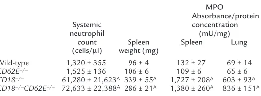
unit–ery-Table 1
Systemic neutrophil counts, spleen weights, and tissue neutrophil infiltration
MPO
Absorbance/protein
Systemic concentration
neutrophil (mU/mg)
count Spleen Spleen Lung
(cells/µl) weight (mg)
Wild-type 1,320 ± 355 96 ± 4 132 ± 27 69 ± 14
CD62E–/– 1,525 ± 136 106 ± 6 109 ± 6 65 ± 6
CD18–/– 61,280 ± 21,623A 339 ± 55A 1,727 ± 208A 603 ± 93A
[image:4.612.273.535.604.701.2]throcyte colony formation was counted on day 7.
Statistics. Average leukocyte rolling velocities, leuko-cyte adhesion, systemic leukoleuko-cyte counts, and differ-entials between groups were compared using one-way ANOVA and the Kruskal-Wallis multiple comparison test. Statistical significance was set at P < 0.05.
Results
Phenotype of CD18–/–CD62E–/–and CD18–/–CD62P–/–mice.
Mice deficient in CD18 and E-selectin (CD18–/–CD62E–/–
mice) were generated by interbreeding CD18 and E-selectin single mutants. CD18–/–CD62E–/–mutants from
CD18+/–CD62E–/– breeders were born at the expected
Mendelian ratios (8 mutants of 30 mice, P = not signif-icant), providing no evidence for in utero lethality.
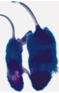
CD18–/–CD62E–/–mice that survived to weaning were
runted compared with littermates (Figure 1). They failed to thrive, reaching maximum body weights of 10–15 grams. CD18–/–CD62E–/–mice required constant
care for survival, including cutting of teeth and being given crushed food and antibiotics. Accurate survival curves could not be obtained because sick mice were killed in accordance with IACUC procedures. Most of the CD18–/–CD62E–/–mice that survived to 6 weeks of
age developed mucocutaneous skin lesions and over-growth of the incisors. They failed to breed and had reduced viability.
In contrast, CD18–/–CD62P–/–mice generated by
inter-breeding single mutants were indistinguishable from lit-termates. CD18–/–CD62P–/–mice showed no differences
in body weights at birth or at weaning compared with lit-termates. CD18–/–CD62P–/–mice did not have weaning
weights that differed from controls (CD18+/–CD62P+/–)
(11.3 ± 1.3 g and 12.1 ± 1.6 g, respectively; mean ± SD).
CD18–/–CD62P–/–mice survived to adulthood (> 90% over
3 months) and bred successfully.
Since CD18 is exclusively expressed on bone mar-row–derived blood cells, and E-selectin is restricted to endothelial cells, mice deficient in CD18 and E-selectin were generated by transplanting lethally irradiated
CD62E–/–mice with CD18–/–bone marrow. As controls,
wild-type, CD62E–/–, and CD18–/–mice were also
gener-ated through the appropriate bone marrow transplan-tation. All nontransplanted mice died within 2 weeks
of irradiation (1.2 Gy). Despite residing in a pathogen-free barrier facility, approximately 50% of the
CD18–/–CD62E–/– mice generated by bone marrow
transplantation died within a few weeks of transplan-tation, compared with less than 5% for wild-type,
CD18–/–, and CD62E–/–mice. CD18–/–CD62E–/–
double-mutant mice became progressively sick, resulting in chronic ulcerative dermatitis and significant weight loss (body weights less than 20 grams, reduced from 25 grams at the time of transplantation). Circulating neu-trophil counts in CD18–/–CD62E–/– mice became
increasingly elevated as a function of time after trans-plantation (reaching 50,000–150,000/µl).
Four weeks after reconstitution, peripheral blood and bone marrow of chimeric mice were analyzed for CD18 expression (Figure 2). CD18 expression was undetectable on bone marrow and blood cells in mice transplanted with CD18–/–bone marrow, confirming that
reconstitu-tion was efficient and complete.
Histopathology. CD18–/–CD62E–/–mice showed
moder-ate to severe ulcerative dermatitis similar to that of
CD18–/–mice (3). Histopathology of the skin revealed
significant hyperplasia of the epithelium, hyperkerato-sis, acute ulceration with bacterial colonies under the ulcer, mild lymphocytic and neutrophilic infiltration throughout the dermis, and a thickened dermis (Figure 3a). Gram-stained histologic sections of
CD18–/–CD62E–/– mice showed Gram-positive cocci
[image:5.612.188.291.54.216.2](Figure 3b). Gram staining of the wild-type skin sec-tions showed no evidence of organisms (data not shown). Bacterial cultures of the lung, liver, spleen, and Figure 1
Mice deficient in CD18 integrins and E-selectin fail to thrive.
CD18–/–CD62E–/– double-mutant
mice are runted at birth and require constant care for survival. A typical
CD18–/–CD62E–/–mutant (left) is
shown with a CD18+/+CD62E–/–
[image:5.612.315.526.440.632.2]lit-termate at 12 days of age. The dou-ble mutant was significantly small-er (2.0 g) than its littsmall-ermate (5.7 g).
Figure 2
Expression of CD18 (mAb C71/16) on unfractionated bone marrow cells 4 weeks after reconstitution. Neutrophils were identified and gated for expression of Gr-1 antigen. (a) Isotype control. Irradiated wild-type mice transplanted with wild-type bone marrow expressed high levels of CD18 (b). Transplantation of CD18–/–mouse bone
marrow into irradiated wild-type mice (c) and irradiated CD62E–/–
blood of six CD18–/–CD62E–/–mice showed one spleen
positive for a Corynebacteriumspecies. None of the four wild-type mice showed positive cultures in any organs.
CD18–/–CD62E–/–mice also displayed splenomegaly,
with an approximately threefold increase in spleen weight due to myeloid hyperplasia within the red pulp consisting of primitive, intermediate, and differentiat-ed cells. Myeloid hyperplasia was also present in the bone marrow of CD18–/–CD62E–/–mice.
To quantify neutrophil content in the spleen, MPO lev-els were measured in wild-type, CD62E–/–, CD18–/–, and
CD18–/–CD62E–/–mice. Spleen weights were increased
threefold in CD18–/–and CD18–/–CD62E–/–mice
com-pared with CD62E–/–and wild-type mice (Table 1).
Con-sistent with extramedullary hematopoiesis, a drastic increase in MPO activity was observed in the spleen of
CD18–/–and CD18–/–CD62E–/–mice compared with
wild-type and CD62E–/– mice (Table 1). In addition,
neu-trophil content in the lungs was elevated in the CD18–/–
and CD18–/–CD62E–/–mice (Table 1), suggesting
abnor-mal neutrophil trafficking and margination.
Analysis of hematopoiesis in CD18–/–CD62E–/–mice.
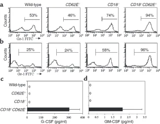
Con-sistent with the highly elevated level of neutrophils in the peripheral blood, a morphological analysis of stained bone marrow cytospins showed a drastic increase in the percentage of neutrophils and neu-trophil precursors compared with wild-type mice (data not shown). Unfractionated bone marrow and periph-eral blood were analyzed for expression of the myeloid marker Gr-1 (31) using flow cytometry (Figure 4, a and b, respectively). CD18–/–mice showed an increase in
bone marrow Gr-1 expression of approximately 75%, compared with approximately 50% in wild-type and
CD62E–/–mice (Figure 4a). Remarkably, the percentage
of Gr-1–expressing cells in the bone marrow and blood of CD18–/–CD62E–/–mice exceeded 90% (Figure 4, a and
b). In vitro hematopoietic progenitor colony assays showed no increase in GM-CFU colony formation at day 7 in CD18–/–CD62E–/–mice compared with
wild-type mice (data not shown).
Serum G-CSF and GM-CSF levels. G-CSF levels in wild-type, CD62E–/–, CD18–/–, and CD18–/–CD62E–/– mice
were measured to investigate the possible role of G-CSF in mediating the significant overproduction of neu-trophils. Peripheral blood neutrophil counts were sig-nificantly elevated in CD18–/–and CD18–/–CD62E–/–
mice. Serum G-CSF levels in CD18–/–CD62E–/–mice
were significantly higher (304 ± 75 pg/ml) than in wild-type, CD62E–/–, and CD18–/–mice (none detected)
(Fig-ure 4c), indicating that G-CSF may mediate the neu-trophilia observed in CD18–/–CD62E–/– mice. To
determine the possible involvement of GM-CSF in the neutrophilia, GM-CSF serum levels were also meas-ured. GM-CSF was elevated in CD18–/–CD62E–/–mice
(2.1 ± 1.2 pg/ml) compared with wild-type, CD62E–/–,
and CD18–/–mice (none detected) (Figure 4d).
Intravital microscopy. Leukocyte rolling and adhesion were investigated in wild-type, CD62E–/–, CD18–/–, and
CD18–/–CD62E–/–mice in the cremaster muscle 2 hours
after injection of TNF-α. During anesthesia and sur-gery in preparation for intravital microscopy experi-ments performed at 4 weeks after transplantation, all
CD18–/–CD62E–/–mice weighing less than 20 grams
died, and were not used for experiments. Therefore, the
CD18–/–CD62E–/–mice used for intravital microscopy
probably represent a subgroup of healthier mice. Leukocyte rolling and adhesion were studied in
hemo-Figure 3
(a) Skin section from the facial area of a CD18–/–CD62E–/–mouse
gen-erated through bone marrow transplantation, stained with hematoxylin and eosin. Section shows hyperplasia of the epithelium, hyperkeratosis, acute ulceration with bacterial colonies in the ulcer, mild lymphocytic and neutrophilic infiltration throughout the dermis, and a thickened dermis. (b) Gram stain of histologic section from the CD18–/–CD62E–/–
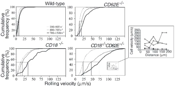
[image:6.612.314.532.168.601.2]dynamically similar venules over a shear-rate range from 100 s–1to approximately 1500 s–1(Table 2).
TNF-α induces the expression of E-selectin and enhances the expression of P-selectin in mouse cremas-ter muscle venules (32). To decremas-termine if CD18 and E-selectin cooperatively mediate efficient slow leukocyte rolling, leukocyte rolling velocities were measured in wild-type, CD62E–/–, CD18–/–, and CD18–/–CD62E–/–mice.
The majority of leukocytes rolled at velocities under 5 µm/s in wild-type mice, resulting in an average leukocyte rolling velocity of 6.9 ±0.2 µm/s (Figure 5). A noticeable shift in the leukocyte rolling velocity distribution (peak at 10–15 µm/s) and an increase in the average rolling velocity (21.1 ±0.5 µm/s) occurred in CD62E–/–mice
(Figure 5). Leukocyte rolling under 5.0 µm/s in CD62E–/–
mice was very rare (1.6% of rolling leukocytes, compared with 50.2% in wild-type mice), confirming that E-selectin is necessary for slow leukocyte rolling (5). The average rolling velocity in CD18–/–mice was increased to 22.7 ±
0.8 µm/s, due to a broadened rolling velocity distribu-tion (leukocyte rolling velocities up to ∼100 µm/s; Fig-ure 5). A further dramatic broadening of the rolling velocity distribution occurred in the CD18–/–CD62E–/–
mice (Figure 5). Very few leukocytes rolled slower than 10 µm/s, but a large population rolled faster than 50
µm/s (36%), shifting the rolling velocity distribution peak to 15–20 µm/s. The average rolling velocity was 50.1 ±1.4 µm/s in CD18–/–CD62E–/–mice, which was
sig-nificantly higher than in either of the single mutants. These findings show that CD18 and E-selectin work cooperatively to efficiently mediate slow leukocyte rolling in TNF-α–treated venules.
[image:7.612.212.536.50.302.2]In many systems, leukocyte rolling has been reported to be influenced by wall shear rate (33, 34). Therefore, we stratified the venules into low, intermediate, and high shear-rate groups (100–400 s–1, 400–700 s–1, and
Figure 4
Expression of Gr-1 antigen on unfraction-ated bone marrow cells (a) and peripher-al blood leukocytes (b). Gr-1 expression in
CD18–/–mice and CD18–/–CD62E–/– mice
[image:7.612.308.531.465.642.2]is dramatically elevated (percent of cells expressing Gr-1 indicated; gate set to exclude 95% of cells stained with isotype control, not shown). (c) Concentration of G-CSF and (d) GM-CSF in plasma as measured by ELISA. Data expressed as mean ± SEM.
Table 2
Hemodynamic data
Wild- CD62E–/– CD18–/– CD18–/–
type CD62E–/–
Number of mice 6 5 4 7
Number of venules 68 58 47 57
Average venule diameter (µm) 47 ± 2 37 ± 1 43 ± 2 51 ± 2 Average wall shear rate (s–1) 470 ± 30 600 ± 40 460 ± 30 580 ± 50
All values expressed as mean ± SEM.
Figure 5
Leukocyte rolling velocity distributions in TNF-α–treated cremaster muscle venules of wild-type, CD62E–/–, CD18–/–, and
CD18–/–CD62E–/–mice. Arrowheads indicate mean leukocyte rolling
velocity. All leukocytes rolling at velocities over 140 µm/s are repre-sented by last bar. Leukocyte rolling velocities in wild-type mice and
CD18–/–CD62E–/–mice were significantly different from those in
700–1500 s–1). Rolling velocity distributions changed lit-tle in wild-type mice as the shear rate was increased (Fig-ure 6). Increasing the shear rate in CD62E–/–mice
pro-duced a noticeable shift in the rolling velocity distribution (Figure 6). A similar shift occurred in the low and medium shear-rate ranges in CD18–/–mice, as
did a broadening of the velocity distribution, as reflect-ed by the decreasreflect-ed slope of the cumulative rolling veloc-ity histograms. A more drastic shift and broadening in rolling velocity distribution was seen in the high shear-rate range in CD18–/–mice. In CD18–/–CD62E–/–mice,
rolling velocity was dramatically elevated over all shear rates (Figure 6). Leukocyte rolling became very unstable in CD18–/–CD62E–/–at high shear rates. The leukocytes
no longer rolled uniformly on the venule surface, but generally skipped along the surface, indicating repetitive capture and detachment events (inset in Figure 6). By contrast, leukocytes in wild-type mice showed slow, steady rolling at high wall shear rates. Table 3 summa-rizes the average leukocyte rolling velocities measured in the three shear-rate ranges. In all cases, the increase in average leukocyte rolling velocity in CD18–/–CD62E–/–
mice was approximately additive of the increases seen in
CD18–/–and CD62E–/–mice.
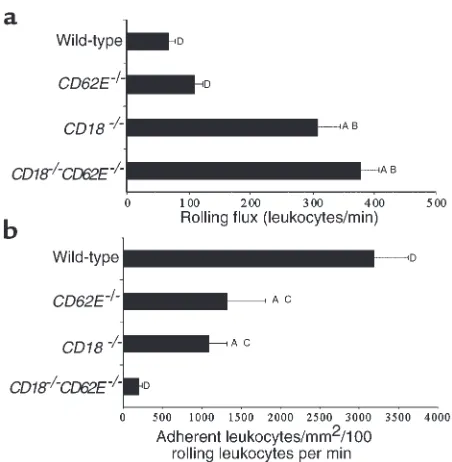
The ability of rolling leukocytes to adhere to TNF-α–stimulated endothelium was studied to determine if leukocyte rolling velocity is an important parameter mediating efficient leukocyte recruitment. Consistent with highly elevated systemic counts, the leukocyte rolling flux was greatly enhanced in CD18–/– and
CD18–/–CD62E–/–mice (Figure 7a). To show how
effi-ciently rolling leukocytes became firmly adhered, leuko-cyte adhesion was normalized for leukoleuko-cyte rolling flux (Figure 7b). In CD18–/–CD62E–/–double-mutant mice,
95% fewer leukocytes became adherent per 100 rolling leukocytes than in wild-type mice (199 ± 42 compared with 3,190 ± 423 in wild-type mice; 1,269 ± 448 in
CD62E–/–mice; and 1,099 ± 218 in CD18–/–mice).
Next, we tested the correlation between leukocyte rolling velocity and the ability of rolling leukocytes to become adherent (Figure 8). Leukocyte adhesion effi-ciency was significantly reduced in each mouse type as the leukocyte rolling velocity increased due to increased
shear rate. Within each shear-rate class, firm leukocyte adhesion was modestly reduced by the absence of E-selectin or CD18, and was drastically reduced in
CD18–/–CD62E–/–mice. The lack of firmly adherent
leukocytes in CD18–/–CD62E–/–mice correlated with the
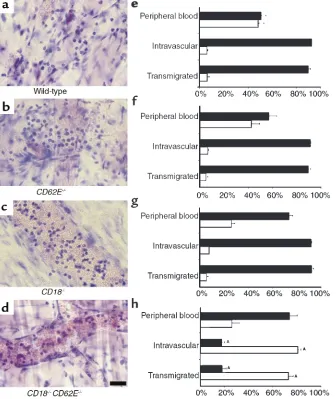
highly elevated leukocyte rolling velocities in all three shear-rate classes. Adhesion efficiency was most severe-ly affected when leukocyte rolling velocity exceeded approximately 20 µm/s, and when transit times fell below about 5 seconds for a 100-µm segment of venule. Giemsa-stained cremaster whole mounts were used to differentiate intravascular and transmigrated leukocytes (Figure 9, a–d). We found a remarkable impairment in the ability of neutrophils to interact successfully with the vessel wall in CD18–/–CD62E–/– mice. Intravascular
(rolling and firmly adhered) and transmigrated neu-trophils were reduced by 80% in CD18–/–CD62E–/–mice
(Figure 9h) compared with wild-type, CD62E–/–, and
CD18–/–mice (Figure 9, e–g). No other adhesion
mole-cule–mutant mouse demonstrated a comparable reduc-tion of neutrophil recruitment in this assay.
Discussion
[image:8.612.244.540.54.201.2]Our data show that mice lacking both CD18 integrins and E-selectin have severely impaired viability. When adult animals are generated by bone marrow trans-plantation, they show the most severe inflammatory defect seen in any adhesion molecule–deficient mouse described to date. Their inability to recruit neutrophils to sites of inflammation results in high systemic neu-trophil counts, which appear to be maintained through Figure 6
Effect of shear rate on leukocyte rolling velocity dis-tributions in TNF-α–treated cremaster muscle venules of wild-type, CD62E–/–, CD18–/–, and
CD18–/–CD62E–/–mice. Cumulative frequency curves
of leukocyte rolling velocities in shear-rate ranges of 100–400 s–1(thin dotted line), 400–700 s–1(thin solid line), and 700–1500 s–1(thick solid line). All leukocytes rolling at velocities over 140 µm/s are rep-resented by last data point. Median rolling velocity for each shear-rate range is indicated by vertical lines. Inset shows rolling velocities of three leukocytes rolling over a 200-µm distance in a venule of a wild-type mouse (closed circles, wall shear rate 754 s–1) and of three leukocytes rolling in a CD18–/–CD62E–/–
[image:8.612.304.538.631.702.2]mouse (open circles, wall shear rate 729 s–1).
Table 3
Average leukocyte rolling velocities (µm/s)
Shear-rate range
100–400 s–1 400–700 s–1 700–1500 s–1
Wild type 4.8 ± 0.2A 7.7 ± 0.3A 9.5 ± 0.7A
CD62E–/– 13.5 ± 0.4B,C 20.7 ± 0.7B,C 29.0 ± 1.3A
CD18–/– 17.9 ± 1.0B,C 22.6 ± 1.1B,C 42.6 ± 3.3A
elevated levels of G-CSF (a key regulator of neutrophil production) and GM-CSF. Using intravital microscopy, we were able to show a strong inverse relationship between the velocity of rolling leukocytes and the effi-ciency of adhesion.
Based on the phenotypes of the CD18 and E-selectin single-mutant mice, it could not be expected that the double mutation would result in severely reduced via-bility. In the first publication to characterize E-selectin–null mice, the mice were described as lacking a clear inflammatory phenotype (9), although defects in leukocyte rolling (5) and adhesion (21), and increased susceptibility to Streptococcus pneumoniae (22) were described later. CD18–/–mice develop skin ulcerations
and have elevated neutrophil counts and immunoglob-ulin levels, increased susceptibility to S. pneumoniae, and a severe defect in leukocyte adhesion and T-cell activa-tion (3). They also have a defect in leukocyte recruitment to peritonitis (35), and a lack of neutrophil recruitment to the skin (36). The unexpected severity of the
CD18–/–CD62E–/–mouse phenotype suggests that CD18
integrins and E-selectin serve a common function at a critical juncture or bottleneck in the leukocyte adhesion cascade, at the transition from rolling to firm adhesion. Although we found many phenotypic differences between CD18–/–CD62E–/–mice and single mutants, our
search for a cause of lethality was not successful. Nei-ther the skin lesions, the neutrophil accumulation in the lungs, nor the hematopoietic changes and
splenomegaly are likely causes for early death and fail-ure to thrive. However, the combination of these defects may well be the cause of the reduced viability. The severe phenotype seen in CD18–/–CD62E–/–mice is
somewhat reminiscent of E- and P-selectin double-mutant (CD62E–/–CD62P–/–) mice. CD62E–/–CD62P–/–
mice develop spontaneous mucocutaneous infections with skin ulcerations, even in a specific pathogen–free environment. They have very high circulating neutrophil counts (elevated 10- to 20-fold), which are thought to result from either an inability to recruit neutrophils to sites of inflammation, elevated circulating inflammato-ry cytokines (11), or alterations in the hematopoietic sys-tem (12, 37). CD62E–/–CD62P–/–mice demonstrate no
leukocyte rolling after tissue trauma or after short-term cytokine treatment (11), but after prolonged (6–8 hour) cytokine treatment (6), these mice show a small number of rolling leukocytes and significant leukocyte recruit-ment. Similar to the current findings, E- and P-selectin were identified as serving an overlapping function at a critical juncture in the inflammatory adhesion cascade, namely, stable leukocyte rolling (11).
Two other mice with mutations in genes encoding for inflammatory adhesion molecules, α4integrins (38) and VCAM-1 (39), have produced a lethal pheno-type. However, in these mice, lethality was not caused by an inflammatory defect, but by developmental defects affecting the formation of the chorioallanto-ic fusion (38, 39), epchorioallanto-icardium, and coronary arteries (38). Although the phenotype of E- and P-selectin double-mutant mice is severe, it did not lead to reduced viability of juvenile mice. Therefore, the
CD18–/–CD62E–/–mouse represents the most severe
inflammatory defect yet described.
One of the striking phenotypic traits of the
CD18–/–CD62E–/–mice is their high circulating
neu-trophil count. In previous studies with several other adhesion molecule–mutant mice, high circulating
neu-Figure 8
Inverse correlation between leukocyte rolling velocity and leukocyte adhesion efficiency in TNF-α–treated cremaster muscle venules of wild-type mice (squares), CD62E–/–mice (triangles), CD18–/–mice
(diamonds), and CD18–/–CD62E–/–mice (circles). Adhesion
[image:9.612.61.287.49.280.2]effi-ciency, represented as the ability of rolling leukocytes to become adherent, was drastically reduced as the leukocyte rolling velocity was increased. Data represented as mean ± SEM (SEM not shown if smaller than symbol).
Figure 7
Leukocyte rolling and adhesion in TNF-α–treated cremaster muscle venules of wild-type, CD62E–/–, CD18–/–, and CD18–/–CD62E–/–mice.
[image:9.612.308.533.511.632.2]trophil counts have been observed. Mild elevations were seen in ICAM-1–/–, LFA-1–/–, and P-selectin–null
mice (7, 18). Severe elevations were found in
CD62E–/–CD62P–/–mice (11, 12) and CD18–/–(3) mice.
Some strains were investigated for possible causes of these elevated counts. In CD62E–/–CD62P–/–mice,
ele-vated levels of GM-CSF and altered hematopoiesis (12) have been described. We also found elevated GM-CSF in CD18–/–CD62E–/–mice. However, GM-CSF is
unlike-ly to be a primary causative factor for elevated neu-trophil counts (40). We hypothesized that G-CSF would be a more likely candidate, based on the pheno-type of the G-CSF–overexpressing transgenic mice and G-CSF–null mice (41), which have severe neutrophilia and neutropenia, respectively. G-CSF is considered to be the major regulator of granulocyte production (42), and our finding that CD18–/–CD62E–/–mice exhibit
increased plasma G-CSF levels associated with neu-trophilia supports this conclusion. The findings of extramedullary hematopoiesis in the spleen and the elevated percentage of neutrophil precursors are both consistent with a severe hematopoietic dysregulation in CD18–/–CD62E–/–mice.
Our intravital microscopy findings in CD18–/–CD62E–/–
mice confirm and extend previous findings made in
CD18–/–mice treated with an E-selectin antibody (23).
We now show that the defect of slow leukocyte rolling is dependent on wall shear rate, and, by inference, wall-shear stress (which is directly related to wall wall-shear rate). In wild-type mice, rolling velocity is almost invariant over a wide range of wall shear rates in TNF-α−induced inflammation; even when E-selectin is missing, the dependence on wall shear rate is moderate, although the rolling velocities are increased throughout. However, eliminating CD18 has a large impact on rolling veloci-ties at high wall shear rates. The shear-rate influence is strongest in the CD18–/–CD62E–/–double-mutant mice.
In CD18–/–CD62E–/–mice, leukocyte recruitment is
[image:10.612.62.398.57.456.2]essen-tially absent in venules with moderate (400–700 s–1) or high (> 700 s–1) shear rates. Only a very modest rate of recruitment is possible in the venules with the slowest flow (wall shear rate < 400 s–1). This is significant for two reasons. First, venules with shear rates above 400 s–1are common in most microcirculatory beds. Although com-plete samples are rarely reported (43, 44), it is estimated that about two-thirds of venules fall in this shear-rate
Figure 9
Differentiation of intravenular leuko-cytes. Left column shows a representa-tive venule for each genotype. Whole mounts were obtained from wild-type (a), CD62E–/– (b), CD18–/– (c), and
CD18–/–CD62E–/–mice (d) and stained
range. Second, leukocyte delivery is directly proportion-al to flow rate, which has a linear dependence on blood-flow velocity and a quadratic dependence on vessel radius. On average, higher wall shear rates are observed more frequently in larger venules, whereas the small postcapillary venules show the lowest wall shear rates. This means that in CD18–/–CD62E–/– mice, the most
important venules, carrying the most leukocytes, are eliminated from contributing to leukocyte recruitment. In conclusion, we have identified an overlapping func-tion of CD18 integrins and E-selectin at a crucial step in the leukocyte adhesion cascade. Apparently, no other molecules can substitute for the essential function of these molecules in converting rolling to firm adhesion. This leads to severe inflammatory impairment and severely reduced viability in CD18–/–CD62E–/–mice.
Acknowledgments
We thank Russell Lindsey (University of Alabama at Birmingham) for the detailed histological analysis. Funding for the study was provided by NIH grants HL-54136 to K. Ley and AI-32177 to A.L. Beaudet. S.B. For-low was supported by NRSA HL-10447.
1. Butcher, E.C. 1991. Leukocyte-endothelial cell recognition: three (or more) steps to specificity and diversity. Cell.67:1033–1036.
2. Kansas, G.S. 1996. Selectins and their ligands: current concepts and con-troversies. Blood.88:3259–3287.
3. Scharffetter-Kochanek, K., et al. 1998. Spontaneous skin ulceration and defective T cell function in CD18 null mice. J. Exp. Med.188:119–131. 4. Arfors, K.E., et al. 1987. A monoclonal antibody to the membrane
glycopro-tein complex CD18 inhibits polymorphonuclear leukocyte accumulation and plasma leakage in vivo. Blood.69:338–340.
5. Kunkel, E.J., and Ley, K. 1996. Distinct phenotype of E-selectin-deficient mice. E-selectin is required for slow leukocyte rolling in vivo. Circ. Res.
79:1196–1204.
6. Jung, U., Ramos, C.L., Bullard, D.C., and Ley, K. 1998. Gene-targeted mice reveal importance of L-selectin-dependent rolling for neutrophil adhesion.
Am. J. Physiol.274:H1785–H1791.
7. Mayadas, T.N., Johnson, R.C., Rayburn, H., Hynes, R.O., and Wagner, D.D. 1993. Leukocyte rolling and extravasation are severely compromised in P selectin-deficient mice. Cell.74:541–554.
8. Tedder, T.F., Steeber, D.A., and Pizcueta, P. 1995. L-selectin-deficient mice have impaired leukocyte recruitment into inflammatory sites. J. Exp. Med.
181:2259–2264.
9. Labow, M.A., et al. 1994. Characterization of E-selectin-deficient mice: demonstration of overlapping function of the endothelial selectins. Immu-nity.1:709–720.
10. Arbones, M.L., et al. 1994. Lymphocyte homing and leukocyte rolling and migration are impaired in L-selectin-deficient mice. Immunity.1:247–260. 11. Bullard, D.C., et al. 1996. Infectious susceptibility and severe deficiency of leukocyte rolling and recruitment in E-selectin and P-selectin double mutant mice. J. Exp. Med.183:2329–2336.
12. Frenette, P.S., Mayadas, T.N., Rayburn, H., Hynes, R.O., and Wagner, D.D. 1996. Susceptibility to infection and altered hematopoiesis in mice deficient in both P- and E-selectins. Cell.84:563–574.
13. Jung, U., and Ley, K. 1999. Mice lacking two or all three selectins demon-strate overlapping and distinct functions for each selectin. J. Immunol.
162:6755–6762.
14. Coxon, A., et al. 1996. A novel role for the β2 integrin CD11b/CD18 in neu-trophil apoptosis: a homeostatic mechanism in inflammation. Immunity.
5:653–666.
15. Schmits, R., et al. 1996. LFA-1-deficient mice show normal CTL responses to virus but fail to reject immunogenic tumor. J. Exp. Med.183:1415–1426. 16. Lu, H., et al. 1997. LFA-1 is sufficient in mediating neutrophil emigration in
Mac-1-deficient mice. J. Clin. Invest.99:1340–1350.
17. Xu, H., et al. 1994. Leukocytosis and resistance to septic shock in intercellu-lar adhesion molecule 1-deficient mice. J. Exp. Med.180:95–109. 18. Sligh, J.E.J., et al. 1993. Inflammatory and immune responses are impaired
in mice deficient in intercellular adhesion molecule 1. Proc. Natl. Acad. Sci. USA.90:8529–8533.
19. Kunkel, E.J., et al. 1996. Absence of trauma-induced leukocyte rolling in mice
deficient in both P-selectin and intercellular adhesion molecule 1. J. Exp. Med.
183:57–65.
20. Bullard, D.C., et al. 1995. P-selectin/ICAM-1 double mutant mice: acute emi-gration of neutrophils into the peritoneum is completely absent but is nor-mal into pulmonary alveoli. J. Clin. Invest.95:1782–1788.
21. Ley, K., Allietta, M., Bullard, D.C., and Morgan, S. 1998. Importance of E-selectin for firm leukocyte adhesion in vivo. Circ. Res.83:287–294. 22. Munoz, F.M., Hawkins, E.P., Bullard, D.C., Beaudet, A.L., and Kaplan, S.L.
1997. Host defense against systemic infection with Streptococcus pneumo-niae is impaired in E-, P-, and E-/P-selectin-deficient mice. J. Clin. Invest.
100:2099–2106.
23. Jung, U., Norman, K.E., Scharffetter-Kochanek, K., Beaudet, A.L., and Ley, K. 1998. Transit time of leukocytes rolling through venules controls cytokine-induced inflammatory cell recruitment in vivo. J. Clin. Invest.
102:1526–1533.
24. Pries, A.R. 1988. A versatile video image analysis system for microcirculato-ry research. Int. J. Microcirc. Clin. Exp.7:327–345.
25. Lipowsky, H.H., and Zweifach, B.W. 1978. Application of the “two-slit” pho-tometric technique to the measurement of microvascular volumetric flow rates. Microvasc. Res.15:93–101.
26. Reneman, R.S., Woldhuis, B., oude Egbrink, M.G.A, Slaaf, D.W., and Tan-gelder, G.J. 1992. Concentration and velocity profiles of blood cells in the microcirculation. In Advances in cardiovascular engineering. N.H.C. Hwang, V.T. Turitto, and M.R.T. Yen, editors. Plenum Publishing Corp. New York, New York, USA. 25–40.
27. Ley, K., et al. 1995. Sequential contribution of L- and P-selectin to leukocyte rolling in vivo. J. Exp. Med.181:669–675.
28. Schwarzenberger, P., et al. 1998. IL-17 stimulates granulopoiesis in mice: use of an alternate, novel gene therapy-derived method for in vivo evaluation of cytokines. J. Immunol.161:6383–6389.
29. Grisham, M.B., Hernandez, L.A., and Granger, D.N. 1986. Xanthine oxidase and neutrophil infiltration in intestinal ischemia. Am. J. Physiol.
251:G567–G574.
30. Hillegass, L.M., Griswold, D.E., Brickson, B., and Albrightson-Winslow, C. 1990. Assessment of myeloperoxidase activity in whole rat kidney. J. Phar-macol. Methods.24:285–295.
31. Fleming, T.J., Fleming, M.L., and Malek, T.R. 1993. Selective expression of Ly-6G on myeloid lineage cells in mouse bone marrow. RB6-8C5 mAb to granulocyte-differentiation antigen (Gr-1) detects members of the Ly-6 fam-ily. J. Immunol.151:2399–2408.
32. Jung, U., and Ley, K. 1997. Regulation of E-selectin, P-selectin, and intercel-lular adhesion molecule 1 expression in mouse cremaster muscle vascula-ture. Microcirculation.4:311–319.
33. Damiano, E.R., Westheider, J., Tozeren, A., and Ley, K. 1996. Variation in the velocity, deformation, and adhesion energy density of leukocytes rolling within venules. Circ. Res.79:1122–1130.
34. Lipowsky, H.H., Scott, D.A., and Cartmell, J.S. 1996. Leukocyte rolling veloc-ity and its relation to leukocyte-endothelium adhesion and cell deformabil-ity. Am. J. Physiol.270:H1371–H1380.
35. Walzog, B., Scharffetter-Kochanek, K., and Gaehtgens, P. 1999. Impairment of neutrophil emigration in CD18-null mice. Am. J. Physiol.
276:G1125–G1130.
36. Mizgerd, J.P., et al. 1997. Neutrophil emigration in the skin, lungs, and peri-toneum: different requirements for CD11/CD18 revealed by CD18-deficient mice. J. Exp. Med.186:1357–1364.
37. Frenette, P.S., Subbarao, S., Mazo, I.B., von Andrian, U.H., and Wagner, D.D. 1998. Endothelial selectins and vascular cell adhesion molecule-1 promote hematopoietic progenitor homing to bone marrow. Proc. Natl. Acad. Sci. USA.
95:14423–14428.
38. Yang, J.T., Rayburn, H., and Hynes, R.O. 1995. Cell adhesion events mediat-ed by alpha 4 integrins are essential in placental and cardiac development.
Development.121:549–560.
39. Gurtner, G.C., et al. 1995. Targeted disruption of the murine VCAM1 gene: essential role of VCAM-1 in chorioallantoic fusion and placentation. Genes Dev.9:1–14.
40. Stanley, E., et al. 1994. Granulocyte/macrophage colony-stimulating fac-tor-deficient mice show no major perturbation of hematopoiesis but develop a characteristic pulmonary pathology. Proc. Natl. Acad. Sci. USA.
91:5592–5596.
41. Lieschke, G.J., et al. 1994. Mice lacking granulocyte colony-stimulating fac-tor have chronic neutropenia, granulocyte and macrophage progenifac-tor cell deficiency, and impaired neutrophil mobilization. Blood.84:1737–1746. 42. Metcalf, D. 1995. The granulocyte-macrophage regulators: reappraisal by
gene inactivation. Exp. Hematol.23:569–572.
43. Pries, A.R., Fritzsche, A., Ley, K., and Gaehtgens, P. 1992. Redistribution of red blood cell flow in microcirculatory networks by hemodilution. Circ. Res.
70:1113–1121.