IL-12 is required for differentiation of
pathogenic CD8
+
T cell effectors that cause
myocarditis
Nir Grabie, … , Jonathan G. Seidman, Andrew H. Lichtman
J Clin Invest.
2003;
111(5)
:671-680.
https://doi.org/10.1172/JCI16867
.
Cardiac antigen–specific CD8
+T cells are involved in the autoimmune component of
human myocarditis. Here, we studied the differentiation and migration of pathogenic CD8
+T
cell effector cells in a new mouse model of autoimmune myocarditis. A transgenic mouse
line was derived that expresses cardiac myocyte restricted membrane-bound ovalbumin
(CMy-mOva). The endogenous adaptive immune system of CMy-mOva mice displays
tolerance to ovalbumin. Adoptive transfer of naive CD8
+T cells from the ovalbumin-specific
T cell receptor–transgenic (TCR-transgenic) OT-I strain induces myocarditis in CMy-mOva
mice only after subsequent inoculation with ovalbumin-expressing vesicular stomatitis virus
(VSV-Ova). OT-I effector T cells derived in vitro in the presence or absence of IL-12 were
adoptively transferred into CMy-mOva mice, and the consequences were compared.
Although IL-12 was not required for the generation of cytolytic and IFN-
g
–producing effector
T cells, only effectors primed in the presence of IL-12 infiltrated CMy-mOva hearts in
significant numbers, causing lethal myocarditis. Furthermore, analysis of OT-I effectors
collected from a mediastinal draining lymph node indicated that only effectors primed in vitro
in the presence of IL-12 proliferated in vivo. These data demonstrate the importance of IL-12
in the differentiation of pathogenic CD8
+T cells that can cause myocarditis.
Article
Autoimmunity
Find the latest version:
Introduction
Myocarditis and dilated cardiomyopathy are associated with enteroviruses and other viral infections. In addi-tion to direct cytopathic effects of cardiotropic viruses, there is compelling evidence that autoimmune respons-es contribute to the heart disease in a significant subset of patients with myocarditis and in several animal mod-els (1–5). Autoantibodies specific for heart antigens are prevalent in patients with myocarditis and dilated car-diomyopathy, and they are more frequently found in asymptomatic relatives of patients with
cardiomyopa-thy than in the general population (6). Evidence for a role of self-reactive T cells in human myocarditis includes the demonstration of myocardial antigen-spe-cific T cells in biopsies from patients with myocarditis and the induction of disease in SCID mice by transfer of blood T cells from patients with myocarditis (7). A path-ogenic role for T cells in virus-induced murine myocarditis is indicated by experiments in which deple-tion of CD4+or CD8+T-cell subsets before infection
with coxsackievirus B3 (CVB3) reduced the severity of disease (8). Furthermore, the myocarditic effects of CVB3 infection are partly ameliorated in CD4 knockout mice, and are further reduced in combined CD4+/CD8+
knockout mice (9). CVB3 myocarditis is also suppressed in lckknockout mice, which have decreased numbers and impaired function of mature T cells (10).
Innate immune responses that accompany viral infec-tions are required for the development of protective adap-tive immune responses and may contribute to the devel-opment of pathological autoimmune responses (11). IL-12, produced mainly by pathogen-stimulated macrophages and dendritic cells, is an important link between innate and adaptive immune responses, and it has been implicated in the pathogenesis of several autoimmune diseases (12–15) as well as myocarditis (16). Experimental autoimmune myocarditis (EAM) is induced by immunization of rodents with cardiac myosin peptides in CFA along with systemic sensitization with
IL-12 is required for differentiation of pathogenic CD8
+T cell effectors that cause myocarditis
Nir Grabie,
1Michael W. Delfs,
1Jason R. Westrich,
1Victoria A. Love,
1George Stavrakis,
2Ferhaan Ahmad,
3Christine E. Seidman,
3Jonathan G. Seidman,
3and
Andrew H. Lichtman
1,21Immunology Research Division and
2Vascular Research Division, Department of Pathology, Brigham and Women’s Hospital and Harvard Medical School,
Boston, Massachusetts, USA
3Department of Genetics, Howard Hughes Medical Institute and Harvard Medical School, Boston, Massachusetts, USA
Cardiac antigen–specific CD8+ T cells are involved in the autoimmune component of human
myocarditis. Here, we studied the differentiation and migration of pathogenic CD8+T cell effector
cells in a new mouse model of autoimmune myocarditis. A transgenic mouse line was derived that expresses cardiac myocyte restricted membrane-bound ovalbumin (CMy-mOva). The endogenous adaptive immune system of CMy-mOva mice displays tolerance to ovalbumin. Adoptive transfer of naive CD8+T cells from the ovalbumin-specific T cell receptor–transgenic (TCR-transgenic) OT-I
strain induces myocarditis in CMy-mOva mice only after subsequent inoculation with ovalbumin-expressing vesicular stomatitis virus (VSV-Ova). OT-I effector T cells derived in vitro in the presence or absence of IL-12 were adoptively transferred into CMy-mOva mice, and the consequences were compared. Although IL-12 was not required for the generation of cytolytic and IFN-γ–producing effector T cells, only effectors primed in the presence of IL-12 infiltrated CMy-mOva hearts in sig-nificant numbers, causing lethal myocarditis. Furthermore, analysis of OT-I effectors collected from a mediastinal draining lymph node indicated that only effectors primed in vitro in the presence of IL-12 proliferated in vivo. These data demonstrate the importance of IL-12 in the differentiation of pathogenic CD8+T cells that can cause myocarditis.
J. Clin. Invest.111:671–680 (2003). doi:10.1172/JCI200316867.
Received for publication September 9, 2002, and accepted in revised form January 7, 2003.
Address correspondence to: Andrew H. Lichtman, Department of Pathology, Brigham and Women’s Hospital, 221 Longwood Avenue, Boston, Massachusetts 02115, USA.
Phone: (617) 732-6532; Fax: (617) 732-5795; E-mail: [email protected].
Conflict of interest: The authors have declared that no conflict of interest exists.
pertussis toxin, and it is largely mediated by CD4+T cells
(17–19). EAM appears to be dependent on IL-12, and the effects of IL-12 in this model are independent of IFN-γ (20, 21). Furthermore, in some strains of mice, the autoimmune component of virally induced myocarditis is dependent on IL-12 (4).
The clonal expansion and differentiation of naive CD8+T cells into effector cytolytic T cells requires at
least two signals provided by APCs, including peptide-MHC complexes that engage the T cell receptor and costimulatory molecules, such as CD80 or CD86, that interact with CD28 on the T cell. Previous studies have shown that IL-12 can enhance cytotoxic T lymphocyte (CTL) responses and suggest that this cytokine pro-vides an obligatory adjuvant-like “third” signal required for the generation of CTL effectors from naive CD8+T cells (22, 23). Since IL-12 is produced as part of
the innate immune response to viral infections (11) and is reported to be important in the development of autoimmune myocarditis (13, 20, 21), we hypothesized that this cytokine may be crucial for the generation of cytopathic CD8+effector cells that play a central role in
myocarditis. We tested this hypothesis using a newly developed transgenic model of myocarditis that per-mits us to examine the pathogenicity of uniform pop-ulations of CD8+T cells specific for a single antigen
expressed in the myocardium.
Methods
Cardiac myocyte restricted membrane-bound ovalbumin trans-genic construct. The DNA sequence corresponding to residues 139–387 of the ovalbumin gene (GenBank accession number V00383.1) was cloned downstream to DNA encoding residues 2–118 of the human transfer-rin receptor (CD71, GenBank accession number XM-052730), similar to a membrane ovalbumin fusion mol-ecule described previously (24). The CD71-ovalbumin sequence was then cloned upstream to the DNA sequence encompassing bases 942–1332 of the pcDNA3.1V5-His6-TOPO plasmid (Invitrogen, Carls-bad, California, USA), which includes polyadenylation signals. The resulting “mOva” cDNA construct was inserted into pcDNA3.1 (Invitrogen) and transfected into NIH-3T3 cells and DO.11 hybridoma cells (Ameri-can Type Culture Collection, Manassas, Virginia, USA), and expression of the encoded protein was verified by immunofluorescence microscopy and flow cytometry using specific antibodies to ovalbumin and the V5 epi-tope (data not shown). Subsequently, this mOva con-struct was cloned directly downstream to a DNA seg-ment encoding the murine cardiac α-myosin heavy-chain promoter (αMyHC-P, GenBank accession number U71441.1), kindly provided by J. Gulick (Chil-dren’s Hospital Medical Center, Cincinnati, Ohio, USA) (25). The final “αMyHC-P–mOva” DNA construct (Fig-ure 1a) was amplified in competent bacteria,linearized with specific endonucleases, separated on agarose gel, extracted on QIAquick columns (Qiagen Inc., Valencia, California, USA), and then fully sequenced.
Mice. All mice used in the current study were bred in the pathogen-free facility at the Braunwald Medical Research Center, in accordance with the guidelines of the committee of animal research at Harvard Medical School and the NIH animal research guidelines. A transgenic mouse line we named Cardiac myocyte restricted membrane-bound ovalbumin (CMy-mOva) carrying the above-described αMyHC-P–mOva trans-gene was trans-generated by a standard pronuclear microin-jection directly into embryonic stem cells derived from C57BL/6 mice. All of the CMy-mOva transgenic ani-mals used for this study originated from the same founder, were maintained on a C57BL/6-Thy 1.2 (CD90) background, and were heterozygous for the transgene. Careful examination indicated that CMy-mOva mice are healthy and have no cardiac functional abnormalities, as determined by ultrasonographic studies, or histological abnormalities for over 2 years of age. The OT-I T cell receptor (TCR) transgenic mouse strain (26) was kindly provided by W. R. Heath and F. Carbone (Walter and Eliza Hall Institute of Medical Research, Melbourne, Australia) and was maintained on a C57BL/6-Thy 1.1 (CD90.1) background. The OT-I TCR is expressed on CD8+T cells and is specific for the
ovalbumin peptide p.257–264 (SIINFEKL) bound to the class I MHC molecule H2-Kb(27). Signal
transduc-er and activator of transcription 4–/–(STAT-4–/–) mice
on a C57BL/6 background (28) were provided by M. Grusby (Harvard School of Public Health, Boston, Massachusetts, USA). C57BL/6 mice used in the study were all purchased from the Jackson Laboratory (Bar Harbor, Maine, USA).
Vesicular stomatitis virus production and inoculations. Vesicular stomatitis virus (VSV) and ovalbumin encod-ing VSV (VSV-Ova) were obtained from L. Lefrancois (University of Connecticut, Storrs, Connecticut, USA). Preparations and titration of VSV and VSV-Ova were performed as previously described (29). Where indi-cated, mice were inoculated with 106PFU of VSV or
VSV-Ova preparations through intravenous injection into the tail vein.
Cell preparations. Spleen and lymph nodes were har-vested from OT-I TCR transgenic mice, single-cell sus-pensions were prepared and treated with Tris-ammo-nium chloride (TAC) buffer to lyse erythrocytes, and CD8+cells were purified using a MACS CD8a (Ly-2)
MicroBeads kit (Miltenyi Biotec Inc., Auburn, Califor-nia, USA). This protocol typically yielded over 95% CD44lowCD8+CD4–cells, as assessed by flow cytometry.
Preparation of OT-I CD8+effector T cells was
per-formed as described previously (30). Briefly, naive CD8+
OT-I cells were placed in culture with mitomycin-C–treated (Sigma-Aldrich, St. Louis, Missouri, USA) APCs prepared from spleens of C57BL/6 mice (31) at a T cell/APC ratio of 1:10, with 10 µM (final concentra-tion) ovalbumin peptide antigen (SIINFEKL), with 2
maintained in RPMI medium (Invitrogen) supple-mented with 10% heat-inactivated FCS (Sigma), 2 mM Na-pyruvate, 100 U/ml penicillin, 100 µg/ml strepto-mycin, and 10 mM HEPES (Invitrogen) in 75-cm2
flasks and were incubated at 37°C (5% CO2). In some
cultures, 10 ng/ml recombinant mouse IL-12 (R&D Systems) was added at the outset, and the resulting effector populations will henceforth be referred to as OT-IIL-12effectors. In some cultures, IL-12 was not
added, but instead 2 µg/ml anti–IL-12 blocking anti-body (BD-Pharmingen) was added, and the resulting effector populations will henceforth be referred to as OT-I0effectors. All cultures were diluted 1:1 with fresh
medium containing 25 U/ml IL-2 (Invitrogen) after 3 days, and cells were harvested for use at day 5. In some experiments, the OT-I cells were stimulated with splenic APCs prepared from C57BL/6 STAT-4–/–mice.
In some experiments, plate-bound anti-CD3 plus solu-ble anti-CD28 (2 µg/ml, both reagents from BD-Pharmingen) were used to stimulate the OT-I cells in the absence of peptide or APCs.
T cell cytolytic activity assays. Cytolytic activity of CD8+
CTL was measured against H-2Kb–expressing EL4 target
cells (ATCC) pulsed with SIINFEKL by a conventional [51Cr]sodium chromate release assay (27) and by the
Live/Dead Cell-Mediated Cytotoxicity Kit (Molecular Probes Inc., Eugene, Oregon, USA) according to the manufacturer’s guidelines.
Flow cytometry analyses. Before analysis, lymphocyte preparations were washed twice in staining buffer (Dulbecco’s phosphate-buffered saline [DPBS] with 1% BSA). For phenotypic analysis of surface markers, 0.5 ×10⁶cells were suspended in 100 µl of staining
buffer containing 1µg of each specific antibody and incubated on ice for 20 minutes followed by washing and fixation with 0.5% paraformaldehyde. Stained-cell preparations were then analyzed by flow cytometry using a FACScalibur instrument and CellQuest soft-ware (Becton Dickinson, San Jose, California, USA). Expression of the OT-I TCR was detected with anti-Vα2 and anti-Vβ5 antibodies (29). Other specific anti-bodies used for flow cytometry were as follows: phyco-erythrin-conjugated anti-mouse CD90.1/Thy 1.1 (clone OX-7), fluorescein-conjugated anti-mouse CD44 (clone IM7), and fluorescein-conjugated anti-mouse CD25 (IL-2R αchain, clone 7D4). All antibod-ies were purchased from BD-Pharmingen.
In vivo proliferation of T cells was followed by flow cytometric analysis of CFSE-stained cells (Molecular Probes) as described (32). Effector cells were harvested from culture at day 5, washed, and rested in media with 10 U/ml IL-2 but no antigen for 24 hours before CFSE labeling and adoptive transfer.
ELISA detection of cytokines, cardiac troponin-T, ovalbumin protein, and specific antibodies. To assay IFN-γproduction, OT-I0or OT-IIL-12cells from differentiation cultures were
washed twice and restimulated in 1-ml polystyrene wells coated with 5 µg of anti-CD3εplus 2 µg/ml anti-CD28 (BD-Pharmingen), and supernatants were collected after
48 hours of culture. Assays for IL-12 were performed on supernatants of OT-I differentiation cultures sampled at 24, 48, and 72 hours. IFN-γ– or IL-12–specific ELISAs were performed using the appropriate antibody pairs (BD-Pharmingen), avidin-conjugated alkaline phos-phatase (Sigma-Aldrich), and SIGMA-FAST pNPP-sub-strate (Sigma-Aldrich). OD measurements at 405 nm were performed on an Emax microplate reader (Molecu-lar Devices, Sunnyvale, California, USA). Calibration curves were generated with recombinant IFN-γor IL-12 standards (BD-Pharmingen).
For analysis of ovalbumin protein, lysates were pre-pared from well-perfused CMy-mOva and C57BL/6 hearts by repetitive freeze-thaw cycles followed by homogenization and centrifugation. Serial dilutions of the lysates were directly applied to ELISA plates, spe-cific detection of ovalbumin protein was performed by incubation with purified anti-ovalbumin IgG, and sub-sequent detection was performed as described above, using alkaline phosphatase–conjugated antibodies.
[image:4.576.304.537.322.637.2]Anti-ovalbumin IgG titers in mouse serum were determined by ELISA on microtiter plates coated with
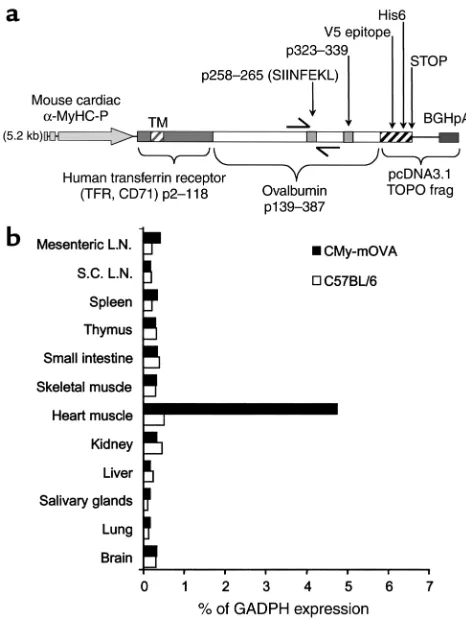
Figure 1
CMy-mOvatransgene construct and expression. (a) A map of the transgene construct used in the CMy-mOva line is shown. The Ova258–265and Ova323–339epitopes are recognized by OT-I and OT-II
ovalbumin or an equal concentration of BSA (Sigma-Aldrich) as controls. Twofold serial dilutions of mouse sera were applied into microwells and incu-bated for 2 hours at 37°C, followed by washing with PBS pH 7.2 containing 0.5% Tween-20 (Sigma-Aldrich). Detection of mouse IgG specifically ad-sorbed to coated microwells was performed using 0.5
µg/ml alkaline phosphatase–conjugated goat anti-mouse IgG (H+L) (Southern Biotechnology Associ-ates, Birmingham, Alabama, USA). Plates were devel-oped as described for IFN-γELISA.
Serum levels of cardiac troponin-T (cTnT) were meas-ured by a clinical quantitative sandwich enzyme immunoassay technique, which cross-reacts with mouse troponin (Troponin T STAT; Roche Diagnostics, Indi-anapolis, Indiana, USA) on the 1010 Elecsys Immuno-analyzer (Roche Diagnostics).
Adoptive transfer of T cells. For adoptive transfer experi-ments, single-cell suspensions of naive or effector Thy 1.1+
OT-I cells in DPBS were injected into the peritoneum of recipient mice in a total volume of 0.5 ml. When indicat-ed, cells were adoptively transferred intravenously through the tail vein in a suspension with a total volume of 100 µl.
Mouse tissue sampling and processing. Each mouse was anesthetized by ketamine injection (0.01 ml per 10 g of body weight), the thorax was opened, the inferior vena cava was nicked, and the animals were lethally exsan-guinated by perfusion of >10 ml DPBS through a 25° needle into the left ventricle. Hearts were then removed, placed in a petri dish with ice-cold RPMI medium, and cut with a scalpel to yield three transverse biventricular sections. The basal section was rapidly frozen in Tissue-Tek OCT compound (Sekura Finetek Inc., Torrance, California, USA) and stored at –80°C for subsequent immunohistochemical staining. The mid portion was fixed with 10% phosphate-buffered formalin, embed-ded in paraffin, and used for preparation of hema-toxylin- and eosin-stained sections. The apical portion was fixed in Trizol reagent for subsequent RNA extrac-tion. Mediastinal lymph nodes, spleen, and subcuta-neous lymph nodes were removed, and single-cell sus-pensions were prepared for flow cytometry studies.
Grading myocarditis. Myocarditis was graded by micro-scopic examination of hematoxylin- and eosin-stained sections in a blinded fashion by a trained pathologist after examination of the entire area of three sections, using a 0–4 scale as follows: 0 indicates no inflamma-tion; 1, one to five distinct mononuclear inflammato-ry foci with involvement of 5% or less of the cross-sec-tional area; 2, more than five distinct mononuclear inflammatory foci, or involvement of over 5% but not over 20% of the cross-sectional area; 3, diffuse mononu-clear inflammation involving over 20% of the area, without necrosis; and 4, diffuse inflammation with sec-ondary necrosis and acute inflammation.
Assessment of cardiac function by ultrasonography and elec-trocardiography. Transthoracic echocardiography, using a 12-MHz probe and an Agilent Sonos 4500 ultra-sonograph (Philips Medical Systems, Bothell,
Wash-ington, USA), and simultaneous electrocardiography were performed as previously described (33, 34).
Immunohistochemistry. Immunohistochemistry was performed as previously described (35). Five-microme-ter-thick cryostat sections of heart were fixed in ace-tone, blocked with 1% BSA in PBS at room temperature, incubated with unlabeled primary antibodies (each at 1–10 µg/ml) at room temperature followed by washing in PBS, and then incubated with biotinylated second-ary antibodies (each at 2.5 or 5 µg/ml) at room temper-ature. Primary rat mouse antibodies included anti-CD4, anti-CD8, anti-Ly6-G (GR-1), anti-CD11b, and anti–class I MHC (all from BD-Pharmingen). Unlabeled isotype-matched antibodies were used as controls. The sections were then incubated with biotinylated goat anti-rat Ig (1:200; Jackson Immuno Research, West Grove, Pennsylvania, USA) at room temperature. CD90.1 (Thy 1.1) was detected using a directly biotiny-lated antibody (BD-Pharmingen). Sections were then blocked with 0.3% hydroperoxide/PBS at room tem-perature and incubated with HRP:avidin:biotin com-plex solutions at a 1:1:100 dilution (Vector Laborato-ries, Burlingame, California, USA). The antibody binding was detected with 3-amino-9-ethylcarbazole (Vector Laboratories) and counterstained with Gill’s number 2 hematoxylin solution (Polysciences Inc., War-rington, Pennsylvania, USA).
Real-time PCR. Quantitative real-time RT-PCR was performed as described elsewhere (36). Briefly, total RNA was isolated from approximately 10 mg of heart tissue, using TRIZOL reagent (Invitrogen) T cells. To eliminate residual traces of DNA, total RNA samples were treated with DNaseI (Invitrogen). For the genera-tion of cDNA, total RNA was quantified by absorbance at 260 nm, and equal amounts from each sample were used as templates for reverse transcription of first-strand cDNA using the ThermoScript RT-PCR system according to the manufacturer’s instructions (Invitro-gen). For quantitative real-time RT-PCR analysis, 50 ng of cDNA were placed into 50-µl reaction wells in a 96-well optical reaction plate (Applied Biosystems, Foster City, California, USA) containing SYBR Green PCR mix (Applied Biosystems) and sequence-specific oligonu-cleotide primers (900 nM each) designed using Primer-Expres software (Applied Biosystems). The thermal cycle conditions used for all reactions were as follows: activation, 50°C for 2 minutes; denaturation, 95°C for 10 minutes; and cycle, 95°C for 15 seconds to 60°C for 1 minute (40 times). Specific primers used for sequence detection were as follows: for detection of message for the mOva transgene, 5′-AGTGGCATCAATGGCTTCT
(sense) and 5′- GTTGATTATACTCTCAAGCTGCTCA (anti-sense); for IFN-γ, 5′-AACGCTACACACTGCATCTTGG
(sense) and 5′-GCCGTGGCAGTAACAGCC(antisense); for CCR3, 5′-TGCTGTGCTACTGAGTCAGCG(sense) and 5′
-CTACAGCCAGGTGGAGCAGG(antisense); for CCR5, 5′
-CCATGCAGGCAACAGAGACTC(sense) and 5′- TCTCTC-CAACAAAGGCATAGATGA (antisense); for CCR7, 5′
CCATC-TGGGCCACTTGGA (antisense); and for GAPDH, 5′
-GGCAAATTCAACGGCACAGT(sense) and 5′- AGATGGT-GATGGGCTTCCC(antisense). All real-time reactions were carried out on an ABI 5700 Sequence Detection System (Applied Biosystems), and analysis was per-formed with the accompanying software. Sequences of amplicons resulting from real-time RT-PCR were veri-fied by dissociation analysis and by sequencing. Levels of IFN-γand mOva gene expression in tissue samples were expressed relative to endogenous levels of GAPDH expression in the same sample. All heart specimens examined in this study had nearly equal levels of GAPDH gene expression that were not affected by the treatments we applied.
Results
Tissue-specific expression of the CMy-mOva transgene. Con-ventional and real-time RT-PCR analysis of RNA col-lected from different tissues indicates that the mOva transgene is selectively expressed only in cardiac tissue in the CMy-mOva transgenic mouse (Figure 1b). This cardiac-specific expression is consistent with other transgenic models using the same αMyHC-P (25, 37). By ELISA, we were able to detect substantial amounts of ovalbumin protein in lysates of CMy-mOva hearts but not in hearts taken from C57BL/6 control animals, and immunofluorescence staining indicated that the transgene was expressed in myocytes (data not shown).
CMy-mOva mice are tolerant to ovalbumin. In order to determine if CMy-mOva mice are tolerant to ovalbu-min as a result of expression of the transgene in the heart, we exposed the animals to exogenous ovalbumin using different routs of immunizations. CMy-mOva mice did not show humoral or cellular
immune responses to ovalbumin injec-tion in CFA or to inoculainjec-tion with VSV-Ova (Table 1), whereas nontransgenic C57BL/6 mice responded strongly. Fur-thermore, these treatments did not lead to any inflammatory consequences in the myocardium of either CMy-mOva or con-trol mice (Table 1).
Adoptive transfer of naive ovalbumin-spe-cific T cells induces myocarditis in CMy-mOva mice only after VSV-Ova infection. Since CMy-mOva mice are tolerant to ovalbu-min expressed in the heart, the effects of
adoptively transferred ovalbumin-specific T cells can be studied in isolation from the endogenous adaptive immune responses. Adoptive transfer of substantial numbers of naive CD8+OT-I cells did not result in any
detectable myocardial inflammatory process in CMy-mOva after 7, 30, or 60 days (Table 2 and data not shown). In contrast, when CMy-mOva mice were injected with naive OT-I cells and then inoculated with VSV-Ova 24 hours later, myocarditis developed and all the mice died within 8 days. Infection of CMy-mOva mice with wild-type VSV after naive OT-I trans-fer did not lead to myocarditis, nor did infection of C57BL/6 mice with either VSV-Ova or VSV (Table 2).
These data show that exposure to a cardiac antigen in the setting of a highly immunogenic viral infection that induces a vigorous innate immune response can prime naive cardiac antigen–specific CD8+T cells to
become pathogenic effectors.
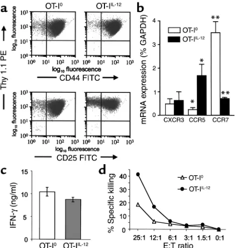
The influence of IL-12 on activation and differentiation of OT-I cells in vitro. We further examined the requirements for the differentiation of pathogenic effector CTLs by activating naive OT-I cells in vitro under different con-ditions before adoptive transfer into CMy-mOva mice. Stimulation of naive OT-I cells in vitro with SIINFEKL, splenic APCs with or without supplementary IL-12 (10 ng/ml), resulted in an approximately 20-fold clonal expansion and differentiation into cells with an activat-ed phenotype (CD44hiCD25+) within 5 days of culture
[image:6.576.250.534.614.685.2](Figure 2a). The degree of clonal expansion and the expression of activation markers CD25 and CD44 were indistinguishable between cultures that were or were not supplemented with exogenous IL-12. By ELISA, we could not detect any endogenous IL-12 in cultures that
Table 1
CMy-mOva mice are tolerant to ovalbumin
Antigenic (Ova) stimulation Readout of immune response Mouse strain C57BL/6 CMy-mOva
Ova/CFA (subcutaneous) Ova-specific Ig titers (day 28) 1:16,384 ± 800 ND
Prevalence of myocarditis (days 7 and 28) 0/12, 0/6 0/12, 0/6
Inoculation with VSV-Ova Ova-specific Ig titers (day 28) 1:12,000 ± 600 ND
Ova-specific cytolytic activity (day 28) 4/4 0/4
Prevalence of myocarditis (days 7 and 28) 0/12, 0/6 0/12, 0/6
ND, none detected.
Table 2
Activation of naive OT-I T cells in CMy-mOva mice by VSV-Ova inoculation
Adoptive transferA Post-transfer Cumulative mortality, Histological score of
treatment mean survival (days)B myocarditis at day 7
(n= 4 per group)C
OT-I — 0/4, >60 0 ± 0
OT-I VSV 0/4, >60 0 ± 0
OT-I VSV-Ova 4/4, 7 ± 1 4 ± 0
were not supplemented with exogenous IL-12 (detec-tion limit, <9 pg/ml) (data not shown). Moreover, there was no observed effect of 5 µg/ml anti IL-12 blocking antibody on cell yield or the phenotype of cells when added to cultures without exogenous IL-12 (data not shown). Interestingly, chemokine receptor expression, as measured by real-time RT-PCR, was different between OT-IIL-12and OT-I0cells. In particular, CCR5 expression
was significantly higher in OT-IIL-12cells, and CCR7
expression was significantly higher in OT-I0cells
(Fig-ure 2b). CXCR3 expression was not significantly differ-ent between the two T-cell populations. Although IL-12 is known to promote differentiation of naive CD4+T
cells into an IFN-γ–producing “Th1” phenotype, the requirement for IL-12 in differentiation of IFN-γ –pro-ducing CD8+effectors is not clear. For example, OT-I
cells differentiated in the absence of IL-12 and the pres-ence of IL-4 still produce IFN-γ (29). When OT-I0and
OT-IIL-12effectors were restimulated using immobilized
anti-CD3 antibody, they were equally capable of secret-ing IFN-γ(Figure 2c). In contrast, when the cytolytic activity of the OT-I populations were compared, we
found that OT-IIL-12effectors were approximately twice
as efficient in killing SIINFEKL-loaded target cells as were OT-I0effectors (Figure 2d).
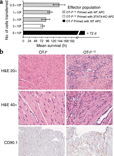
In vivo behavior of activated OT-I cells in CMy-mOva mice: priming in the presence of IL-12 is required for generation of pathogenic effectors. Adoptive transfer of OT-IIL-12
effec-tors into CMy-mOva mice consistently caused myocarditis that led to death at 96–144 hours, depend-ing on the dose of T cells transferred (Figure 3a). The severity of myocarditis, evaluated by histological grad-ing of hearts sampled at 72 hours after transfer, was a function of the number of T cells transferred (data not shown). Inflammation and damage to the hearts was characterized by intense lymphocytic infiltration, numerous lymphoblastic mitotic figures, myocyte apoptosis and necrosis, as well as secondary infiltration of macrophages and neutrophils (Figure 3b). No inflammation was seen in skeletal muscle or other tis-sues in the CMy-mOva mice with myocarditis, nor in hearts of nontransgenic C57BL/6 mice injected with the same OT-IIL-12cells (data not shown).
Immunohis-tochemical analysis confirmed the abundant presence of Thy1.1+cells, which specifically mark the adoptively
transferred OT-I cells in the Thy1.2 hosts (Figure 3b). By immunohistochemistry, there was a paucity of CD4+T cells present, but foci of macrophages and
granulocytes were present (data not shown). Real-time RT-PCR analysis of myocardial tissue indicated abun-dant IFN-γexpression in the inflamed hearts that cor-related with the histological score (Figure 4a). Several chemokine mRNAs, including IP-10, MCP-1, Mig, Mip-1α, Mip-1β, and RANTES were also elevated in inflamed hearts as compared with hearts from untreat-ed CMy-mOva mice (data not shown). Class I MHC was detected by immunohistochemistry on myocytes from CMy-mOva hearts, and there was increased expression in the setting of myocarditis (data not shown). In addi-tion to the histology, myocyte damage could also be demonstrated by a significant increase in the serum levels of cTnT in the CMy-mOva animals that received OT-IIL-12effectors (Figure 4b). Functional studies
indi-cated that the mice with myocarditis had diminished contractile functioning (fractional shortening) and bradycardia with irregular rhythm. These changes cor-related with the presence of T-cell infiltration and cTnT in the serum.
The results of adoptive transfer of OT-I0were
strik-ingly different from those obtained with OT-IIL-12cells.
Transfer of OT-I0effector cells (activated in the absence
of IL-12) did not result in lethal myocarditis in CMy-mOva mice (Figure 3a). There were rare small foci of T-cell infiltration in the hearts of animals that received these cells, but there was no histological evidence of myocyte damage or secondary inflammation (Figure 3b). Furthermore, IFN-γmRNA expression was not ele-vated in the hearts of these animals (Figure 4a), and these findings also correlated with the absence of cTnT in the serum (Figure 4b). Even though there was no his-tological evidence of significant OT-I0cell accumula-Figure 2
Phenotype of OT-I effector cells differentiated in vitro in the absence or presence of exogenous IL-12. Culture conditions are described in Methods. T cells were harvested from the differentiation cultures without added IL-12 (OT-I0) or with exogenously added 10 ng/ml
IL-12 (OT-IIL-12) on day 5 for analysis. (a) Thy1.1, CD25, and CD44
[image:7.576.59.293.54.301.2]tion in the hearts or myocardial damage, real-time RT-PCR analyses did indicate indirectly that at least some OT-I0T cells had entered the heart and had some
local effects. Specifically, as compared with hearts from CMy-mOva animals that did not receive T-cell trans-fers, hearts removed from CMy-mOva mice at72 hours after OT-I0transfer had elevated levels of IP-10 and Mig
mRNAs, similar to animals receiving OT-IIL-12 cells
(data not shown). Unlike OT-IIL-12recipients, OT-I0–
recipient hearts did not have elevated levels of MCP-I, Mip-1α, Mip-1β, or RANTES mRNAs.
To determine if IL-12 was directly acting on the T cells during the differentiation of pathogenic effec-tors or was acting indirectly through APCs, we gener-ated OT-IIL-12cells using splenic APCs from C57BL/6
STAT-4–/–mice, which have impaired responses to
IL-12. The effector T cells derived under these
condi-tions were as potent in inducing lethal myocarditis as were OT-IIL-12effectors generated in the presence of
STAT-4+/+C57BL/6 APCs (Figure 3a). We also found
that IL-12 supported the differentiation of patho-genic effectors when CD8+OT-I T cells were
stimu-lated by the combination of immobilized anti-CD3 and soluble anti-CD28 in the absence of APCs and antigen (data not shown). Together, these findings further indicate that IL-12 acts directly on T cells rather than through effects on APCs.
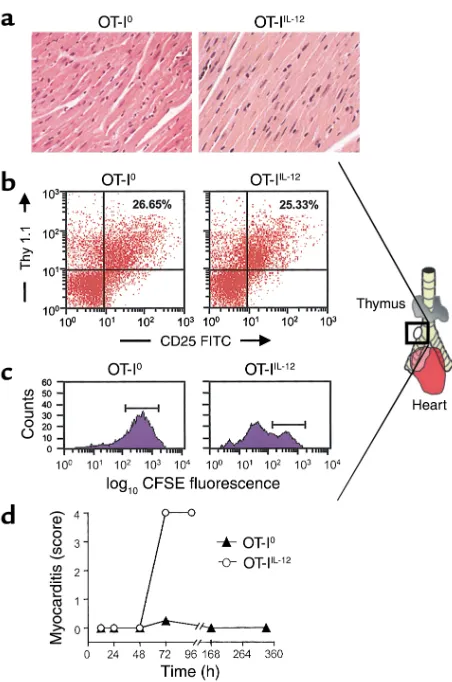
In vivo proliferation and migration of OT-I effector cells. There was no detectable myocarditis in CMy-mOva hearts up to 48 hours after adoptive transfer of 3 ×10⁶
OT-IIL-12 or OT-I0 cells (Figure 5a). Nonetheless,
enlarged lymph nodes could be consistently identified in the mediastinum adjacent to the right mainstem bronchus by 48 hours after adoptive transfer of either type of OT-I effector T cell (Figure 5b). Flow cytomet-ric analysis of cell suspensions recovered from these nodes indicated that they contained activated OT-I cells (Figure 5b). Both OT-I0and OT-IIL-12effectors were
[image:8.576.60.291.57.372.2]found with equivalent frequency and comparable CD25 expression in the mediastinal lymph nodes of
Figure 3
OT-I T-cell–mediated myocarditis in CMy-mOva mice. (a) Mice were injected with indicated numbers of OT-I0or OT-IIL-12effector T cells,
whose phenotypes are described in Figure 2, or with OT-IIL-12cells
dif-ferentiated in the presence of C57BL/6 STAT-4–/–splenic APCs. Mean
survival is shown for each group. Data represent means ± SD (n= 4 per group). Additional studies with more than 16 mice confirmed that CMy-mOva mice survived longer than 72 days after adoptive transfer of 5 ×10⁶OT-I0. (b) Hearts were harvested 72 hours after adoptive
transfer of 106OT-I effector T cell populations (described in Figure 2).
[image:8.576.335.487.496.731.2]Low- and high-power photomicrographs of hematoxylin- and eosin-stained sections and immunohistochemical stains for CD90.1 (Thy 1.1), which specifically marks the OT-I cells, are shown.
Figure 4
Cardiac IFN-γmRNA expression and serum troponin T levels in CMy-mOva mice. (a) Total RNA was isolated from hearts removed from untreated CMy-mOva mice (labeled None) or from hearts removed 72 hours after adoptive transfer of 2.5 ×10⁶OT-I effectors, as
indi-cated. Real-time RT-PCR quantification of IFN-γmRNA was per-formed. Data represent means ± SD (n= 4 mice per group). (b) Tro-ponin T was measured by ELISA in serum samples collected from CMy-mOva mice 72 hours after adoptive transfer of 2.5 ×10⁶OT-I
CMy-mOva mice at 48 hours. The presence of OT-I cells in the lymph nodes before any evidence of myocar-dial inflammation in mice receiving OT-IIL-12cells
sug-gests that these T cells first home to the draining lymph nodes of the heart before they migrate to the myocardium. OT-I cells could not be found in extra-mediastinal lymph nodes (data not shown). Because both OT-I0 and OT-IIL-12 T cells found in the
peri-bronchial lymph node at 48 hours had activated phe-notypes, we wished to determine the proliferative sta-tus of these two populations. Using CFSE-labeled OT-I cells, we were able to determine that in the
peri-bronchial lymph node, OT-IIL-12cells had undergone
significant proliferation before 72 hours, whereas OT-I0cells had not (Figure 5c). Histological grading of
myocarditis at sequential time points revealed that OT-IIL-12–mediated myocarditis developed between 48
and 72 hours after adoptive transfer. In contrast, after transfer of OT-I0effectors, a transient infiltrate was
observed in the heart at 72 hours, but there was no infiltrate seen at later time points (Figure 5d).
Discussion
This study utilizes a newly developed model of T-cell myocarditis that has allowed us to compare the in vivo behavior of different populations of T cells specific for the same defined myocardial antigen. A transgenic model of myocarditis has been described using mice expressing bacterial β-galactosidase (β-gal) under the control of a smooth-muscle cell–specific promoter (38). In those animals, myocarditis restricted to the right ventricle and accompanied by pulmonary and systemic arterial inflammation can be induced by repetitive immunization with β-gal peptide–pulsed dendritic cells. The model described here is based on transgenic models of organ-specific immunity in which OT-I TCR transgenic T cells are adoptively transferred into ani-mals expressing transgenic ovalbumin in pancreatic islets and kidney (24, 39, 40) or intestine (41). The fact that the endogenous immune system of the CMy-mOva mice is tolerant to ovalbumin simplifies the interpretation of adoptive transfer experiments of oval-bumin-specific T cells, because secondary activation of the host adaptive immune system does not play a sig-nificant role in the pathologic processes. Adoptively transferred naive ovalbumin-specific CD8+T cells do
not spontaneously cause myocarditis in the CMy-mOva mouse, and this is consistent with the tolerant behavior of naive OT-I cells transferred to mice express-ing ovalbumin in pancreatic islets or intestine (39, 41). Naive OT-I cells were activated and became pathogen-ic effectors in the CMy-mOva mouse after VSV-Ova inoculations, consistent with the hypothesis that innate immune responses associated with viral infec-tion provide signals that overcome self-tolerance and lead to the generation of pathogenic effector T cells. Recent studies using peptide immunization–induced EAM clearly indicate the importance of IL-12 in the development of autoimmune disease. Nonetheless, the influence of IL-12 on the development of pathogenic CD8+T cells is not clarified by those studies, since EAM
is predominantly a CD4+T-cell–mediated process. The
adaptive immune response to viral infections, including myocarditic viruses, includes a predominant CD8+T
cell response. Furthermore, IL-12 secretion is one com-ponent of the innate immune response to viral infec-tions that influences the type of viral-specific adaptive immune response that will develop (42, 43). It is there-fore important to know how IL-12 may influence the differentiation of pathogenic CD8+effector T cells that
[image:9.576.61.287.49.390.2]can cause myocarditis. The data presented in this paper
Figure 5
In vivo migration and proliferation of OT-I effector cells. (a) Hema-toxylin- and eosin-stained sections were prepared from hearts har-vested at 48 hours after T cell transfer of 2.5 ×10⁶OT-I0or OT-IIL-12
effector cells. (b) Enlarged peribronchial lymph nodes, consistently found in the illustrated location in CMy-mOva mice with myocardi-tis, were harvested 48 hours after adoptive transfer of 2.5 ×10⁶
OT-I0or OT-IIL-12effectors, and FACS analysis was performed as
described in Methods. (c) CFSE-labeled OT-I effector cells were transferred into CMy-mOva mice, and peribronchial lymph nodes were harvested after 72 hours for flow cytometric analysis. The his-tograms show CFSE fluorescence intensity in Thy 1.1+–gated cells.
indicate that IL-12 is not required for the differentiation of IFN-γ–producing CD8+effector T cells, but it is
required for the differentiation of CD8+effector cells
that can cause cardiac damage. Although OT-I effector T cells produced abundant IFN-γin vitro whether or not they differentiated in the presence of IL-12, there was abundant IFN-γexpression only in the hearts of CMy-mOva mice that received OT-IIL-12cells. This
prob-ably reflects the abundance of the OT-IIL-12cells and the
scarcity of the OT-I0cells in the hearts. Although recent
reports suggest that IFN-γis protective in EAM (21, 44), it does not appear to be protective in OT-IIL-12
–mediat-ed myocarditis in the CMy-mOva mice, since there is abundant IFN-γexpression in the hearts of animals with lethal disease.
The ability to identify and analyze draining medi-astinal lymph nodes in the CMy-mOva mice repre-sents a significant advance that, to our knowledge, has not been reported in previous studies of murine autoimmune myocarditis. The unexpected data that adoptively transferred OT-IIL-12effector T cells home
to and accumulate in draining lymph nodes before they are detectable in the heart (Figure 5) suggests that these T cells must undergo further lymph node–based activation before they are competent to infiltrate and cause damage to the heart. Further-more, OT-IIL-12cells proliferate in the host mice after
adoptive transfer. The nonpathogenic OT-I0cells also
accumulate in the draining lymph nodes to the same extent as the OT-IIL-12cells; however, they do not
pro-liferate. These data indicate that OT-I0cells are viable
and activated in vivo yet fail to proliferate and migrate in significant numbers into the myocardium.
In summary, it is likely that the IL-12–induced phe-notype of CD8+T cells includes several components
that render these cells more effective mediators of antimicrobial immunity and more pathogenic effectors of autoimmunity. The relatively greater pathogenicity of OT-IIL-12 cells as compared with OT-I0 cells may
reflect more than one of these IL-12–induced changes. First, we find that there is a proliferative block of OT-I0
but not OT-IIL-12cells in draining lymph nodes. Second,
OT-IIL-12cells exhibit enhanced cytolytic activity relative
to OT-I0cells, which is consistent with the results of
other studies with CD8+T cells. However, reduced lytic
ability cannot explain the minimal infiltration of the cells into the heart. Third, our finding that OT-IIL-12cells
express more CCR5 and less CCR7 than OT-I0cells may
explain the relative retention of OT-I0cells in lymph
nodes and the propensity of OT-IIL-12cells to home to
heart. CCR5 binds Mip-1α, Mip-1β, and RANTES, all of which are expressed in the myocarditic hearts. CCR7 binds CCL19 and CCL21, which are present in T-cell zones of lymph nodes. The possibility of other IL-12– dependent changes in the migratory phenotype of CD8+
effectors is currently being investigated. The fact that a few OT-I0T cells are transiently detectable in
CMy-mOva hearts, and that some chemokine mRNAs are ele-vated in the hearts of OT-I0recipients, suggests that the
OT-I0T cells that do get into the heart mount a weak
response that cannot be sustained. Therefore, IL-12 may have additional effects, including reducing T-cell sus-ceptibility to negative regulatory mechanisms.
Acknowledgments
This work was supported by NIH grants HL36028 (to A. H. Lichtman and N. Grabie) and HL56985 (to A. H. Lichtman). We thank Arlene Sharpe, director of the Brigham and Women’s Hospital Transgenic Core Facil-ity, and Lina Du for pronuclear injections and deriva-tion of the CMy-mOva transgenic founder. We thank Gary Bradwin of the Clinical and Epidemiologic Research Laboratory, Children’s Hospital, Boston, for technical assistance with the serum troponin T assays.
1. Huber, S.A. 1997. Autoimmunity in myocarditis: relevance of animal models. Clin. Immunol. Immunopathol.83:93–102.
2. Huber, S.A. 1997. Coxsackievirus-induced myocarditis is dependent on distinct immunopathogenic responses in different strains of mice. Lab. Invest.76:691–701.
3. Fairweather, D., Kaya, Z., Shellam, G.R., Lawson, C.M., and Rose, N.R. 2001. From infection to autoimmunity. J. Autoimmun.16:175–186. 4. Hill, S.L., and Rose, N.R. 2001. The transition from viral to
autoim-mune myocarditis. Autoimmunity.34:169–176.
5. Penninger, J.M., and Bachmaier, K. 2000. Review of microbial infec-tions and the immune response to cardiac antigens. J. Infect. Dis.
181(Suppl. 3):S498–S504.
6. Caforio, A.L., Goldman, J.H., Haven, A.J., Baig, K.M., and McKenna, W.J. 1996. Evidence for autoimmunity to myosin and other heart-spe-cific autoantigens in patients with dilated cardiomyopathy and their relatives. Int. J. Cardiol.54:157–163.
7. Schwimmbeck, P.L., Badorff, C., Rohn, G., Schulze, K., and Schultheiss, H.P. 1996. The role of sensitized T-cells in myocarditis and dilated car-diomyopathy. Int. J. Cardiol.54:117–125.
8. Henke, A., Huber, S., Stelzner, A., and Whitton, J.L. 1995. The role of CD8+T lymphocytes in coxsackievirus B3-induced myocarditis. J. Virol.
69:6720–6728.
9. Opavsky, M.A., et al. 1999. Susceptibility to myocarditis is dependent on the response of αβT lymphocytes to coxsackieviral infection. Circ. Res.85:551–558.
10. Liu, P., et al. 2000. The tyrosine kinase p56lck is essential in coxsack-ievirus B3-mediated heart disease. Nat. Med.6:429–434.
11. Xing, Z., Zganiacz, A., Wang, J., Divangahi, M., and Nawaz, F. 2000. Th1-type immune responses to respiratory viral infection: require-ment of IL-18 for IFN-γrelease in the lung but not for the
differ-entiation of viral-reactive Th1-type lymphocytes. J. Immunol.
164:2575–2584.
12. Leonard, J., Waldburger, K., and Goldman, S. 1995. Prevention of experimental autoimmune encephalomyelitis by antibodies against interleukin 12. J. Exp. Med.181:381–386.
13. Tarrant, T.K., et al. 1999. Interleukin 12 protects from a T helper type 1-mediated autoimmune disease, experimental autoimmune uveitis, through a mechanism involving interferon γ, nitric oxide, and apop-tosis. J. Exp. Med.189:219–230.
14. Neurath, M., Fuss, I., Kelsall, B., Stuber, E., and Strober, W. 1995. Anti-bodies to interleukin 12 abrogate established experimental colitis in mice. J. Exp. Med.182:1281–1290.
15. McIntyre, K.W., et al. 1996. Reduced incidence and severity of collagen-induced arthritis in interleukin-12-deficient mice. Eur. J. Immunol.
26:2933–2938.
16. Okura, Y., et al. 1998. Recombinant murine interleukin-12 facilitates induction of cardiac myosin-specific type 1 helper T cells in rats. Circ. Res.82:1035–1042.
17. Pummerer, C., et al. 1996. Cardiac myosin-induced myocarditis: target recognition by autoreactive T cells requires prior activation of cardiac interstitial cells. Lab. Invest.74:845–852.
18. Neumann, D.A., et al. 1992. In vivo deposition of myosin-specific autoantibodies in the hearts of mice with experimental autoimmune myocarditis. J. Immunol.148:3806–3813.
19. Bachmaier, K., et al. 1999. Chlamydia infections and heart disease linked through antigenic mimicry. Science.283:1335–1339. 20. Afanasyeva, M., et al. 2001. Interleukin-12 receptor/STAT4 signaling
2001. Dual role of the IL-12/IFN-γaxis in the development of
autoim-mune myocarditis: induction by IL-12 and protection by IFN-γ.
J. Immunol.167:5464–5469.
22. Schmidt, C.S., and Mescher, M.F. 2002. Peptide antigen priming of naive, but not memory, CD8 T cells requires a third signal that can be provided by IL-12. J. Immunol.168:5521–5529.
23. Kieper, W.C., Prlic, M., Schmidt, C.S., Mescher, M.F., and Jameson, S.C. 2001. IL-12 enhances CD8 T cell homeostatic expansion. J. Immunol.
166:5515–5521.
24. Kurts, C., et al. 1996. Constitutive class I-restricted exogenous presen-tation of self antigens in vivo. J. Exp. Med.184:923–930.
25. Gulick, J., Subramaniam, A., Neumann, J., and Robbins, J. 1991. Isola-tion and characterizaIsola-tion of the mouse cardiac myosin heavy chain genes. J. Biol. Chem.266:9180–9185.
26. Hogquist, K.A., et al. 1994. T cell receptor antagonist peptides induce positive selection. Cell.76:17–27.
27. Carbone, F.R., and Bevan, M.J. 1989. Induction of ovalbumin-specific
cytotoxic T cells by in vivo peptide immunization. J. Exp. Med.
169:603–612.
28. Chitnis, T., et al. 2001. Effect of targeted disruption of STAT4and STAT6
on the induction of experimental autoimmune encephalomyelitis. J. Clin. Invest.108:739–747. doi:10.1172/JCI200112563.
29. Kim, S.K., et al. 1998. Generation of mucosal cytotoxic T cells against soluble protein by tissue-specific environmental and costimulatory sig-nals. Proc. Natl. Acad. Sci. U. S. A.95:10814–10819.
30. Dobrzanski, M.J., Reome, J.B., and Dutton, R.W. 2000. Type 1 and type
2 CD8+effector T cell subpopulations promote long-term tumor
immunity and protection to progressively growing tumor. J. Immunol.
164:916–925.
31. Delfs, M.W., Furukawa, Y., Mitchell, R.N., and Lichtman, A.H. 2001.
CD8+T cell subsets TC1 and TC2 cause different histopathologic
forms of murine cardiac allograft rejection. Transplantation.
71:606–610.
32. Mintern, J., et al. 1999. The use of carboxyfluorescein diacetate suc-cinimidyl ester to determine the site, duration and cell type
responsi-ble for antigen presentation in vivo. Immunol. Cell. Biol.77:539–543. 33. McConnell, B.K., et al. 1999. Dilated cardiomyopathy in homozygous myosin-binding protein-C mutant mice. J. Clin. Invest.104:1235–1244. 34. Fatkin, D., et al. 2000. An abnormal Ca2+response in mutant
sarcom-ere protein-mediated familial hypertrophic cardiomyopathy. J. Clin. Invest.106:1351–1359.
35. Buono, C., et al. 2002. Influence of C3 deficiency on atherosclerosis.
Circulation.105:3025–3031.
36. Wagers, A.J., Waters, C.M., Stoolman, L.M., and Kansas, G.S. 1998. Interleukin 12 and interleukin 4 control T cell adhesion to endothelial selectins through opposite effects on α1,3-fucosyltransferase VII gene expression. J. Exp. Med.188:2225–2231.
37. Bryant, D., et al. 1998. Cardiac failure in transgenic mice with myocar-dial expression of tumor necrosis factor-α. Circulation.97:1375–1381. 38. Ludewig, B., et al. 2000. Linking immune-mediated arterial inflam-mation and cholesterol-induced atherosclerosis in a transgenic mouse model. Proc. Natl. Acad. Sci. U. S. A.97:12752–12757.
39. Kurts, C., et al. 1997. CD4+T cell help impairs CD8+T cell deletion
induced by cross-presentation of self-antigens and favors autoimmu-nity. J. Exp. Med.186:2057–2062.
40. Kurts, C., Kosaka, H., Carbone, F.R., Miller, J.F.A.P., and Heath, W.R. 1997. Class I-restricted cross-presentation of exogenous self-antigens leads to deletion of autoreactive CD8+T cells. J. Exp. Med.186:239–245.
41. Vezys, V., Olson, S., and Lefrancois, L. 2000. Expression of intestine-specific antigen reveals novel pathways of CD8 T cell tolerance induc-tion. Immunity.12:505–514.
42. Biron, C.A., and Gazzinelli, R.T. 1995. Effects of IL-12 on immune responses to microbial infections: a key mediator in regulating disease outcome. Curr. Opin. Immunol.7:485–496.
43. Romani, L., Puccetti, P., and Bistoni, F. 1997. Interleukin-12 in infec-tious diseases. Clin. Microbiol. Rev.10:611–636.
44. Henke, A., Zell, R., Ehrlich, G., and Stelzner, A. 2001. Expression of immunoregulatory cytokines by recombinant coxsackievirus B3 vari-ants confers protection against virus-caused myocarditis. J. Virol.