Resequencing Microarray for Detecting and Genotyping Equine
Arteritis Virus
Aymeric Hans,aDelphine Gaudaire,aJean-Claude Manuguerra,bAlbertine Leon,cAntoine Gessain,d,eClaire Laugier,a Nicolas Berthet,d,e*Stephan Zientaraf
ANSES-Dozulé Laboratory for Equine Diseases, Virology Unit, Goustranville, Francea; Institut Pasteur, Unité Environnement et Risques Infectieux, Cellule d’Intervention Biologique d’Urgence (CIBU), Paris, Franceb; Frank Duncombe Laboratory, Animal Health Department, IFR146 ICORE, Caen, Francec; Institut Pasteur, Unité d’Epidémiologie et Physiopathologie des Virus Oncogènes, Paris, Franced; Centre National de la Recherche Scientifique, UMR 3569, Paris, Francee; Université Paris-Est, Anses Maisons-Alfort Laboratory for Animal Health, UMR1161 Virologie, Maisons-Alfort, Francef
This study shows that an unbiased amplification method applied to equine arteritis virus RNA significantly improves the sensi-tivity of the real-time reverse transcription-quantitative PCR (RT-qPCR) recommended by the World Organization for Animal Health. Twelve viral RNAs amplified using this method were hybridized on a high-density resequencing microarray for effective viral characterization.
E
quine arteritis virus (EAV), the causative agent of equine viral arteritis (EVA), is a member of theArteriviridaefamily. Its genome is made up of a single positive-strand RNA molecule (1, 2). EAV infectsEquidaeand may be transmitted through the re-spiratory and venereal routes. EAV can persist in the reproductive tract of stallions only (3). Following the primary infection, up to 70% of stallions can be persistently infected and shed the virus in their semen, sometimes for life. These “shedder” stallions spread the virus in the horse population during breeding or when their semen is used for artificial insemination (4). The financial conse-quences are potentially huge, since shedder stallions lose their value. Moreover, infection may cause abortion in pregnant mares and the death of young foals (5). The World Organization for Animal Health (OIE) prescribes viral isolation (VI) on cell culture to detect EAV for international trade (6). However, a recent study showed that under certain conditions, the real-time reverse tran-scription-quantitative PCR (RT-qPCR) assay developed by Balasuriya et al. in 2002 (7) is as sensitive as VI for detecting EAV in semen (8). Therefore, RT-qPCR targeting open reading frame 7 (ORF7) of the EAV genome, encoding the viral nucleoprotein, is increasingly used for diagnosis.The main challenge to EAV surveillance is detecting EAV in the semen of shedder stallions and the organs of aborted fetuses to prevent costly outbreaks. In particular, when the viral load is very low and thus the amount of viral nucleotides targeted is limited (9). The aim of our study was to increase the sensitivity of the OIE-recommended RT-qPCR method (6) by combining it with an unbiased amplification method using the Phi29 polymerase coupled to a high-density resequencing microarray (RMA) to ge-notype the viruses detected.
Sixty different samples were used in this study. Of the 48 EAV-positive samples, 31 were from semen (Table 1), 12 were from virus isolation cell culture supernatants, and 5 were tissue samples from the lungs, spleen, or liver of 1 aborted foetus, 3 young foals, and 1 adult. Viral RNA was extracted from semen, cell culture supernatant, or 1 g of tissue sample (previously homogenized in 5 ml of cell medium) with the QIAmp viral RNA minikit (Qiagen, Valencia, CA) following the manufacturer’s instructions.
In order to evaluate the gain in sensitivity of the newly
devel-oped method, extracted and purified viral RNA was amplified in triplicate using three different protocols (Fig. 1). First, RNA sam-ples were amplified using the one-step RT-qPCR (assay 1) method recommended by the OIE (7). In parallel, the extracted RNA was treated with Turbo DNase (Life Technologies, Inc., Carlsbad, CA, USA) and then retrotranscribed into cDNA using the SuperScript III first-strand synthesis kit (Life Technologies, Inc.) in the pres-ence of random hexamers. These cDNA samples were then either amplified using qPCR (assay 2) with the same program and the same primers and probe sequences as those used in assay 1 or subjected to random unbiased isothermal Phi29 amplification us-ing the QuantiTect whole transcriptome kit (Qiagen, Valencia, CA), as described by Berthet et al. (10), prior to qPCR amplifica-tion (assay 3).
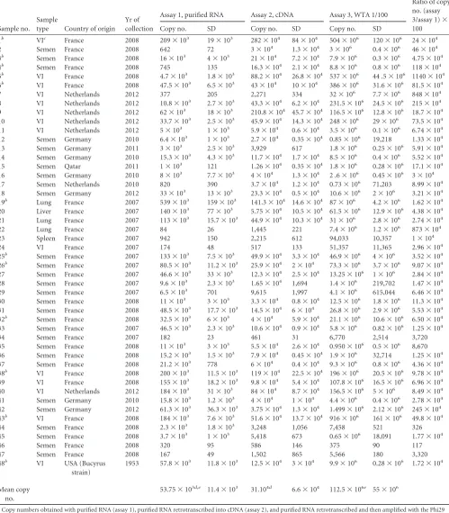
Table 1presents the results obtained with assays 1,2, and 3. To ensure that the comparisons between the assays were normalized and standardized, the RNA samples used in each of the three as-says were from the same extraction process. Interestingly, all pos-itive samples were detected by all three assays, indicating that iso-thermal amplification with Phi29 polymerase did not change the specificity of the OIE-recommended RT-qPCR (6). Moreover, no PCR signals were detected from the 12 negative samples, indicat-ing that the assay was highly specific for EAV detection. Further-more, the analytical sensitivity of each assay was evaluated and
Received8 July 2014 Returned for modification11 August 2014
Accepted10 October 2014
Accepted manuscript posted online22 October 2014
CitationHans A, Gaudaire D, Manuguerra J-C, Leon A, Gessain A, Laugier C,
Berthet N, Zientara S. 2015. Combination of an unbiased amplification method and a resequencing microarray for detecting and genotyping equine arteritis virus. J Clin Microbiol 53:287–291.doi:10.1128/JCM.01935-14.
Editor:A. M. Caliendo
Address correspondence to Aymeric Hans, [email protected].
* Present address: Nicolas Berthet, CIRMF, Unité Zoonose et Maladies Emergentes, Franceville, Gabon.
Copyright © 2015, American Society for Microbiology. All Rights Reserved.
doi:10.1128/JCM.01935-14
on May 16, 2020 by guest
http://jcm.asm.org/
TABLE 1The 48 positive EAV samples tested with their origin, year of collection, and copy numbersa
Sample no. Sample
type Country of origin Yr of collection
Assay 1, purified RNA Assay 2, cDNA Assay 3, WTA 1/100
Ratio of copy no. (assay 3/assay 1)⫻ 100 Copy no. SD Copy no. SD Copy no. SD
1b VIc France 2008 209⫻103 19⫻103 282⫻104 84⫻104 504⫻106 120⫻106 24⫻104
2 Semen France 2008 642 72 3⫻104 1.3⫻104 3⫻106 0.4⫻106 46⫻104
3b Semen France 2008 16⫻103 4⫻103 21⫻104 7.2⫻104 7.9⫻106 0.3⫻106 4.75⫻104
4b Semen France 2008 745 135 16.3⫻104 2.1⫻104 8.8⫻106 0.8⫻106 118⫻104
5b VI France 2008 4.7⫻103 1.8⫻103 88.2⫻104 26.8⫻104 537⫻106 44 .5⫻106 1140⫻104
6b VI France 2008 47.5⫻103 6.5⫻103 43⫻104 10⫻104 386⫻106 31.6⫻106 81.5⫻104
7 VI Netherlands 2012 377 205 2,271 334 32⫻106 7.7⫻106 848⫻104
8 VI Netherlands 2012 10.8⫻103 2.7⫻103 43.3⫻104 6.2⫻104 231.5⫻106 24.5⫻106 215⫻104
9 VI Netherlands 2012 62⫻103 18⫻103 210.8⫻104 45.7⫻104 116.5⫻106 12.8⫻106 18.7⫻104
10 VI Netherlands 2012 33.7⫻103 2.5⫻103 45.9⫻104 14.3⫻104 248⫻106 29⫻106 73.5⫻104
11 VI Netherlands 2012 5⫻103 1⫻103 5.9⫻104 0.6⫻104 3.5⫻106 0.1⫻106 6.74⫻104
12 Semen Germany 2010 6.4⫻103 1⫻103 2.7⫻104 0.35⫻104 0.85⫻106 19,218 1.33⫻104
13 Semen Germany 2011 3⫻103 2.5⫻103 3,929 617 1.8⫻106 0.25⫻106 5.91⫻104
14 Semen Germany 2010 15.3⫻103 4.3⫻103 11.7⫻104 1.7⫻104 8.5⫻106 0.4⫻106 5.52⫻104
15 Semen Qatar 2011 1⫻103 121 1.26⫻104 0.35⫻104 1.8⫻106 0.28⫻106 17.1⫻104
16 Semen Germany 2010 8⫻103 7.7⫻103 4⫻104 1.3⫻104 2 .6⫻106 0.45⫻106 3⫻104
17 Semen Netherlands 2010 820 390 3.7⫻104 1.2⫻104 0.73⫻106 71,203 8.99⫻104
18 Semen Germany 2012 33⫻103 13⫻103 23.3⫻104 0.5⫻104 10.6⫻106 2⫻106 3.21⫻104
19b Lung France 2007 539⫻103 159⫻103 141.3⫻104 14.6⫻104 87⫻106 4.2⫻106 1.62⫻104
20 Liver France 2007 140⫻103 77⫻103 5.75⫻104 10.5⫻104 61.5⫻106 12.9⫻106 4.38⫻104
21 Lung France 2007 113⫻103 15.7⫻103 44.9⫻104 10.3⫻104 31⫻106 2.8⫻106 2.74⫻104
22 Lung France 2007 84 26 1,445 221 7.4⫻106 1.2⫻106 873⫻104
23 Spleen France 2007 942 150 2,215 612 94,033 10,357 1⫻104
24 VI France 2007 174 48 517 133 51,357 11,365 2.96⫻104
25b Semen France 2007 133⫻103 7.5⫻103 49.9⫻104 3.3⫻104 46.9⫻106 4⫻106 3.52⫻104
26b Semen France 2007 80.5⫻103 11.2⫻103 25.9⫻104 2⫻104 73.3⫻106 3.7⫻106 9.07⫻104
27 Semen France 2007 46.6⫻103 33⫻103 12.3⫻104 2.5⫻104 13.25⫻106 1⫻106 2.84⫻104
28 Semen France 2007 9.6⫻103 2.3⫻103 1.65⫻104 1,694 1.4⫻106 219,702 1.47⫻104
29 Semen France 2007 6.5⫻103 701 9,615 1,997 4.1⫻106 615,044 6.46⫻104
30 Semen France 2008 11⫻103 3⫻103 3.3⫻104 0.8⫻104 12.5⫻106 1.8⫻106 11.3⫻104
31 Semen France 2008 48.5⫻103 17.7⫻103 14.5⫻104 6⫻104 26.8⫻106 2.9⫻106 5.53⫻104
32b Semen France 2008 32.5⫻103 6⫻103 4⫻104 5.9⫻104 21.1⫻106 10.6⫻106 6.50⫻104
33 Semen France 2007 46.5⫻103 2.3⫻103 10.6⫻104 0.9⫻104 5.8⫻106 0.82⫻106 1.25⫻104
34 Semen France 2007 182 23 461 31 6,770 2,514 3,720
35 Semen France 2008 11⫻103 3⫻103 5.5⫻104 2.6⫻104 0.950⫻106 0.5⫻106 8,670
36 Semen France 2008 15.2⫻103 1.5⫻103 7.9⫻104 0.45⫻104 1.9⫻106 32,714 1.25⫻104
37 Semen France 2008 21.2⫻103 778 6⫻104 0.4⫻104 9.3⫻106 0.8⫻106 4.36⫻104
38b VI France 2008 200⫻103 11.5⫻103 119⫻104 22.5⫻104 196⫻106 20.5⫻106 9.78⫻104
39 VI France 2008 155⫻103 18.2⫻103 9.8⫻104 5.4⫻104 107.8⫻106 16.5⫻106 6.96⫻104
40 VI Netherlands 2012 184⫻103 31⫻103 84⫻104 8.7⫻104 156.5⫻106 5⫻106 8.49⫻104
41 Semen Germany 2010 15.8⫻103 1.2⫻103 4⫻104 1⫻104 4.4⫻106 0.4⫻106 2.78⫻104
42 Semen Germany 2012 61.3⫻103 36.3⫻103 3.75⫻104 1.3⫻104 1.499⫻106 2.12⫻106 245⫻104
43b VI France 2008 184⫻103 7.6⫻103 51.6⫻104 13.7⫻104 916⫻106 161⫻106 49.8⫻104
44 Semen France 2008 2.3⫻103 1.8⫻103 3,248 1,056 7,458 521 326
45 Semen France 2008 3.7⫻103 1⫻103 5,418 673 0.65⫻106 18,091 1.77⫻104
46 Semen France 2008 320 95 586 146 375 90 117
47 Semen France 2008 167 49 1,502 865 5,566 180 3,320
48b VI USA (Bucyrus
strain)
1953 57.8⫻103 11.8⫻103 12.5⫻104 3⫻104 9.9⫻106 0.28⫻106 1.72⫻104
Mean copy no.
53.75⫻103d,e 11.4⫻103 31.104d 6.6⫻104 112.5⫻106e 55⫻106
a
Copy numbers obtained with purified RNA (assay 1), purified RNA retrotranscribed into cDNA (assay 2), and purified RNA retrotranscribed and then amplified with the Phi29 polymerase (assay 3) are shown. The gains in sensitivity between assays 1 and 3 are also shown (ratio of copy numbers).
b
Samples tested on the high-density resequencing DNA microarray.
cVI, culture fluid from virus isolation performed on RK-13 cells (ATCC CCL-37), as described in reference6. d
Among these two values,P⬍0.05 (using Student’s t test).
eAmong these two values,P⬍0.05 (using Student’s t test).
on May 16, 2020 by guest
http://jcm.asm.org/
varied markedly depending on the assay performed. The mean copy numbers of viral genome for the 48 positive samples ob-tained with assays 1, 2, and 3 were 53.75⫻103⫾11.4⫻103, 31⫻
104⫾6.6⫻104, and 112.5⫻106⫾55⫻106, respectively. Our results show that isothermal amplification with Phi29 polymerase significantly increased the ratio of amplification compared to as-say 1. Indeed, the detected amount of DNA with asas-say 3 was 102to
107times the amount detected using assay 1 (Table 1). Therefore, compared to the OIE-recommended RT-qPCR, the analytical sensitivity of assay 3 is significantly enhanced (Student’s t test,P⬍
0.05) (Table 1). These findings were obtained regardless of the sample’s origin (culture fluid, raw semen, or homogenized or-gans).
In addition to enhancing the sensitivity of the current diagnos-tic method, the second objective of our study was to rapidly geno-type the viruses detected. We therefore decided to combine the unbiased amplification of nucleic acids with a high-density rese-quencing microarray. The two EAV sequences tiled on the mi-croarray cover a region of 267 bp (Table 2) located in the ORF 1 region encoding the nonstructural protein 9 (nsp9) that is in-volved in RNA synthesis (11). Amplified DNA samples were frag-mented and labeled using the GeneChip resequencing assay kit (Affymetrix, Inc., Santa Clara, CA). After overnight hybridization at 45°C, the RMA was washed, stained, and scanned according to the manufacturer’s instructions. The raw image file (.DAT)
ob-tained after scanning was transformed into a fluorescence inten-sity file (.CEL) using GeneChip operating software (GCOS) (Af-fymetrix, Inc.). Bases were called using a derivative of the ABACUS base-calling algorithm (12). Sequences were output in FASTA format, and for each sequence obtained, the call rate value was calculated as the ratio of the number of determined bases (A, T, C, or G) to a sequence length of 243 bp (Table 3). Sequences retrieved from the microarray were used to perform an evolution-ary study. The phylogenetic trees were constructed using the max-imum-likelihood method, and the tree’s statistical robustness was assessed by bootstrap resampling (100 data sets) of the multiple alignments. Phylogenetic reconstructions were performed using SeaView software, version 4 (13). In the literature, EAV isolates have been classified into two distinct groups: the North American group (NA) and the European group (EU) (14). The EU group can be subdivided into two subgroups known as European sub-group 1 (EU-1) and European subsub-group 2 (EU-2) (14). This clas-sification was obtained by aligning and comparing either the 3-kb sequences covering the ORF2-to-ORF7 region of the EAV ge-nome, which encodes the structural protein, or a 518-nucleotide portion of ORF5, from positions 11,296 to 11,813, encoding viral glycoprotein 5 (14). A phylogenetic analysis with EAV sequences, retrieved from either GenBank or the 12 samples in our study (samples 1, 3, 4, 5, 6, 19, 25, 26, 32, 38, 43, and 48), was performed according to the internationally recommended rules described above. A phylogenetic tree constructed with the 518-nucleotide sequence obtained following ORF5 sequencing (Fig. 2a) con-firmed the classification of known EAV strains. Moreover, sam-ples 1 to 6, 19 to 38, and 43 to 48 were classified as being in the EU-1, EU-2, and NA groups, respectively (Fig. 2b). The products obtained after isothermal amplification from these 12 samples were hybridized on the RMA (Table 1). Surprisingly, the phyloge-netic tree obtained with the nsp9 nucleotide sequences retrieved from the microarray separated the strains into the NA and EU groups and divided the EU group into subgroups EU-1 and EU-2 (Fig. 2b). Thus, this result indicates that the 243-nucleotide se-quence from the ORF1 region amplified in our study could also be used for genotyping EAV.
This study is the first in the scientific literature to use an unbi-ased isothermal amplification method using the Phi29 polymerase in combination with a high-density resequencing microarray for diagnosing nidoviruses. Furthermore, amplifying EAV RNA by
FIG 1Schematic view of EAV detection by qPCR and RT-qPCR using three different assays (assays 1, 2, and 3).
TABLE 2EAV partial ORF1 sequences of two 267-nucleotide strains from theArteriviridaefamily tiled on the resequencing microarray Viral strain
Location along ORF 1
GenBank Accession no.
CW 96 6579–6845 AY349167
Bucyrus (EAV ATCC VR-796) 6575–6841 DQ846750
TABLE 3Call rate value for each of the 12 strains tested on the RMAa
Sample no.
Call rate value
(%) Phylogenetic group
1 34.69 European subgroup 1
3 34.69 European subgroup 1
4 34.69 European subgroup 1
5 51.43 European subgroup 1
6 50.61 European subgroup 1
19 56.33 European subgroup 2 25 52.65 European subgroup 2 26 45.97 European subgroup 2
32 60 European subgroup 2
38 71.43 European subgroup 2 43 73.47 North American group 48 90.2 North American group aCall rate value is the ratio of the number of determined bases (A, T, C, or G) to a
sequence length of 243 bp.
on May 16, 2020 by guest
http://jcm.asm.org/
whole transcription amplification, which requires an additional time of 10 h, provides much greater sensitivity than the OIE-rec-ommended RT-qPCR method developed in 2002 by Balasuriya et al. (6, 7). Its association with a resequencing microarray has greatly reduced the time taken for genotyping even if it remains more expensive than reference RT-qPCR and Sanger sequencing of PCR amplicons. However, the resequencing microarray is less expensive than next-generation sequencing for EAV genotyping. This method can be recommended for the detection of EAV in semen and aborted fetuses, especially when the viral load is very low. In addition, this study validated the usefulness of the high-density resequencing DNA microarray for the diagnosis of equine viral diseases and the genotyping of RNA viruses such as equine arteritis virus.
ACKNOWLEDGMENTS
This study was supported by European Commission funding for the Ref-erence Laboratory for Equine Diseases other than African Horse Sickness, by the Institut Pasteur‘s transverse research programs grant 246 (Detec-tion of Emerging Viral Agents [DEVA] program), and by l’Institut Fran-çais du Cheval et de l’Equitation (IFCE), France.
We thank the platform “Genotyping of Pathogens and Public Health” and G. Coralie for use of the Affymetrix station and J. Tapprest (head of the necropsy center at the Anses laboratory for equine diseases) for pro-viding organs.
The funders had no role in the study design, data analysis, or prepa-ration of the manuscript.
REFERENCES
1.Cavanagh D.1997.Nidovirales: a new order comprisingCoronaviridae
andArteriviridae. Arch Virol142:629 – 633.
2.Snijder EJ, Kikkert M, Fang Y.2013. Arterivirus molecular biology and pathogenesis. J Gen Virol94:2141–2163.http://dx.doi.org/10.1099/vir.0 .056341-0.
3.Timoney PJ, McCollum WH.1993. Equine viral arteritis. Vet Clin North Am Equine Pract9:295–309.
4.Timoney PJ, McCollum WH, Roberts AW, Murphy TW.1986. Dem-onstration of the carrier state in naturally acquired equine arteritis virus infection in the stallion. Res Vet Sci41:279 –280.
5.Miszczak F, Legrand L, Balasuriya UB, Ferry-Abitbol B, Zhang J, Hans A, Fortier G, Pronost S, Vabret A.2012. Emergence of novel equine arteritis virus (EAV) variants during persistent infection in the stallion: origin of the 2007 French EAV outbreak was linked to an EAV strain present in the semen of a persistently infected carrier stallion. Virology
423:165–174.http://dx.doi.org/10.1016/j.virol.2011.11.028.
6.World Organization for Animal Health. 2012. Equine viral arteritis (EVA). In OIE manual of diagnostic tests and vaccines for terrestrial ani-mals, vol 2. World Organization for Animal Health, Paris, France. 7.Balasuriya UB, Leutenegger CM, Topol J B, McCollum WH, Timoney
PJ, MacLachlan NJ.2002. Detection of equine arteritis virus by real-time TaqMan reverse transcription-PCR assay. J Virol Methods 101:21–28. http://dx.doi.org/10.1016/S0166-0934(01)00416-5.
8.Miszczak F, Shuck KM, Lu Z, Go YY, Zhang J, Sells S, Vabret A, Pronost S, Fortier G, Timoney PJ, Balasuriya UB.2011. Evaluation of two magnetic-bead-based viral nucleic acid purification kits and three real-time reverse transcription-PCR reagent systems in two TaqMan as-says for equine arteritis virus detection in semen. J Clin Microbiol49:
3694 –3696.http://dx.doi.org/10.1128/JCM.01187-11.
9.Pronost S, Pitel PH, Miszczak F, Legrand L, Marcillaud-Pitel C, Hamon M, Tapprest J, Balasuriya UB, Freymuth F, Fortier G.2010. Description of the first recorded major occurrence of equine viral ar-teritis in France. Equine Vet J42:713–720.http://dx.doi.org/10.1111/j .2042-3306.2010.00109.x.
10. Berthet N, Reinhardt AK, Leclercq I, van Ooyen S, Batejat C, Dickinson P, Stamboliyska R, Old IG, Kong KA, Dacheux L, Bourhy H, Kennedy GC, Korfhage C, Cole ST, Manuguerra JC. 2008. Phi29 polymerase based random amplification of viral RNA as an alternative to random RT-PCR. BMC Mol Biol 9:77.http://dx.doi.org/10.1186/1471-2199-9-77. 11. Beerens N, Selisko B, Ricagno S, Imbert I, van der Zanden L, Snijder EJ, Canard B.2007.De novoinitiation of RNA synthesis by the arterivirus RNA-dependent RNA polymerase. J Virol81:8384 – 8395.http://dx.doi .org/10.1128/JVI.00564-07.
FIG 2Phylogenetic analysis of the partial ORF5 nucleotide sequences (518 nucleotides in length) (a) and partial ORF1 nucleotide sequences (243 nucleotides in length) (b) of 27 EAV strains. Twelve isolates are specific to this study, and 15 are from previously published EAV strains. Neighbor-joining and maximum-likelihood trees were constructed using SeaView version 4, and equivalent results were obtained. Only the maximum-maximum-likelihood trees are shown. The percentages of trees,⬎50 only, in which the associated taxa clustered together are shown next to the branches. The North American group and European subgroups 1 and 2 are indicated.
on May 16, 2020 by guest
http://jcm.asm.org/
[image:4.585.43.542.76.286.2]12. Cutler DJ, Zwick ME, Carrasquillo MM, Yohn CT, Tobin KP, Kashuk C, Mathews DJ, Shah NA, Eichler EE, Warrington JA, Chakravarti A.2001. High-throughput variation detection and geno-typing using microarrays. Genome Res11:1913–1925.
13. Gouy M, Guindon S, Gascuel O.2010. SeaView version 4: a multi-platform graphical user interface for sequence alignment and
phyloge-netic tree building. Mol Biol Evol27:221–224. http://dx.doi.org/10 .1093/molbev/msp259.
14. Zhang J, Miszczak F, Pronost S, Fortier C, Balasuriya UB, Zientara S, Fortier G, Timoney PJ.2007. Genetic variation and phylogenetic analysis of 22 French isolates of equine arteritis virus. Arch Virol152:1977–1994. http://dx.doi.org/10.1007/s00705-007-1040-z.