C A S E R E P O R T
Open Access
Evaluation of bilateral central retinal artery
occlusions with optical coherence
tomography-based microangiography: a
case report
Aaron Y. Lee
1†, Qinqin Zhang
2†, Douglas M. Baughman
1, Raghu Mudumbai
1, Ruikang K. Wang
1,2and Cecilia S. Lee
1*Abstract
Background:We report a case of bilateral central retinal artery occlusion and the evaluation of retinal vasculature and capillaries by using optical coherence tomography angiography.
Case presentation:A 75-year-old white man presented with central retinal artery occlusion in one eye and underwent a carotid angioplasty. Upon discontinuing anticoagulant, he had a subsequent central retinal artery occlusion in the other eye. Optical coherence tomography angiography images were obtained to compare the retinal microvasculature in both eyes.
Conclusions:Atrophy of the involved retina continues for several weeks after central retinal artery occlusion but the loss of retinal capillaries is immediate and stable over time. The presence of cilioretinal arteries that perfuse the central macula can prevent profound vision loss.
Keywords:Central retinal artery occlusion, Optical coherence tomography angiography (OCTA), Optical coherence tomography-based microangiography (OMAG), Case report
Background
Central retinal artery occlusion (CRAO) is an important vascular cause of serious vision loss. The incidence of non-arteritic CRAO is estimated to be 1 in 100,000 people [1] and most result in profound vision loss [2]. Despite being a well-recognized ophthalmic emergency [3], limited progress has been made in diagnostic evalua-tions. Studies suggest that CRAO lasting approximately 240 minutes results in irreparable retinal damage [3, 4].
Fluorescein angiography (FA) has been a standard method in assessing retinal vasculature and diseases such as CRAO [5]. Sodium fluorescein is given intraven-ously and the fluorescence emitted from retinal and choroidal circulation is detected on angiography. CRAO can be diagnosed clinically with common funduscopic
findings such as diffuse retinal edema,“cherry-red spot,”
“box-carring,” or attenuation of the vessels [6, 7]. The delayed filling of the affected vessels on FA confirms the diagnosis. However, FA is invasive and relatively contra-indicated in pregnancy or chronic kidney failure. In addition, the visualizations of retinal vasculature are lim-ited with this imaging [5]. Recently, the introduction of optical coherence tomography angiography (OCTA) has shown promising results in allowing noninvasive func-tional imaging of retinal vasculature [8]. With this im-aging modality, both structural and functional changes of the retina can be assessed.
Optical coherence tomography-based microangiography (OMAG) is an OCTA algorithm and has been used for evaluation of various retinal diseases [9, 10]. With this technology, the retinal microvasculature can be visualized, quantified, and dissected in various layers. There has been increasing interest in evaluating the area of retinal ische-mia following retinal arterial occlusions using structural
* Correspondence:[email protected]
†Equal contributors
1Department of Ophthalmology, University of Washington, Seattle, WA, USA Full list of author information is available at the end of the article
images of spectral domain OCT (SD-OCT) without the angiography component [5, 11]. Using OMAG, the super-ficial, intermediate, and deeper retinal capillary plexus can be visualizedin vivoin addition to structural images. The purpose of this case report is to describe key retinal struc-ture changes and vasculastruc-ture findings in a case of bilateral CRAOs using fundus photo, SD-OCT, and OMAG.
Case presentation Clinical details
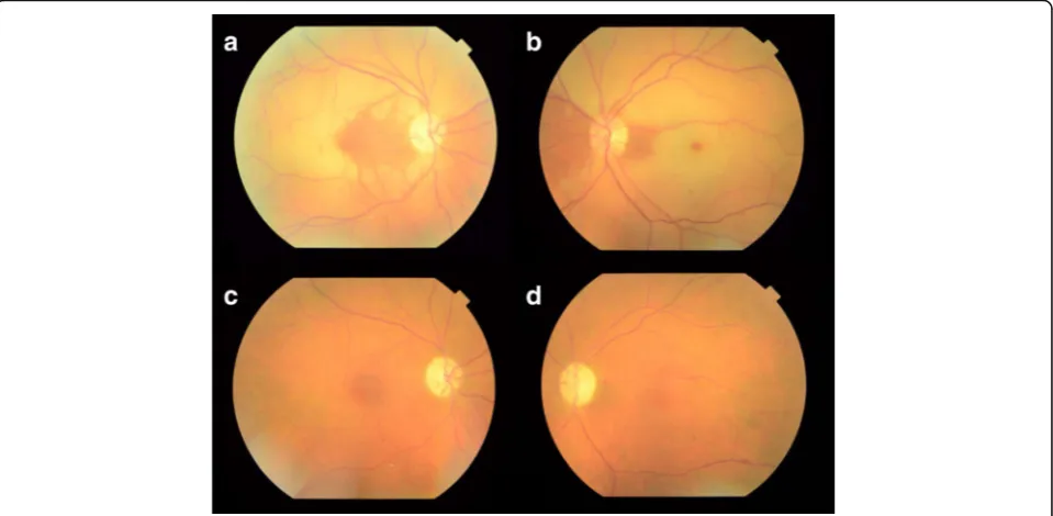
A 75-year-old white man with a history of ischemic car-diomyopathy status post-heart transplant presented with acute loss of vision in his right eye for 1 week, no history of polymyalgia rheumatica, and no scalp tenderness, jaw claudication, proximal muscle weakness, or temporal headache. His best-corrected visual acuity was 20/25 eccentrically in his right eye and 20/25 in his left eye. Fundus imaging showed several Hollenhorst plaques and diffuse whitening of his retina except for the nasal macula (Fig. 1a). FA was not obtained due to severe chronic renal insufficiency.
He underwent a stroke workup including a complete blood count (CBC), erythrocyte sedimentation rate (ESR), C-reactive protein (CRP), carotid Doppler, echo-cardiogram, and magnetic resonance imaging/angiog-raphy (MRI/MRA) of his brain. Of note, his platelet count was 148×103/uL, ESR 21, and CRP was 7.4 mg/L. An approximate 90 % stenosis at the origin of his innominate artery with 30 % stenosis of the right
carotid bulb was found. He underwent balloon angio-plasty and stenting of his innominate artery and was placed on a daily dose of aspirin (325 mg) and clopido-grel (75 mg); however, he stopped the medicine due to poor tolerance.
Four months after the CRAO in his right eye and a few weeks after discontinuing clopidogrel, he had another episode of acute painless vision loss involving his left, pre-viously unaffected, eye. At this time, his visual acuity was 20/30 eccentrically in his right eye and 20/400 in his left eye. A funduscopic examination 4 days after the episode of his left eye revealed diffuse whitening of his retina with a cherry red spot (Fig. 1b). A repeat workup was unre-markable and he was restarted on clopidogrel. Fundus photographs were repeated three months after the CRAO in each eye (Fig. 1c, d).
Investigations
He underwent a comprehensive ophthalmologic examin-ation and color fundus imaging (Topcon, Tokyo, Japan) tests and SD-OCT imaging (Heidelberg, Heidelberg, Germany) tests whenever possible during his initial visit and follow-up visits. In addition, OMAG imaging was obtained subsequently using a modified SD-OCT (CIRRUS prototype provided by Carl Zeiss Meditec Inc., Dublin, CA, USA) with a central wavelength of 850 nm and a speed of 100,000 A-scans per second.
The details of OMAG scanning protocol and algorithm have been described previously [9]. The complex version
[image:2.595.59.539.449.684.2]of the OMAG algorithm was applied to the images that were extracted. The retinal images were segmented into three different retinal layers using a semi-automated algo-rithm: an inner retinal layer from the ganglion cell layer to the inner plexiform layer, a middle retinal layer from the inner nuclear layer to the outer plexiform layer, and an outer retinal layer from the external limiting membrane and the outer nuclear layer. The three-dimensional struc-ture of the retina was projected using Matlab (The Math-Works, Inc., Natick, MA, USA).
The mean retinal thickness was measured by calculat-ing the average of five consecutive scans. The macula was divided into nine zone Early Treatment Diabetic Retinopathy Study (ETDRS) grids: parafovea, inner/outer nasal, inner/outer temporal, inner/outer superior, and inner/outer inferior. Perfusion index was defined as the percent coverage of the area by retinal vessels with flow. For his right eye, the perfusion indices were compared between two subsequent visits following the foveal-sparing CRAO. For his left eye, the perfusion indices before and after CRAO were compared.
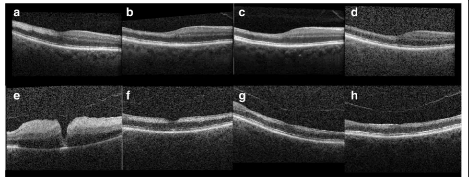
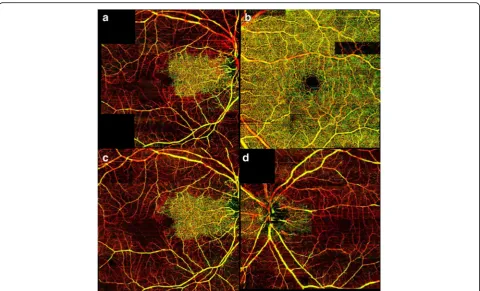
SD-OCT imaging was obtained at 1, 3, 5, and 7 months after the foveal-sparing CRAO in his right eye (Fig. 2a–d). Both superficial and deep retinal layer thick-ness decreased in the involved temporal macula com-pared to the nasal macula. Mean retinal thickness temporal to fovea (500μm away from the foveal center) in his right eye at months 1, 3, 5, and 7 was 250μm, 204 μm, 194μm, and 191μm, respectively. OMAG images of his right eye at 1 and 3 months are shown in Fig. 3a, c. OMAG images of his left eye prior to the CRAO and 1 month after the CRAO are shown In Fig. 3b, d.
SD-OCT imaging was obtained on day 4 and at 1 month, 3 months, and 5 months after the CRAO in his
left eye. (Fig. 2e–h) On day 4, there was diffuse edema involving both superficial and deep retinal layers. Mean retinal thickness temporal to fovea (660 μm away from the foveal center) was 504μm. Follow-up images of the same area revealed thicknesses of 246 μm, 215μm, and 196μm, at months 1, 3, and 5, respectively.
Perfusion indices showed no significant difference during two consecutive visits after the CRAO in his right eye (Fig. 4). The perfusion indices in all zones were below 40 except for the inner and outer nasal ETDRS zones which were unaffected by the CRAO. There was a statistically significant difference in the perfusion indices of all nine zones before and after the CRAO in his left eye (p-value <0.001, Fig. 4). The average perfusion index in the eight non-central zones prior to the CRAO was 40 (range 39 to 45) while the average perfusion index after the CRAO in the non-central zones was 20 (range 10 to 30).
Outcomes
The retinal vasculature loss following CRAO was acute and permanent, but the retinal atrophy progressed over several months. In our patient, the thickness of inner retina continued to decrease until month 5, but then it stabilized on follow-up visits. However, the structural architecture of nasal retina remained undisrupted by SD-OCT throughout the follow-up period. OMAG images showed severe loss of all retinal microvasculature involving intermediate and deep plexus more than the superficial plexus. The en face OMAG images ob-tained at months 1 and 3 demonstrated visible absence of the retinal microvasculature in his right eye except for the nasal macula (Fig. 3a, c). The en face OMAG image obtained at month 1 after the CRAO in the left
[image:3.595.59.539.512.693.2]eye revealed loss of retinal capillary plexus in the entire macula (Fig. 3d).
Even though the FA is a standard imaging modality in evaluating retinal vascular diseases, FA is often not pos-sible due to time commitment and patient factors such as renal insufficiency. Although FA would have been diagnostic in our patient, it was deferred due to chronic renal insufficiency. Contraindications for FA include fluorescein allergy, renal failure, pregnancy, moderate-severe asthma, and significant cardiac cases [12]. The noninvasive nature of OCTA is advantageous in many of our patients with multiple comorbidities. In addition, the image quality can vary due to media opacity such as cataract and patient cooperation. Smaller capillaries and deeper retinal plexus are not visible with FA. In contrast, OCTA provides a safe and potentially superior alterna-tive method of assessing total retinal vasculature.
Recently, retinal artery occlusions were characterized as superficial and/or intermediate and deep capillary plexus ischemia using SD-OCT images [11]. In particular, the ischemia within the intermediate and deeper retinal capil-lary plexus were described as a hyper-reflective lesion at the level of the inner nuclear layer and referred to as
paracentral acute middle maculopathy [13]. The character-istic ischemia in the inner retina cannot be presumed based only on the change in the reflective pattern seen on SD-OCT, but OCTA allows for direct visualization of perfusion and quantification. Thus, the vascular change evident on OCTA such as OMAG could become an important clinical endpoint in future studies.
[image:4.595.58.539.86.377.2]the perfusion percentage within independently subsam-pled areas of the retina. This method allowed the quanti-fication of the OCTA signal per each ETDRS zone leading to a more robust quantification.
To the best of our knowledge, this is the first descrip-tion of OCTA imaging in bilateral CRAO and compari-son of the OCTA imaging before and after CRAO. OCTA provides important information regarding vascu-lar changes including hypoperfusion, ischemia, and ab-normal structure. However, the lack of standardizations, the lack of full understanding of the artifacts, and the lack of large trials to determine the clinical relevance of image findings are the main current limitations. Despite these challenges, OCTA such as OMAG will probably become an effective adjunct modality in diagnosing and following retinal vascular diseases.
Conclusions
CRAO is a relatively common sight-threatening condition. Using OCTA, we demonstrate that the atrophy of the
involved retina continues for several weeks following CRAO but the loss of retinal capillaries is immediate and stable over time. The presence of cilioretinal arteries that perfuse the central macula can prevent profound vision loss.
Abbreviations
CBC:Complete blood count; CRAO: Central retinal artery occlusion; CRP: C-reactive protein; ESR: Erythrocyte sedimentation rate; ETDRS: Early Treatment Diabetic Retinopathy Study; FA: Fluorescein angiography; MRI/MRA: Magnetic resonance imaging/angiography; OCTA: Optical coherence tomography angiography; OMAG: Optical coherence tomography-based microangiography; SD-OCT: Spectral domain optical coherence tomography
Acknowledgements
Not applicable.
Funding
The study was supported by the following grants: K23EY024921 (CSL), R01EY024158 (RKW), Research to Prevent Blindness (CSL, AYL, RKW). Dr Wang received research support from Carl Zeiss Meditec, Inc.
Availability of data and materials
The data that support the findings of this study are available from the corresponding author upon reasonable request.
Authors’contributions
Conception and design (CSL, AYL, RKW); analysis and interpretation (CSL, AYL, QZ, DB); writing the article (CSL, AYL); critical revision of the article (QZ, DB, RM, RKW); final approval of the article (CSL, AYL, QZ, DB, RM, RKW); data collection (QZ, RM); statistical expertise (AYL); and literature search (CSL). All authors read and approved the final manuscript.
Competing interests
Dr Wang and the Oregon Health & Science University co-own a patent that is licensed to Carl Zeiss Meditec, Inc. All other authors declare that they have no competing interests.
Consent for publication
Written informed consent was obtained from the patient for publication of this case report and any accompanying images. A copy of the written consent is available for review by the Editor-in-Chief of this journal.
Ethics approval and consent to participate
The Institutional Review Board of the University of Washington approved the study and our patient gave informed consent to participate. The study was performed in accordance with the tenets of the Declaration of Helsinki.
Author details
1Department of Ophthalmology, University of Washington, Seattle, WA, USA. 2Department of Bioengineering, University of Washington, Seattle, WA, USA.
Received: 30 March 2016 Accepted: 10 October 2016
References
1. Rumelt S, Dorenboim Y, Rehany U. Aggressive systematic treatment for central retinal artery occlusion. Am J Ophthalmol. 1999;128(6):733–8. 2. Varma DD, Cugati S, Lee AW, Chen CS. A review of central retinal artery
occlusion: clinical presentation and management. Eye (Lond). 2013;27(6):688–97. 3. Hayreh SS, Zimmerman MB. Central retinal artery occlusion: visual outcome.
Am J Ophthalmol. 2005;140(3):376–91.
4. Hayreh SS, Zimmerman MB, Kimura A, Sanon A. Central retinal artery occlusion. Retinal survival time. Exp Eye Res. 2004;78(3):723–36. 5. Yu S, Wang F, Pang CE, Yannuzzi LA, Freund KB. Multimodal imaging
findings in retinal deep capillary ischemia. Retina (Philadelphia, Pa). 2014; 34(4):636–46.
6. Zhou A, Weizblit N, Noble J. Can you identify this condition? Can Fam Physician. 2008;54(12):1699–700.
[image:5.595.58.290.86.400.2]7. Hayreh SS, Zimmerman MB. Fundus changes in central retinal artery occlusion. Retina. 2007;27(3):276–89.
8. Spaide RF, Klancnik JM, Cooney MJ. Retinal vascular layers imaged by fluorescein angiography and optical coherence tomography angiography. JAMA Ophthalmol. 2015;133(1):45–50.
9. Huang Y, Zhang Q, Thorell MR, et al. Swept-source OCT angiography of the retinal vasculature using intensity differentiation-based optical
microangiography algorithms. Ophthalmic Surg Lasers Imaging Retina. 2014;45(5):382–9.
10. Thorell MR, Zhang Q, Huang Y, et al. Swept-source OCT angiography of macular telangiectasia type 2. Ophthalmic Surg Lasers Imaging Retina. 2014; 45(5):369–80.
11. Yu S, Pang CE, Gong Y, et al. The spectrum of superficial and deep capillary ischemia in retinal artery occlusion. Am J Ophthalmol. 2015;159(1):53–63. e1–2. 12. Bowling B. Kanski’s Clinical Ophthalmology: A Systemic Approach. 8th ed.
China: Elsevier; 2016. p. 587.
13. Rahimy E, Sarraf D. Paracentral acute middle maculopathy spectral-domain optical coherence tomography feature of deep capillary ischemia. Curr Opin Ophthalmol. 2014;25(3):207–12.
14. Justice J, Lehmann RP. Cilioretinal arteries. A study based on review of stereo fundus photographs and fluorescein angiographic findings. Arch Ophthalmol. 1976;94(8):1355–8.
• We accept pre-submission inquiries
• Our selector tool helps you to find the most relevant journal
• We provide round the clock customer support
• Convenient online submission
• Thorough peer review
• Inclusion in PubMed and all major indexing services
• Maximum visibility for your research
Submit your manuscript at www.biomedcentral.com/submit