Biology Theses Department of Biology
7-13-2009
To Degrade or Not to Degrade: The Role of P300/CBP-Associated
To Degrade or Not to Degrade: The Role of P300/CBP-Associated
Factor (PCAF) in Ciita Stability and Ubiquitination
Factor (PCAF) in Ciita Stability and Ubiquitination
Jeanne Kaye Brooks
Follow this and additional works at: https://scholarworks.gsu.edu/biology_theses
Part of the Biology Commons
Recommended Citation Recommended Citation
Brooks, Jeanne Kaye, "To Degrade or Not to Degrade: The Role of P300/CBP-Associated Factor (PCAF) in Ciita Stability and Ubiquitination." Thesis, Georgia State University, 2009.
https://scholarworks.gsu.edu/biology_theses/21
This Thesis is brought to you for free and open access by the Department of Biology at ScholarWorks @ Georgia State University. It has been accepted for inclusion in Biology Theses by an authorized administrator of
(PCAF) IN CIITA STABILITY AND UBIQUITINATION
by
JEANNE K. BROOKS
Under the Direction of Dr. Susanna F. Greer
ABSTRACT
The ubiquitin-proteasome pathway plays vital roles in multiple cellular processes
including protein turnover and transcription regulation. The fate of a ubiquitinated protein is
determined by the number of ubiquitin molecules added and the site to which they are added.
Monoubiquitinated proteins are stabilized and often activated, while polyubiquitinated proteins
are rapidly targeted for degradation. Major histocompatibility complex class II (MHC II)
molecules are a vital part of the immune response and are responsible for presenting antigens to
CD4+ T cells. The class II transactivator (CIITA) is the master regulator of MHC II
transcription and has been shown to have increased transactivity when monoubiquitinated. The
focus of this thesis is on the impact of ubiquitination on CIITA stability and MHC II gene
(PCAF) IN CIITA STABILITY AND UBIQUITINATION
by
JEANNE K. BROOKS
A Thesis Submitted in Partial Fulfillment of the Requirements for the Degree of
Master of Science
in the College of Arts and Sciences
Georgia State University
Copyright by Jeanne K. Brooks
(PCAF) IN CIITA STABILITY AND UBIQUITINATION
by
JEANNE K. BROOKS
Committee Chair: Dr. Susanna F. Greer
Committee: Dr. Richard Dix
Dr. Zehava Eichenbaum
Electronic Version Approved:
DEDICATION
Once upon a time, in a land far, far away there was a little girl. This little girl would
spend hours asking her father questions; questions about everything from why the sky is blue to
what made her look like him. He patiently answered all of her questions to the best of his
knowledge and always told her that one day she would grow up to be a scientist. She eventually
began her college career to be an elementary school teacher, far from the scientist dreams her
father had for her. One day she started her first biology class, and as that class went on she
realized how fascinating and amazing the world could be through the eyes of a scientist. She
thought back to what her father had been saying for so many years, and finally decided that
maybe he was right. She changed her major and the rest is history.
This thesis is dedicated to the memory of my father who never got to see me become his
“little scientist” as he always dreamed. His guidance, patience, and support of my learning and
development throughout my childhood have made me the curious and questioning individual that
I am today. Without his support and influence on my life I would not be the individual that I am.
While my father was the biggest influence throughout my childhood and my journey towards
science I would not be where I am today without the support of many family members and
friends; especially my husband who has put up with many moods and stressful times, and has
ACKNOWLEDGEMENTS
I would like to take this opportunity to thank the many people who have contributed to
this research. First and foremost my fearless leader, Susanna Greer, who took a chance on me in
allowing me to join her lab, her no nonsense style and upfront personality have been invaluable,
and her passion for science and research are truly an inspiration. She has supported my ideas,
and when needed steered me in the right direction, she has taught me to think like a scientist and
for this I thank you. I would like to thank Richard Dix for his encouragement, and gentle
reminder to quantify my data, and Zehava Eichenbaum for providing me a new perspective on
my research.
Also, my lab mates, how long the days would be without all of you there to share the
joys, frustrations, and sometimes downright insanity of work in a laboratory. We have had some
rough times, and some good ones and hopefully we’ve learned a little bit of science while we
were at it. You are all so willing to help, and have helped me really learn and grow during our
time together. I look forward to our future, not as fellow students, but as colleagues and friends.
My mom Denise, for always being there when I needed an ear to talk to and a little bit of
encouragement; and of course my siblings, Andy, Adam, and Jessica, all of which have listened
to my frustrations, and offered their version of encouragement, my husband Dustin for his
unwavering support and encouragement throughout this. And finally my grandparents, Max and
Betty, who still “don’t have a clue what I do” but have supported me both materially and through
TABLE OF CONTENTS
ACKNOWLEDGEMENTS v
LIST OF FIGURES viii
CHAPTER
1. INTRODUCTION 1
Innate Immunity 1
Adaptive Immunity 2
T Cells 3
Major Histocompatibility Complex 5
Variations of MHC II 6
Activation by MHC 6
MHC II Expression 7
CIITA Regulation 9
Ubiquitin-Proteasome System 11
The Effect of Ubiquitination on Transcription 14
pCAF and Its Role in Ubiquitination 14
2 . MATERIALS AND METHODS 22
Maintenance of Cell Line 22
Plasmids and Reagents 22
Transient Transfection and Luciferase Reporter Assays 22
Knockdown Luciferase Reporter Assay 23
Half Life Assays 23
Densitometry 24
RNA Expression 24
Knockdown RNA Expression 25
Chromatin Immunoprecipitation 26
Co-immunoprecipitation 27
3. RESULTS 29
pCAF Drives CIITA Transactivity 29
CIITA Is Stabilized by the Presence of WT pCAF 30
Knockdown of pCAF Leads to a Decrease in CIITA Transactivity 31
Knockdown of pCAF Decreases the Expression of MHC II 31
Knockdown of pCAF Leads to a Reduction of CIITA Binding at the MHC II Promoter
32
CIITA Ubiquitination Is Increased in the Presence of pCAF 32
MHC II Expression is Dependent on pCAF E3 Ligase Activity 33
CIITA Is Destabilized in the Presence of ∆E3 pCAF 33
4. DISCUSSION 43
REFERENCES 49
Figure 1. CIITA Enhanceosome Complex 16
Figure 2. Functional Domains of CIITA 17
Figure 3. The Ubiquitin-Proteasome Pathway Enzyme Cascade 18
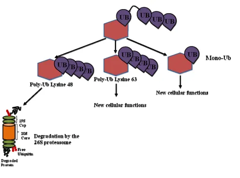
Figure 4. Ubiquitination Patterns 19
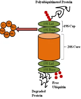
Figure 5. The 26S Proteasome 20
Figure 6. Functional Domains of pCAF 21
Figure 7. Transactivation and Stabilization of CIITA 36
Figure 8. pCAF Knockdown Decreases CIITA Transactivation and Promoter Binding 38
Figure 9. CIITA Ubiquitination Is Enhanced by pCAF 39
Figure 10. pCAF E3 Ligase Activity Affects MHC II Gene Expression 40
CHAPTER 1.
INTRODUCTION
Innate Immunity
The immune system of vertebrates is made of two distinct branches, innate immunity and
adaptive immunity (Medzhitov and Janeway 1998; Janeway, Travers et al. 2005). While innate
immunity is evolutionarily ancient and found in most multicellular organisms, adaptive
immunity is only found in higher order vertebrates. Both branches contain however specific
cells and tissues that perform distinct roles in host defense against pathogens (Medzhitov and
Janeway 1998; Janeway and Medzhitov 2002; Janeway, Travers et al. 2005). The innate branch
of the immune system is the first line of defense against infection and is vital in controlling
infections until and if an adaptive response is mounted. In addition, innate immunity also plays
critical roles in activating the adaptive immune response (Medzhitov and Janeway 1998;
Janeway and Medzhitov 2002; Janeway, Travers et al. 2005). The innate immune system
initiates immediate inflammatory responses to infection, in which macrophages, neutrophils,
basophils, eosinophils, and mast cells act rapidly in order to control infection (Janeway and
Medzhitov 2002; Drozina, Kohoutek et al. 2005; Janeway, Travers et al. 2005). If a pathogen
eludes these responses, the adaptive immune response is activated by the presentation of antigens
from pathogens that have infiltrated secondary lymphatic tissue (Janeway and Medzhitov 2002;
Drozina, Kohoutek et al. 2005; Janeway, Travers et al. 2005).
While the innate immune system is a fundamental component of host defense, it lacks
two key features that make the adaptive immune response critical for survival; specificity for the
pathogens that it encounters, and immunologicalmemory of these pathogens (Medzhitov and
Janeway, Travers et al. 2005). The importance of these key factors, the ability to respond
quickly to antigens, and the ability to provide long term protection from pathogens makes the
adaptive immune response absolutely critical for survival (Farber, Acuto et al. 1997; Janeway
2001).
Adaptive Immunity
Once activated by pathogen, the adaptive immune response relies on specific effector
cells which allow for precise pathogen recognition through a repertoire of receptors (Farber,
Acuto et al. 1997). While the innate immune system also contains receptors effective at
recognizing pathogens, these receptors are germline encoded, and are most effective at
distinguishing between self and non-self (Drozina, Kohoutek et al. 2005; Janeway, Travers et al.
2005). The effector cells of the adaptive immune system, B-cell and T-cells, are collectively
called lymphocytes (Medzhitov and Janeway 1998; Janeway and Medzhitov 2002; Drozina,
Kohoutek et al. 2005; Janeway, Travers et al. 2005). Lymphocytes provide specific pathogen
recognition though their receptors, which are generated though a series of genetic
rearrangements to allow for the generation of receptors capable of recognizing any antigen
presented to the host (Janeway and Medzhitov 2002; Janeway, Travers et al. 2005). Antigen
presenting cells play a major role in the adaptive immune response, by presenting antigens to
lymphocyte receptors and thus activating the adaptive immune response (Abbas and Janeway
2000; Janeway 2001; Janeway, Travers et al. 2005). Following the activation of a B or T cell,
these cells clonally divide and generate a vast number of daughter cells, each of which contains
the specific receptor needed to eliminate the pathogen (Janeway, Travers et al. 2005).
B cell receptors can occur either as cell bound receptors or as secreted antibodies
immune response to pathogens. Antibodies serve two functions to recognize and bind
specifically to antigenic molecules, and to recruit additional immune cells to elicit specific
effector functions at the site of infection (Janeway, Travers et al. 2005). The cell bound B cell
receptor is able to recognize a wide variety of antigenic macromolecules and chemicals which
allows the B cell to internalize, process, and present antigenic peptides to T cells (Abbas and
Janeway 2000; Janeway 2001; Janeway, Travers et al. 2005). T cell receptors occur only in a
cell bound form, a unique feature of the T cell receptor is that it does not bind to antigens
directly, but rather antigens must be presented to the T cell receptor (Abbas and Janeway 2000;
Janeway, Travers et al. 2005). An additional feature of T cells that makes them distinct from B
cells is that there are several different types of T cells, each serving a unique function in the
adaptive immune response.
T Cells
There are several distinct types of T cells that have been well defined with specific
known effector functions (Janeway, Travers et al. 2005). Natural killer T cells are considered
part of the innate immune system, but are an important link between the innate and adaptive
immune responses (Janeway, Travers et al. 2005). These cells do not have antigen specific
receptors but recognize abnormal cells, and following this recognition, can release cytokines and
lytic granules to kill the abnormal cell (Janeway, Travers et al. 2005). Regulatory T cells or Tregs
serve to suppress autoreactive T cells and thus play a large part in preventing autoimmune
diseases. Accordingly, the loss of Tregs has been shown to be linked to a higher instance of
autoimmune diseases including rheumatoid arthritis and multiple sclerosis (Abbas and Janeway
2000; Marzo, Kinnear et al. 2000; Janeway, Travers et al. 2005; Swanberg, Lidman et al. 2005).
specifically viral or tumor antigens (Abbas and Janeway 2000; Janeway, Travers et al. 2005).
These cells play a fundamental role in controlling infection, because they attempt to prevent the
spread of infection by killing cells that are abnormal (Abbas and Janeway 2000; Janeway,
Travers et al. 2005)
The remaining subset of T cells are the helper CD4+ T cells which are further divided
into two classes: TH1 and TH2 helper cells. Both classes of CD4+ T are responsible for activating
additional cells of the immune system through the secretion of specific cytokines (Abbas and
Janeway 2000; Janeway and Medzhitov 2002). CD4+ T
H1 cells are activated by intracellular
antigens or antigens that have been ingested by phagocytic cells. The T cells then release
interferon-γ to activate the phagocytic cells to kill the ingested pathogen. In addition CD4+ TH1
cells can also activate B cells to stimulate endocytosis of pathogens (Abbas and Janeway 2000;
Janeway, Travers et al. 2005). CD4+ TH2 helper cells are specific in activating B cells and
stimulating antibody production against antigens (Abbas and Janeway 2000; Janeway, Travers et
al. 2005). CD4+ TH2 helper cells do not recognize antigens directly but rather recognize major
histocompatibility complex II (MHC II) molecules that process and present antigens on both
antigen presenting cells and nucleated cells activated during an inflammatory response
(Medzhitov and Janeway 1998; Abbas and Janeway 2000; Janeway 2001; Janeway, Travers et al.
2005). Following activation of both CD8+ and CD4+ T cells, memory T cells will be created,
these cells are a fundamental part of the adaptive immune response and are where the important
immunological memory of the adaptive immune response is derived (Abbas and Janeway 2000;
Janeway, Travers et al. 2005). These memory cells remain in the system after an infection is
cleared and upon re-infection of the host are able to mount a rapid and specific immune response
memory also applies to B cells (Janeway, Travers et al. 2005). This interaction between T cells
and MHC molecules is crucial for the activation and maintenance of the adaptive immune
response.
Major Histocompatibility Complex
As stated previously T cells do not recognize antigens directly, instead antigenic peptides
are presented to T cells by MHC molecules (Drozina, Kohoutek et al. 2005; Janeway, Travers et
al. 2005). Cells infected with a pathogen will display these molecules on their surface to alert T
cells that an infectious agent is present (Abbas and Janeway 2000; Janeway, Travers et al. 2005).
There are two classes of MHC molecules, MHC I and MHC II; while similar, each class
performs a specific function in the adaptive immune response (Medzhitov and Janeway 1998;
Janeway and Medzhitov 2002; Drozina, Kohoutek et al. 2005; Janeway, Travers et al. 2005).
The MHC complexes vary in the type of cell that presents them, in the antigen they can present
and the type of effector cell that they are able to activate (Janeway 2001; Janeway, Travers et al.
2005). MHC I molecules are responsible for presenting exogenous peptides from the cytosol,
such as viral or tumor antigens, to CD8 T cells, and are therefore expressed on all nucleated cells
(Abbas and Janeway 2000; Janeway, Travers et al. 2005). MHC II molecules are responsible for
presenting exogenous peptides from intracellular compartments to CD4+ T cells, and are
expressed constitutively on the antigen presenting cells of the immune system: B cells, dendritic
cells, and macrophages (Abbas and Janeway 2000; LeibundGut-Landmann, Waldburger et al.
2004; Janeway, Travers et al. 2005). Expression of MHC II molecules on other nucleated cells
types can be induced by the inflammatory cytokine interferon-γ, and this inducible expression
plays a major role in altering T cell response to infections (LeibundGut-Landmann, Waldburger
Variations of MHC II
MHC II molecules are heterodimeric, transmembrane glycoproteins which are
constitutively expressed on the surface of antigen presenting cells and their expression is further
inducible by interferon-γ on all nucleated cells (Waldburger, Masternak et al. 2000; Drozina,
Kohoutek et al. 2005; Janeway, Travers et al. 2005). The genes that encode MHC II complexes
are located on chromosome six, and are extremely variable (Drozina, Kohoutek et al. 2005;
Janeway, Travers et al. 2005). This variation results in different MHC II isotypes that can be
expressed on the surface of cells. The main isotypes of MHC II are the HLA-DP, HLA-DQ, and
HLA-DR; two additional isotypes, HLA-DM and HLA-DO are considered non-classical and are
not expressed on the surface of cells but, are instead involved in the cytoplasmic loading of
peptides (Waldburger, Masternak et al. 2000; Drozina, Kohoutek et al. 2005). Together these
different MHC II isotypes are able to present different peptides to T cells, which in turn further
increases the diversity of peptides that the immune system can recognize (Waldburger,
Masternak et al. 2000; Drozina, Kohoutek et al. 2005).
Activation by MHC
Both classes of MHC molecules activate the adaptive immune response. MHC I
presentation activates CD8+ T cells to kill the infected cell which is presenting the exogenous
peptide (Abbas and Janeway 2000; Janeway, Travers et al. 2005). MHC II presentation activates
CD4+ T cells, which leads to further activation of the adaptive immune response, specifically
macrophage phagocytosis and B cell antibody production (Abbas and Janeway 2000; Janeway,
Travers et al. 2005). The expression of MHC II genes is critical for the initiation, maintenance,
therefore the expression and activation of these genes is of specific importance and is tightly
regulated (Abbas and Janeway 2000; Janeway 2001; Jabrane-Ferrat, Nekrep et al. 2003; Drozina,
Kohoutek et al. 2005).
MHC II Expression
MHC II molecules present antigens to CD4+ T cells play a major role in the regulation of
the adaptive immune response, and are therefore highly regulated. Dysregulation of MHC II
expression has dire results because of the vital role that CD4+ T cells play in activating the
adaptive immune response. Deficiencies in MHC II molecules result in a rare but fatal disease
called Bare Lymphocyte Syndrome (BLS) (Reith and Mach 2001; Drozina, Kohoutek et al.
2005). BLS is characterized by the lack of constitutive and inducible MHC II expression
(Drozina, Kohoutek et al. 2005; Otten, Leibundgut-Landmann et al. 2006). In comparison, over
expression of MHC II is associated with the majority of autoimmune diseases, including
diabetes, arthritis and multiple sclerosis (Swanberg, Lidman et al. 2005). MHC II molecules also
play a role in the immune response to tumors, as tumors often down regulate MHC II expression
to evade the immune system (Guy, Krajewski et al. 1986; Wang 2003).
MHC II expression, both constitutive and inducible, is therefore tightly regulated, and
this regulation occurs primarily at the level of transcription (Waldburger, Masternak et al. 2000).
Multiple proteins orchestrate the transcriptional regulation of MHC II molecules (Waldburger,
Masternak et al. 2000; Jabrane-Ferrat, Nekrep et al. 2003; Schnappauf, Hake et al. 2003). The
proximal promoter region of MHC II genes is a highly conserved region that contains several
cis-acting regulatory elements, the S, X, X2, and Y boxes (Waldburger, Masternak et al. 2000;
Jabrane-Ferrat, Nekrep et al. 2003; Drozina, Kohoutek et al. 2005; Zika and Ting 2005). These
transcription of MHC II genes. Regulatory factor X, (RFX), is a complex of three subunits
(RFX5, RFX-AP, RFX-ANK), which binds to the S and X boxes (Jabrane-Ferrat, Fontes et al.
1996; Waldburger, Masternak et al. 2000; Jabrane-Ferrat, Nekrep et al. 2003; Drozina, Kohoutek
et al. 2005; Zika and Ting 2005). Nuclear factor Y, (NFY), a complex of three subunits (NFYA,
NFYB, and NFYC) binds to the Y box (Jabrane-Ferrat, Fontes et al. 1996; Waldburger,
Masternak et al. 2000; Jabrane-Ferrat, Nekrep et al. 2003; Drozina, Kohoutek et al. 2005; Zika
and Ting 2005). Finally the cyclic adenosine monophosphate, (c-AMP), responsive element
binds at the X2 box (Jabrane-Ferrat, Fontes et al. 1996; Waldburger, Masternak et al. 2000;
Jabrane-Ferrat, Nekrep et al. 2003; Drozina, Kohoutek et al. 2005; Zika and Ting 2005).
Each of these factors is ubiquitously expressed and absolutely required for MHC II
expression; however their expression and interactions with the proximal promoter regions of
MHC II genes are insufficient for activation of MHC II transcription (Jabrane-Ferrat, Nekrep et
al. 2003; Drozina, Kohoutek et al. 2005). These factors, in addition to other basal transcription
factors, combine at the proximal promoter region to form a binding platform termed the
enhanceosome, which recruits the master regulator of MHC II, the class II transactivator
(CIITA) (Jabrane-Ferrat, Fontes et al. 1996; Waldburger, Masternak et al. 2000; Harton, Zika et
al. 2001; Jabrane-Ferrat, Nekrep et al. 2003; LeibundGut-Landmann, Waldburger et al. 2004;
Muhlethaler-Mottet, Krawczyk et al. 2004; Drozina, Kohoutek et al. 2005; Zika and Ting 2005;
Otten, Leibundgut-Landmann et al. 2006). CIITA is absolutely critical for the expression of
MHC II genes. Although other factors are expressed and bind constitutively to the MHC II
promoter, transcription is halted until CIITA is expressed and binds to the MHC II promoter,
Jabrane-Ferrat, Nekrep et al. 2003; LeibundGut-Landmann, Waldburger et al. 2004; Drozina,
Kohoutek et al. 2005).
Following the recruitment of CIITA to the enhanceosome, CIITA binds to all 7 subunits
of the enhanceosome complex. Chromatin remodeling enzymes are also recruited to the
promoter to “open” chromatin structure and allow transcription to occur (Harton, Zika et al.
2001; Drozina, Kohoutek et al. 2005). Two histone acetyltransferases, (HATs), CREB binding
protein (CBP/p300), and p300/CBP-associated factor, (pCAF), have been shown to be recruited
to and to remodel the chromatin structure of the MHC II promoter both prior to and in the
presence of CIITA (Harton, Zika et al. 2001; Zika and Ting 2005). In addition CIITA has been
shown to possess intrinsic HAT activity (Harton, Zika et al. 2001). Once the chromatin structure
is opened, CIITA promotes transcription elongation of MHC II genes through interactions with
positive transcription elongation factor b, (pTefb), and RNA polymerase II, (pol II), (Drozina,
Kohoutek et al. 2005; Kohoutek, Blazek et al. 2006). The final step in MHC II gene activation is
the recruitment of cyclin-dependent kinases, (CDKs), to the promoter by CIITA to phosphorylate
the C-terminal domain of RNA pol II, all of which increases the rate of transcription elongation
(Drozina, Kohoutek et al. 2005) (Figure 1).
CIITA Regulation
CIITA is a non-DNA binding protein which binds to the enhanceosome complex on the
MHC II promoter to activate transcription of MHC II genes (Jabrane-Ferrat, Fontes et al. 1996;
Jabrane-Ferrat, Nekrep et al. 2003; Drozina, Kohoutek et al. 2005). CIITA is 1130 amino acids
in length, and can be divided into multiple functional domains. The first of these domains is the
activation domain (AD); located in the N-terminal region, (amino acids 30-61), of CIITA which
et al. 1997; Kretsovali, Agalioti et al. 1998; Camacho-Carvajal, Klingler et al. 2004; Drozina,
Kohoutek et al. 2005). The second domain is a proline/serine/threonine (P/S/T) domain, (amino
acids 163 to 322), which is involved in CIITA’s ability to interact with basal transcription factors
to initiate MHC II transcription (Chin, Li et al. 1997; Camacho-Carvajal, Klingler et al. 2004;
Drozina, Kohoutek et al. 2005). The P/S/T domain has also been shown to contain a degron
sequence, (amino acids 275-305), which contains proteolytic signal sequences that destabilize
CIITA and lead to its degradation (Schnappauf, Hake et al. 2003; Drozina, Kohoutek et al.
2005). The GTP-binding domain (GBD), (amino acids 421-461), and four leucine-rich repeat
domains (LRR), have been shown to be required for the localization of CIITA into the nucleus
which is critical for its function in the transactivation of MHC II genes (Chin, Li et al. 1997;
Linhoff, Harton et al. 2001; Camacho-Carvajal, Klingler et al. 2004; Drozina, Kohoutek et al.
2005). (Figure 2)
Post-translational modifications to CIITA also play a major role in controlling its
functions. CIITA is post-translationally modified in several different ways, including
phosphorylation, acetylation, and ubiquitination (Harton, Zika et al. 2001; Greer, Harton et al.
2004; Drozina, Kohoutek et al. 2005). These modifications precisely regulate CIITA’s location,
function and stability inside the cell. Phosphorylation and acetylation have been shown to
increase CIITA activity at the MHC II promoter (Masternak, Muhlethaler-Mottet et al. 2000;
Spilianakis, Papamatheakis et al. 2000; Harton, Zika et al. 2001; Tosi, Jabrane-Ferrat et al. 2002;
Jabrane-Ferrat, Nekrep et al. 2003; Sisk, Nickerson et al. 2003; Wright and Ting 2006). Both
pCAF and CBP/p300 have been shown to be responsible for acetylation of CIITA, and it has
been suggested that acetylation may also assist in the ubiquitination of CIITA (Spilianakis,
Ubiquitination has also been shown to be a vital post-translational modification of
CIITA, and it has been well demonstrated that monoubiquitination of CIITA increases its
stability and activation at the MHC II promoter (Greer, Zika et al. 2003). However the
mechanisms of how CIITA is stabilized and maintained at the MHC II promoter throughout an
immune response remain unclear. Understanding how post-translational modifications of CIITA
regulate its stability and maintenance at the promoter will help to clarify the molecular
mechanisms that govern MHC II expression and thus the adaptive immune response.
Ubiquitin-Proteasome System
Ubiquitination plays an important role in both the degradation and the stabilization of
proteins, and therefore is a tightly regulated process. Ubiquitin molecules are conjugated to
lysine residues in the substrate through a series of enzymatic reactions which allow the formation
of multi-ubiquitin chains, consisting of mono, di, tri, and poly species of ubiquitinated target
proteins (Adams 2003; Pickart 2004; Taylor and Jobin 2005; Sun 2006). Ubiquitination
reactions rely on three types of enzymes; termed E1, E2, and E3 (Scheffner, Nuber et al. 1995;
Adams 2003; Pickart 2004; Taylor and Jobin 2005; Sun 2006). The first step in the
ubiquitination cascade involves ATP hydrolysis and is catalyzed by E1, the ubiquitin activating
enzyme, which activates the carboxyl terminal end of ubiquitin (Scheffner, Nuber et al. 1995;
Pickart 2004; Taylor and Jobin 2005; Sun 2006). In the next reaction, one of several E2,
(ubiquitin conjugating enzymes), transfers activated ubiquitin from E1 to the active site of E2
forming an E2-ubiquitin thiol ester intermediate (Scheffner, Nuber et al. 1995; Ciechanover,
Orian et al. 2000; Pickart 2004; Taylor and Jobin 2005; Sun 2006). In the majority of cases the
ubiquitin-proteasome pathway then utilizes an E3 ubiquitin ligase enzyme to catalyze the
1995; Ciechanover, Orian et al. 2000; Pickart 2004; Taylor and Jobin 2005; Sun 2006; Arama,
Bader et al. 2007). In a few cases, ubiquitin may also be transferred directly from E2 to the
protein substrate (Scheffner, Nuber et al. 1995; Ciechanover, Orian et al. 2000; Pickart 2004;
Taylor and Jobin 2005; Sun 2006; Arama, Bader et al. 2007).
The ubiquitin cascade has a high level of specificity and regulation due to its hierarchical
structure; there is only one known E1 enzyme which recognizes E2 enzymes and there are a
limited number of E2 enzymes (Scheffner, Nuber et al. 1995; Hersko and Ciechanover 1998;
Ciechanover, Orian et al. 2000; Adams 2003; Pickart 2004; Taylor and Jobin 2005; Sun 2006;
Arama, Bader et al. 2007). Hundreds of E3 ligases have been identified, and each E3 ligase has
a limited number of substrate proteins that it is capable of interacting with (Scheffner, Nuber et
al. 1995; Hersko and Ciechanover 1998; Ciechanover, Orian et al. 2000; Adams 2003; Pickart
2004; Taylor and Jobin 2005; Sun 2006; Arama, Bader et al. 2007). E3 ligases are capable of
recognizing not only E2 enzymes, but also the protein substrate that they are interacting with
(Scheffner, Nuber et al. 1995; Hersko and Ciechanover 1998; Ciechanover, Orian et al. 2000;
Adams 2003; Pickart 2004; Taylor and Jobin 2005; Sun 2006; Arama, Bader et al. 2007) (Figure
3). The addition of the first ubiquitin to the substrate by the E2 or E3 ligase results in
monoubiquitination of the protein substrate (Adams 2003; Taylor and Jobin 2005). Subsequent
addition of ubiquitin to a lysine residue of the first ubiquitin leads to di, tri, and
poly-ubiquitination (Adams 2003; Taylor and Jobin 2005). Following the formation of a
monoubiquitinated protein substrate, proteins are stabilized in a monoubiquitinated state or
polyubiquitin chains are formed through the subsequent reactions through the E1-E2-E3 cascade
(Scheffner, Nuber et al. 1995; Ciechanover, Orian et al. 2000; Adams 2003; Pickart 2004; Taylor
Recent work has indicated that the formation of polyubiquitin chains also occurs by an
E3 transferring a preformed ubiquitin chain to the protein substrate (Li, Tu et al. 2007). In some
instances an E4 enzyme, the ubiquitin chain assembly factor, is required to promote the
degradation of certain substrates (Hatakeyama and Nakayama 2003; Kaneko, Hatakeyama et al.
2003; Kaneko-Oshikawa, Nakagawa et al. 2005). These reports make clear that there remains a
great deal to be characterized regarding both the mechanism of ubiquitination and the enzymes
involved.
It is well accepted that the fate of the protein is determined by both the number of
ubiquitin molecules and the site to which they are added (Taylor and Jobin 2005; Sun 2006).
Ubiquitination can occur in two unique ways, via a lysine 48 linked chain on the protein
substrate, or via a lysine 63 linked ubiquitin chain (Taylor and Jobin 2005; Sun 2006). A lysine
48 liked ubiquitin chain targets the protein substrate for degradation by the 26S proteasome
(Taylor and Jobin 2005; Sun 2006). Proteins that are polyubiquitinated via a lysine 63 chain
have been shown to be involved in new cellular functions (Taylor and Jobin 2005; Sun 2006).
(Figure 4) Proteins that are monoubiquitinated, by the addition of a single ubiquitin molecule to
one or more lysine residues of the substrate protein, have been demonstrated to be involved in
cellular trafficking, histone modifications, DNA repair, and transcriptional activation (Brooks, Li
et al. 2004; Taylor and Jobin 2005; Sun 2006). Addition of di- and tri- species of ubiquitin has
no known biological function, while polyubiquitinated proteins are targeted to the 26S
proteasome for degradation (Taylor and Jobin 2005; Sun 2006). In order for a polyubiquitinated
protein to be degraded, the 19S ATPase subunits of the lid of the 26S proteasome bind to and
recognize the poly-ubiquitin chain, which is then cleaved from the protein and free ubiquitin
and Upadhya 2006; Sun 2006). The protein substrate is denatured and shuttled into the 20S
proteolytic core of the proteasome, where the protein is cleaved into small peptides (Adams
2003; Taylor and Jobin 2005; Hegde and Upadhya 2006; Sun 2006) (Figure 5).
The Effect of Ubiquitination on Transcription
While ubiquitination is traditionally thought of as a tag which targets proteins for
degradation by the 26S proteasome, transcriptional regulation by ubiquitin has also been
demonstrated. Recently, mono-ubiquitination of transcriptional activators in yeast has been
implicated in promoting transcription by protecting transcription factors from degradation
(Archer, Burdine et al. 2008). Ubiquitination has also been implicated in playing an important
role in both the stability and activation of CIITA (Greer, Zika et al. 2003; Schnappauf, Hake et
al. 2003). CIITA has been shown to have increased promoter association in the presence of
monoubiquitin, and to have an increase in MHC II transactivation (Greer, Zika et al. 2003).
pCAF and Its Role in Ubiquitination
pCAF is traditionally known as a chromatin remodeling enzyme and as a histone
acetylatransferase. pCAF has been shown to be not only recruited to the MHC II promoter by
CIITA, but to activate transcription at the MHC II promoter (Drozina, Kohoutek et al. 2005;
Wright and Ting 2006). pCAF has also recently been shown to have intrinsic E3 properties
(Linares, Kiernan et al. 2007). pCAF’s intrinsic E3 properties are responsible for controlling the
expression of human double minute 2, (Hdm2); which is a protein that controls p53 stability
(Linares, Kiernan et al. 2007). The ability of pCAF to ubiquitinate Hdm2 was also shown to be
to some extent dependent upon its HAT domain (Linares, Kiernan et al. 2007). It has been
auto-ubiquitination and the other responsible for the auto-ubiquitination of Hdm2 (Linares, Kiernan et al.
2007). (Figure 6)
Previous studies have demonstrated that pCAF must be localized to the promoter for
transcription of MHC II genes to occur, that pCAF cooperates with CIITA at the MHC II
promoter, and that the interaction of pCAF and CIITA is not dependent upon pCAF’s HAT
domain (Harton, Zika et al. 2001). Studies have further shown that CIITA’s ubiquitination status
is increased in the presence of pCAF (Greer, Zika et al. 2003). The ubiquitin ligase for CIITA is
unknown and pCAFs potential role in regulating CIITA ubiquitination remains to be explored.
We hypothesize that pCAF is playing a novel role as an E3 ligase for CIITA in addition to its
traditional role as a HAT. In this thesis we show pCAF has a profound effect on CIITA
ubiquitination and stability. Ubiquitination is increased in the presence of WT pCAF, but is
decreased in the presence of an E3 ligase mutant pCAF. In addition, we demonstrate that
knocking down pCAF results in a decrease of CIITA binding at the MCH II promoter. Further
CIITA has an extended half life and is stabilized in the presence of WT pCAF, but a shortened
half life and is destabilized CIITA in the presence of a pCAF E3 ligase mutant. Taken together,
these results demonstrate that pCAF plays a vital role in the stability of CIITA, and thus the
expression of MHC II genes. The identification of an E3 ligase responsible for the
ubiquitination of CIITA will further our understanding of the principles that govern the
expression of CIITA and of MHC II genes. These studies will enhance our ability to develop
therapeutic approaches to autoimmune disease and tumors through the manipulation of the
Figure 1: CIITA Enhanceosome Complex. The CIITA enhanceosome complex at the
CHAPTER 2.
MATERIALS AND METHODS
Maintenance of Cell Line
HeLa (human epithelial) cells from ATCC (Manassas, VA) were maintained with
Dulbecco’s modified Eagle’s medium (Mediatech, Inc. Herndon, VA) supplemented with 10%
fetal calf serum, 5mM L-glutamine, and 5 mM penicillin-streptomycin at 37°C with 5% carbon
dioxide.
Plasmids and Reagents
HLA-DRA Luciferase and flag tagged CIITA were previously described (Cressman, Chin
et al. 1999; Cressman, O'Connor et al. 2001; Greer, Zika et al. 2003). Flag tagged pCAF
constructs, WT pCAF, and E3 ligase mutant pCAF (∆E3) were a generous gift from Dr. O.
Coux, and Dr. M. Benkirane (Linares, Kiernan et al. 2007). Monoclonal anti-CIITA was
obtained from Rockland (Gilberstville, PA), anti-ubiquitin was obtained from Biomol (Plymouth
Meeting, PA), and anti-pCAF was obtained from Santa Cruz Biotechnology (Santa Cruz, CA).
Polyclonal anti-flag was obtained from Sigma-Aldrich (Saint Louis, MO). Small interfering
RNA (siRNA) for pCAF was obtained from Santa Cruz Biotechnology (Santa Cruz, CA), control
siRNA was obtained from Qiagen (Valencia, CA).
Transient Transfection and Luciferase Reporter Assays
HeLa cells were plated in 6 well tissue culture plates at a density of 5 × 10⁵. Following
adhesion, cells were transfected with 100ng HLA-DRA, 3.33ng Renilla, 25ng pcDNA or 25ng
CIITA, and where indicated 100ng of WT pCAF, and ∆E3 pCAF. Transfection was carried out
using Fugene 6 (Roche, Indianapolis, IN) according to the manufacturer’s protocol.
(Promega, Madison, WI). Following lysis luciferase assays were performed following the
manufacturer’s protocol.
Knockdown Luciferase Reporter Assay
HeLa cells were plated in 6 well tissue culture plates at a density of 5 × 10⁴. Following
adhesion cells were transfected with 1.0µg of siRNA, diluted in buffer ECR (Qiagen), using the
RNAi transfection reagent (Qiagen) according to the manufacturer’s protocol. Cells were
transfected with a pCAF specific pooled siRNA (Santa Cruz) or with control scrambled sequence
siRNA (Qiagen). Thirty hours after siRNA treatment cells were transfected with 100ng
HLA-DR, 3.33ng Renilla, 25ng pcDNA or 25ng CIITA, and where indicated 100ng of WT pCAF.
Twenty-four hours post transfection cells were lysed as above, and luciferase assays were
performed.
Half Life Assays
HeLa cells were plated in 10cm tissue culture plates at a density of 8 × 10⁵. Following
cell adhesion cells were transfected with 5µg of flag-CIITA, and where indicated 3µg of either
flag-WT pCAF or flag-∆E3 pCAF, using Fugene 6 (Roche, Indianapolis, IN) according to the
manufacturer’s protocol. Twenty-four hours post transfection cells were treated with
cycloheximide (Sigma, Saint Louis, MO) at the indicated time points to inhibit protein
biosynthesis. Cells were also treated with proteasome inhibitor MG132 (EMD Biosciences, San
Diego, CA) as indicated. Cells were then harvested and lysed in 1% NP-40 (1 M Tris [pH 8.0], 1
M KCl, 10% NP-40, 0.5 M EDTA, 5 M NaCl, 1 M DTT, distilled water [dH2O]). Lysates were
centrifuged, normalized for protein concentration, and denatured with Leammli buffer (Bio-Rad,
Hercules, CA), boiled and separated by SDS polyacrylamide gel electrophoresis. Gels were
Knockdown Half Life Assays
HeLa cells were plated in 10cm tissue culture plates at a density of 8 × 10⁵. Following
adhesion cells were transfected as indicated with 1.0µg of siRNA, diluted in buffer ECR
(Qiagen), using the RNAi transfection reagent (Qiagen) according to the manufacturer’s
protocol. Cells were transfected with a pCAF specific pooled siRNA (Santa Cruz) and with a
control scrambled sequence siRNA (Qiagen). Thirty hours after siRNA treatment cells were
transfected with 5µg of flag-CIITA. Half life assays were then performed as described above.
Densitometry
Developed immunoblots were analyzed using MultiGauge software (Version 3.1,
Fujifilm). Quantification mode was used to select the area of the band to be measured; a
background area was also selected to be subtracted from the bands measured. Half life readings
were normalized to the zero hour time point. Ubiquitination readings were normalized to CIITA
expression.
RNA Expression
HeLa cells were plated in 10cm tissue culture plates at a density of 8 × 10⁵. Following
attachment cells were transfected with 3µg of flag-WT pCAF and/or flag-∆E3 pCAF, where
indicated, using Fugene 6 (Roche, Indianapolis, IN) according to the manufacturer’s protocol.
Cells were stimulated with 500 U/ml IFN-γ. Twenty-four hours following stimulation cells were
harvested, washed with PBS, and centrifuged. Total RNA was prepared from the samples with
1ml of TRIzol reagent (Invitrogen, Carlsbad, CA) according to the manufacturer’s instructions.
RNA was resuspended in 60µl DEPC water (Qiagen) and then stored at −80 ◦C.
Omniscript reverse transcription kit (Qiagen) was used to make 20µl of cDNA from 1µg
transcription (RT). PCR was done in an eppendorf microcycler, according to the manufacturer’s
protocol (Qiagen). Real-time PCR was done with an ABI prism 7900 (Applied Biosystems,
Foster City,CA). MHC II probe was labeled 5’ with 6-carboxyfluorescein (FAM) reporter dye
and 3’ with N,N,N,N-tetramethyl-6-carboxyrhodamine (TAMRA) quencher dye. Primer and
probe sequences are as follows: MHC II sense sequence, 5’-AA GCCAACCTGGAAATCA-3’;
antisense sequence, 5’-GGCTGTTCGTGAGCACAGTT-3’; probe sequence, 5’-6
FAM-CTCCGATCACCAATGTACCTCCAGA-TAMRA-3’. Housekeeping gene GAPDH RNA was
used to normalize values. The GAPDH primers and probe used were previously described
(Medhurst, Harrison et al. 2000). Values presented are from real time PCR reactions and were
calculated based on standard curves generated for each gene, and were analyzed using the SDS
2.0 program.
Knockdown RNA Expression
HeLa cells were plated in 10cm tissue culture plates at a density of 8 × 10⁵. Following
attachment cells were transfected with 1.0µg of siRNA, diluted in buffer ECR (Qiagen), using
the RNAi transfection reagent (Qiagen) according to the manufacturer’s protocol. Cells were
transfected with a pCAF specific pooled siRNA (Santa Cruz) or with control scrambled sequence
siRNA (Qiagen). Thirty hours after siRNA treatment cells were stimulated with 500 U/ml
IFN-γ. Cells were harvested and 10% of the total cell volume was lysed in 1% NP-40 (1 M Tris [pH
8.0], 1 M KCl, 10% NP-40, 0.5 M EDTA, 5 M NaCl, 1 M DTT, distilled water [dH2O]), with
protease inhibitors and were analyzed by western blot for pCAF knockdown as above. The
Chromatin Immunoprecipitation
HeLa cells were plated in 10cm tissue culture plates at a density of 8 × 10⁵. Following
adhesion cells were transfected with 1.0µg of siRNA, diluted in buffer ECR (Qiagen), using the
RNAi transfection reagent (Qiagen) according to the manufacturer’s protocol. Cells were
transfected with a pCAF specific pooled siRNA (Santa Cruz) or with a control scrambled
sequence siRNA (Qiagen). Thirty hours after siRNA treatment cells were stimulated with 500
U/ml IFN-γ. 18 hours after stimulation cells were harvested 10% of the total cell volume was
lysed in 1% NP-40 (1 M Tris [pH 8.0], 1 M KCl, 10% NP-40, 0.5 M EDTA, 5 M NaCl, 1 M
DTT, distilled water [dH2O]), with protease inhibitor and analyzed by Western blotting for
pCAF knockdown using anti-pCAF Santa Cruz Biotechnology (Santa Cruz, CA). The remaining
fraction of cells were subjected to a ChIP assay. Briefly, cells were cross-linked with 1%
formaldehyde for 10 minutes at room temperature. Crosslinking was stopped by the addition of
0.125M glycine for 5 minutes at room temperature. Cells were lysed in SDS lysis buffer
(1%SDS, 10mM EDTA, 50mM Tris [pH 8], dH20) with protease inhibitor for 20 minutes on ice
and were sonicated to generate sheared DNA an average of 500 to 750 base pairs in length.
Samples were precleared with salmon sperm coated agarose beads (Upstate), and half of the
lysate was immunoprecipitated with 5µg of polyclonal anti-CIITA (Rockland) at 4°C. The
second half of the lysate was immunoprecipitated with 5µg of an isotype control antibody
(Upstate). After the overnight incubation 60µl of salmon sperm coated agarose beads were
added to the samples and immunoprecipitated for an additional 2 hours. Then samples were
washed for 5 min at 4°C with the following buffers: low-salt buffer (0.1% SDS, 1% Triton
X-100, 2 mM EDTA, 20 mM Tris [pH 8.0], 150 mM NaCl, dH2O), high-salt buffer (0.1% SDS,
buffer (0.25MLiCl, 1% NP-40, 1% deoxycholate, 1 mM EDTA, 10 mM Tris [pH
8.0], dH2O), and 1 X Tris-EDTA buffer and were then eluted with SDS elution buffer
(1% SDS, 0.1 M NaHCO3, dH2O). After elutions, cross-links were reversed overnight with 5M
NaCl at 65°C and the immunoprecipitated DNA was isolated using a phenolchloroform-
isoamyl alcohol mix (Invitrogen) as per the manufacturer’s instructions. The isolated DNA was
analyzed via real time PCR using primers spanning the W–X–Y box of the MHC II HLA-DRA
promoter. MHC II promoter primers and probe sequences are as follows: MHC II probe 5’-6
FAMCTGGACCCTTTGCAAGAACCCTTCCC-TAMRA-3’; sense primer, 5’
-TCCAATGAACGGAGTATCTTGTG T-3’; and antisense primer, 5’
-TGAGATGACGCATCTGTTGCT-3’. Values were calculated based on the standard curves
generated.
Co-immunoprecipitation
HeLa cells were plated in 10cm tissue culture plates at a density of 8 × 10⁵. Following cell
adhesion cells were transfected with 5µg of myc-CIITA and where indicated 3µg of flag-WT
pCAF, using Fugene 6 (Roche, Indianapolis, IN) according to the manufacturer’s protocol.
Twenty-four hours after transfection cells were harvested and lysed in 1% NP-40 (1 M Tris [pH
8.0], 1 M KCl, 10% NP-40, 0.5 M EDTA, 5 M NaCl, 1 M DTT, distilled water [dH2O]).
Lysates were centrifuged, normalized for protein concentration, and precleared with 50µl mouse
IgG beads (Sigma-Aldrich, St. Louis, MO). Lysates were then immunoprecipitated with 50µl
anti-myc agarose beads (Sigma-Aldrich). Immune complexes were then denatured with
Leammli buffer Bio-Rad, Hercules, CA), boiled and separated by SDS- polyacrylamide gel
anti-ubiquitin (Biomol). Equal loading was confirmed with immunoblotting with anti-flag
CHAPTER 3.
RESULTS
MHC II molecules present exogenous peptides to CD4+ T cells and play a major role in the
regulation of the adaptive immune response. Deficiencies in MHC II molecules result in a rare
fatal disease called Bare Lymphocyte Syndrome (BLS) (Reith and Mach 2001; Drozina,
Kohoutek et al. 2005). The expression of MHC II molecules is controlled by the master
regulator CIITA (Jabrane-Ferrat, Fontes et al. 1996; Waldburger, Masternak et al. 2000; Harton,
Zika et al. 2001; Jabrane-Ferrat, Nekrep et al. 2003; LeibundGut-Landmann, Waldburger et al.
2004; Muhlethaler-Mottet, Krawczyk et al. 2004; Drozina, Kohoutek et al. 2005; Zika and Ting
2005; Otten, Leibundgut-Landmann et al. 2006). Post-translational modifications to CIITA,
including ubiquitination, are critical to its function, but remain to be well characterized.
pCAF Drives CIITA Transactivity
pCAF is traditionally known for its HAT activities, directed both toward chromatin
remodeling and as a histone acetyltransferase or multiple transcription factors and co-activators.
pCAF has recently been shown to have intrinsic E3 ligase properties in addition to its known
HAT activity (Linares, Kiernan et al. 2007). pCAF is recruited to the MHC II promoter, and is
known to participate in MHC II transcription (Drozina, Kohoutek et al. 2005; Wright and Ting
2006). CIITA is the master regulator of MHC II gene expression as CIITA expression and
binding at the MHC II promoter is critical for the expression of MHC II genes (Waldburger,
Masternak et al. 2000; Jabrane-Ferrat, Nekrep et al. 2003; LeibundGut-Landmann, Waldburger
et al. 2004; Drozina, Kohoutek et al. 2005). To first determine the effect of pCAF on CIITA
transactivation, luciferase reporter assays were performed with HeLa cells transfected with
100ng of pCAF (Figure 7). HLA-DRA expression in the presence and absence of CIITA and
pCAF are indicated in Figure 7A. In the presence of overexpressed CIITA and pCAF, CIITA
transactivation at the MHC II promoter HLA-DRA promoter is increased 6 fold over cells
expressing only CIITA.
CIITA Is Stabilized by the Presence of WT pCAF
CIITA transactivation is regulated by multiple post translational modifications including
monoubiquitination which has been shown to enhance CIITA binding to the MHC II promoter
(Greer, Zika et al. 2003). While monoubiquitination has been well demonstrated to stabilize
CIITA binding and activity at the MHC II promoter, the mechanisms that govern CIITA
ubiquitination remain unclear (Greer, Zika et al. 2003; Drozina, Kohoutek et al. 2006). As
CIITA transactivation was enhanced by pCAF, we next sought to determine the effect of pCAF
on CIITA stability. To determine CIITA stability in the presence of overexpressed pCAF, we
performed half life assays in HeLa cells. HeLa cells were transfected with CIITA and
flag-pCAF as indicated (Figure 7B) and were treated with cycloheximide to inhibit protein
biosynthesis. As a control one sample was treated with a proteasome inhibitor MG132, as
indicated, which resulted in the accumulation of CIITA. Cells were harvested at indicated times
points and lysates subjected to SDS-gel electrophoresis and immunoblotting with anti-flag
antibodies to determine CIITA half life in the absence (Figure 7B, top panel) and in the presence
of overexpressed pCAF (Figure 7B, bottom panel). Immunoblots were further analyzed using
densitometry to indicate changes in CIITA half life (Figure 7C). CIITA transactivation is
enhanced and CIITA half life is extended in the presence of pCAF; these data indicate that pCAF
Knockdown of pCAF Leads to a Decrease in CIITA Transactivity
Monoubiquitination has previously been shown to increase CIITA transactivity at the
MHC II promoter (Greer, Zika et al. 2003). pCAF has been shown to be recruited to the MHC II
promoter by CIITA, and to activate transcription at the MHC II promoter (Drozina, Kohoutek et
al. 2005; Wright and Ting 2006). pCAF has also recently been shown to have intrinsic E3 ligase
properties (Linares, Kiernan et al. 2007). Because we saw an increase in CIITA transactivity and
a stabilization of CIITA expression in the presence of expressed pCAF, we next sought to
determine if reducing pCAF expression would also affect CIITA transactivity. To investigate
the effects of reducing pCAF expression in the regulation of CIITA transactivation, we utilized
control or pCAF specific siRNA duplexes to modulate expression of endogenous pCAF in HeLa
cells (Figure 8A) and assayed CIITA activity with luciferase assays. Luciferase assays to
determine CIITA transactivation demonstrated a significant decrease in CIITA transactivity in
cells transfected with pCAF siRNA compared to that of control siRNA (Figure 8B). Importantly
CIITA activity is reconstituted in pCAF knockdown cells transfected with WT pCAF,
demonstrating the specificity of the pCAF siRNA construct (Figure 8B).
Knockdown of pCAF Decreases the Expression of MHC II
We have demonstrated that expressed pCAF increases both the transactivation and the
stability of CIITA and that in the presence of reduced pCAF expression, CIITA transactivation is
decreased. We next sought to investigate the effects of decreased levels of pCAF on the
expression of MHC II genes. To investigate effects of pCAF on MHC II gene expression, we
used control or pCAF specific siRNA duplexes to modulate pCAF expression, and then assayed
the absence of pCAF, indicating that the ability of pCAF to modulate CIITA regulation plays a
major role MHC II expression.
Knockdown of pCAF Leads to a Reduction of CIITA Binding at the MHC II Promoter
How CIITA is recruited to and stabilized at the MHC II promoter through the duration of
an immune response remains largely unknown, though monoubiquitination has been shown to
increase CIITA stability at the MHC II promoter (Greer, Zika et al. 2003; Drozina, Kohoutek et
al. 2006). As changing levels of endogenous pCAF reduced both CIITA transactivation and
MHC II gene expression, we were interested in the effects of pCAF knockdown on the ability of
CIITA to stably bind to the MHC II proximal promoter. To determine pCAF’s role in recruiting
and/or stabilizing CIITA at the MHC II promoter, we utilized control or pCAF specific siRNA
duplexes to specifically knock down pCAF expression in HeLa cells and then performed ChIP
experiments to determine the level of CIITA binding at the MHC II promoter (Figure 8D). ChIP
experiments demonstrate an almost total lack of CIITA binding at the MHC II promoter in the
absence of pCAF (Figure 8D).
CIITA Ubiquitination Is Increased in the Presence of pCAF
Mono-ubiquitination has been shown to increase the transactivity of CIITA (Greer, Zika
et al. 2003). As expressed pCAF stabilizes CIITA and pCAF knockdown dramatically decreases
CIITA binding to the MHC II proximal promoter, we next sought to determine pCAF’s role in
CIITA ubiquitination. To determine if pCAF regulates the ubiquitination status of CIITA, we
performed co-immunoprecipitation (IP) experiments to assay CIITA ubiquitination in the
presence of WT pCAF. HeLa cells were transfected with myc-CIITA and flag-pCAF.
Polyclonal antibody was used to isolate CIITA associated immune complexes. Immune
ubiquitination was enhanced in the presence of expressed pCAF (Figure 9A, top panel). Equal
transfection of CIITA and pCAF was confirmed by immunoblot analysis of lysates (Figure 9A,
lower panels). The immunoblots were analyzed using densitometry software (Figure 9B).
Increased CIITA ubiquitination is seen in the presence of overexpressed pCAF.
MHC II Expression Is Dependent on pCAF E3 Ligase Activity
We have shown that pCAF increases the transactivation, stability and ubiquitination of
CIITA; we have further shown that a knockdown of pCAF leads to a decrease in MHC II
expression. We next sought to determine if the effect of pCAF on MHC II gene expression is
due to pCAF’s E3 ligase activity. To investigate effects of the E3 ligase domain of pCAF on
MHC II gene expression, we assayed MHC II message levels in HeLa cells which were
transfected WT pCAF or ∆E3 pCAF as indicated (Figure 10). The message level of MHC II is
significantly decreased in the presence of ∆E3 pCAF, indicating that pCAF E3 ligase activity to
modulates CIITA activation and MHC II expression.
CIITA Is Destabilized in the Presence of ∆E3 pCAF
As pCAF’s E3 ligase activity is important for MHC II gene expression, we next sought to
determine if the E3 ligase domain of pCAF is important for stable CIITA expression. To
determine if CIITA is destabilized in the presence of ∆E3 pCAF, we performed half life assays
in HeLa cells. HeLa cells were transfected with flag-CIITA and flag-∆E3 pCAF as indicated
and were then treated with cycloheximide to inhibit protein biosynthesis (Figure 11). Cells were
harvested at indicated times points and lysates subjected to SDS gel electrophoresis and
immunoblotting with anti-flag (Figure 11A). To quantitate CIITA expression, the immunoblots
decreased in the presence of ∆E3 pCAF, indicating that the ability of pCAF to modulate CIITA
Figure 7: Transactivation and Stabilization of CIITA. (A) Enhanced CIITA
Transactivation in the presence of pCAF. HeLa cells were transfected with 100ng of MHC II
HLA-DRA-Luc reporter construct, 3.33ng of Renilla, 25ng of CIITA, and 100ng of pCAF.
Luciferase assays were performed in triplicate in three independent experiments and data are presented as fold increase in the luciferase activity. Results are standardized to Renilla values and represent the mean ± S.D. * P<.05. (B) Stabilization of CIITA by pCAF. HeLa cells were transfected with 5µg of flag-CIITA and 3µg of flag-pCAF as indicated. Twenty-four hours post transfection HeLa cells were treated with cycloheximide to inhibit protein synthesis.
Positive control sample seven was treated with proteasome inhibitor MG132. Cells were harvested at indicated time points, and analyzed via Western blotting for flag-CIITA. (C)
Figure 8: pCAF Knockdown Decreases CIITA Transactivation and Promoter Binding (A)
pCAF siRNA specifically decreases pCAF protein expression. HeLa cells were transfected with control or pCAF specific siRNA duplexes, harvested and subjected to Western blot analysis using anti-pCAF mAb (top blot). Specificity of pCAF siRNA is shown by subjecting lysates to Western blot analysis using anti-tubulin mAb (bottom blot). Data presented are representative data of three experiments. (B) CIITA transactivity is decreased in the absence of pCAF.
HeLa cells were transfected with MHC II HLA-DRA-Luc reporter construct (DR), Renilla,
CIITA, pcDNA and WT pCAF and were treated with control or pCAF specific siRNA duplexes. Reduced expression of pCAF, (compare black and light gray bars), results in decreased CIITA transactivity while the addition of WT pCAF, (compare black and dark gray bars), restores CIITA transactivity. Luciferase assays were performed in triplicate in two independent
Figure 9: CIITA Ubiquitination Is Enhanced by pCAF. (A)Myc-CIITA was co-transfected without or with WT pCAF in HeLa cells, CIITA was immunoprecipitated and immune
CHAPTER 4.
DISCUSSION
MHC II complexes are responsible for presenting exogenous antigens to CD4+ T cells and
thus stimulating an adaptive immune response (Abbas and Janeway 2000;
LeibundGut-Landmann, Waldburger et al. 2004; Janeway, Travers et al. 2005). MHC II complexes are
constitutively expressed on antigen presenting cells including B cells, dendritic cells, and
machrophages and are inducibly expressed on all nucleated cells by the inflammatory cytokine
interferon-γ. Both constitutive and inducible expression of MHC II plays a key role in altering T
cell response to infections (Abbas and Janeway 2000; LeibundGut-Landmann, Waldburger et al.
2004; Janeway, Travers et al. 2005). Deficiencies in MHC II expression result in the fatal
immune disease Bare Lymphocyte Syndrome (BLS), while over expression of MHC II
complexes is associated with the majority of autoimmune diseases, including diabetes, arthritis,
and multiple sclerosis (LeibundGut-Landmann, Waldburger et al. 2004; Drozina, Kohoutek et al.
2005; Swanberg, Lidman et al. 2005; Otten, Leibundgut-Landmann et al. 2006). MHC II
expression is therefore tightly regulated at the level of transcription through the regulated
expression of CIITA, the master regulator of MHC II gene expression (Waldburger, Masternak
et al. 2000; Jabrane-Ferrat, Nekrep et al. 2003; Schnappauf, Hake et al. 2003).
Following stimulation with inflammatory cytokines including INF-γ, activation of the
JAK-STAT signal transduction cascade promotes the inducible expression of CIITA (Morris,
Beresford et al. 2002; Pattenden, Klose et al. 2002; Ni, Karaskov et al. 2005). Once CIITA is
expressed, its activities within the cell are tightly regulated. Post-translational modifications,
such as phosphorylation, acetylation, and ubiquitination of CIITA have been shown to regulate
Drozina, Kohoutek et al. 2006). Several studies have indicated that the acetylation and
phosphorylation of CIITA aid in its ubiquitination (Drozina, Kohoutek et al. 2005; Drozina,
Kohoutek et al. 2006). Ubiquitination has been demonstrated to be a critical regulating
post-translational modification of CIITA, as monoubiquitination of CIITA increases CIITA stability
and transactivation at the MHC II promoter (Greer, Zika et al. 2003; Drozina, Kohoutek et al.
2006).
The stable expression and promoter binding of CIITA is vital for an effective immune
response as MHC II genes are not expressed until the master regulator CIITA binds to the MHC
II enhanceosome complex to initiate transcription (Jabrane-Ferrat, Fontes et al. 1996;
Waldburger, Masternak et al. 2000; Harton, Zika et al. 2001; Jabrane-Ferrat, Nekrep et al. 2003;
LeibundGut-Landmann, Waldburger et al. 2004; Muhlethaler-Mottet, Krawczyk et al. 2004;
Drozina, Kohoutek et al. 2005; Zika and Ting 2005; Otten, Leibundgut-Landmann et al. 2006).
Monoubiquitination has been previously shown to play be a post translational modification
capable of stabilizing proteins, specifically transcription factors, while polyubiquitination leads
to protein and transcription factor degradation (Greer, Zika et al. 2003; Pickart 2004; Taylor and
Jobin 2005; Drozina, Kohoutek et al. 2006; Sun 2006). While the mechanisms that govern the
expression of CIITA have been described, how CIITA is stabilized and maintained at the MHC
II promoter throughout an immune response remains unclear. Understanding how
post-translational modifications to CIITA regulate CIITA stability and maintenance at the MCH II
promoter will clarify the molecular mechanisms governing MHC II expression and initiation,
maintenance and termination of the adaptive immune response.
In order to understand the mechanisms regulating stable binding of CIITA at the MHC II
monoubiquitination versus polyubiquitination, characterized. The identification of the E3
ubiquitin ligase for CIITA has several important implications. The aberrant expression of MHC
II genes has been connected to many diseases, from BLS and tumor growth, to autoimmune
disease (Reith and Mach 2001; Drozina, Kohoutek et al. 2005; Otten, Leibundgut-Landmann et
al. 2006). Monoubiquitination regulates the stability and function of CITIA; determination of
the ligase responsible for CIITA monoubiquitination will enable the ability to alter the
ubiquitination status of CIITA and may lead to new therapies targeting conditions which result
from CIITA misregulation.
pCAF is known as a chromatin remodeling enzyme and as a histone acetylatransferase.
pCAF has previously been demonstrated to be recruited to and activate transcription of the MHC
II promoter, but the effects of pCAF on MHC II transcription were attributed to its HAT
activities (Drozina, Kohoutek et al. 2005; Wright and Ting 2006). It has recently been
demonstrated that pCAF has an intrinsic ubiquitination domain that plays a role in the
ubiquitination and stability of critical cell cycle proteins (Linares, Kiernan et al. 2007). Our
research is novel as it indicates that pCAF, in addition to its role as a HAT, contributes to the
regulation of CIITA transactivation via its E3 ligase activity, and that it can affect both the
ubiquitination pattern and stability of CIITA.
To determine if pCAF affects the stability and transactivation of CIITA, we performed
luciferase assays, a knockdown of endogenous pCAF, a ChIP, and a gene expression assay. We
show that pCAF drives CIITA transactivity and that the expression of WT pCAF increases
CIITA stability. We next showed that a knockdown of endogenous pCAF leads to a decrease in
CIITA transactivity, a decrease in MHC II RNA expression, and to a substantial decrease in
specific and can be reversed through the over expression of WT pCAF. Together these results
argue that pCAF is playing an important role in the transactivation and stabilization of CIITA.
Based on the above observations we next sought to determine if pCAF affects the
ubiquitination status of CIITA. pCAF is known to function as a HAT, but recent studies have
shown that it also contains an E3 ubiquitin ligase domain (Linares, Kiernan et al. 2007). In
addition pCAF has been shown to affect the ubiquitination status of the critical cell cycle protein,
p53 (Linares, Kiernan et al. 2007). We first showed that in the presence of WT pCAF, CIITA
ubiquitination is increased. We further demonstrate that expression of MHC II is increased in
the presence of WT pCAF, but decreased in the presence of ∆E3 pCAF, indicating pCAF
ubiquitination results in stable CIITA promoter binding. Finally we demonstrate that in the
presence of ∆E3 pCAF, CIITA is destabilized. In summary these data indicate pCAF is
regulating the monoubiquitination status of CITIA through its E3 ubiquitin ligase domain.
Ubiquitination plays a role in both the degradation and the stabilization of proteins.
Ubiquitin molecules are conjugated to lysine residues, and are able to form both mono and
multi-ubiquitin chains on target proteins (Adams 2003; Pickart 2004; Taylor and Jobin 2005; Sun
2006). The fate of a protein is determined by both the number of ubiquitin molecules added and
the site to which they are added (Taylor and Jobin 2005; Sun 2006). Polyubiquitinated proteins
via a lysine 48 chain are targeted to the 26S proteasome for degradation (Taylor and Jobin 2005;
Sun 2006). Proteins that are polyubiquitinated via a lysine 63 chain have been shown to be
involved in new cellular functions (Taylor and Jobin 2005; Sun 2006). Proteins that are
monoubiquitinated have been demonstrated to be involved in cellular trafficking, histone
modifications, DNA repair, and transcriptional activation (Taylor and Jobin 2005; Sun 2006).
stability are increased in the presence of expressed pCAF, while a knockdown of pCAF
decreases CIITA transactivity, stability, and binding at the MHC II promoter and ultimately
leads to a decrease in MHC II gene expression.
pCAF was first demonstrated to be recruited to the MHC II promoter to function as a
HAT. pCAF has been shown to acetylate CIITA, which enhances CIITA transactivation
(Spilianakis, Papamatheakis et al. 2000; Zika and Ting 2005). This acetylation has been
hypothesized to aid in the ubiquitination of CIITA (Drozina, Kohoutek et al. 2005). pCAF has
also been demonstrated to play a role in remodeling the chromatin structure at the MHC II
promoter through the acetylation of histones (Spilianakis, Papamatheakis et al. 2000; Drozina,
Kohoutek et al. 2005; Zika and Ting 2005). Several recent studies have indicated that pCAF
may be playing an additional role at the MHC II promoter (Harton, Zika et al. 2001; Drozina,
Kohoutek et al. 2005). One study demonstrated that pCAF’s interaction with CIITA is
independent of its HAT domain, and that pCAF increases CIITA transactivity independent of its
HAT domain (Harton, Zika et al. 2001). Another study implicated pCAF in playing a role in the
ubiquitination of CIITA by demonstrating that CIITA’s ubiquitination pattern is increased in the
presence of pCAF (Greer, Zika et al. 2003). Together these data support our novel observations
that pCAF is the E3 ligase for CIITA.
Furthering our understanding of the regulation of CIITA through the identification of the
E3 ubiquitin ligase for CIITA has profound implications for therapeutic treatments for conditions
resulting from the aberrant expression of MHC II molecules. The potential to control the
stability and transactivation of CIITA through regulating its ubiquitination status may allow
researchers to manipulate the immune systems of patients with tumors, BLS, and autoimmune