Copyright © 2003, American Society for Microbiology. All Rights Reserved.
Molecular Analysis of
Cryptococcus neoformans
Mitochondrial
Cytochrome
b
Gene Sequences
Swarajit Kumar Biswas,† Li Wang,‡ Koji Yokoyama,* and Kazuko Nishimura
Research Center for Pathogenic Fungi and Microbial Toxicoses, Chiba University,Chuo-ku, Chiba 260-8673, Japan
Received 13 May 2003/Returned for modification 4 July 2003/Accepted 20 September 2003
Mitochondrial cytochromebgenes (cyt b) of 40 strains ofCryptococcus neoformanswere partially sequenced to determine the genetic relations. With the exception of the type strain ofC. neoformansvar. neoformans, all strains contained introns in their sequences. Analysis of 386 bp of coding sequence from each strain under investigation revealed a total of 27 (6.99%) variable nucleotide sites and categorized isolates ofC. neoformans
into ninecyt btypes.C. neoformansvar.gattiiincludedcyt btypes I to V, andC. neoformansvar. neoformans
comprised types VI to IX.cyt btypes were correlated with serotypes. All strains withcyt btypes I, IV, and V were serotype B. All other strains except IFM 5878 (serotype B) withcyt btypes II and III were serotype C. Serotype D strains hadcyt btypes VI and IX, and serotype A strains werecyt btype VIII. Of four serotype AD strains, one wascyt btype VII and the remaining three were type VIII. The phylogenetic tree based on deduced amino acid sequences divided the strains only intoC. neoformansvar.neoformansandC. neoformansvar.gattii. These results indicate thatcyt bsequences are effective for DNA typing as well as phylogenetic analysis ofC.neoformans.
Cryptococcus neoformans is an encapsulated basidiomyce-tous yeast and is the causative agent of cryptococcosis. The incidence of cryptococcosis, which was formerly a relatively rare disease, has increased markedly in recent years because of the increases in the numbers of AIDS patients and organ transplantation patients. Meningitis and, to a lesser extent, pneumonia are the most frequent life-threatening manifesta-tions of cryptococcosis (6, 15, 18, 21).
Isolates ofC. neoformansfrom patients with cryptococcosis
have been divided into five serotypes, A, B, C, D, and AD, on the basis of the immunologic properties of the capsular poly-saccharides (9, 15, 16, 20). These five serotypes have been
grouped into two separate varieties: C. neoformansvar.
neo-formans(serotypes A, D, and AD) andC. neoformansvar.gattii
(serotypes B and C) (11–13, 23).
The issue ofC. neoformansnomenclature is still unsettled.
Franzot et al. (7) proposed thatC. neoformansvar.neoformans
strains be subdivided into two varieties, C. neoformans var.
neoformans(serotype D) andC. neoformansvar.grubii
(sero-type A), on the basis of sequence fingerprint data for theC.
neoformansrepetitive element 1 and nucleotide sequence
anal-yses of theURA5gene. Sequence analysis of theCAP59gene
(19) revealed a phylogenetic separation between serotypes A and D. The two serotypes can also be differentiated by analysis
of mating type (MAT) genes,MAT␣andMATa(3). However,
analysis of the D1/D2 region of the large-subunit ribosomal DNA (rDNA), which is widely used for phylogenetic analysis,
revealed that the sequences of serotype A (CBS 132; DDBJ/ EMBL/GenBank accession no. AF075484) and serotype D (CBS 882; DDBJ/EMBL/GenBank accession no. AF189845) strains were identical. There is only one nucleotide difference
between the D1/D2 sequences of Fidobasidiella neoformans
var.neoformansandF. neoformansvar.bacillispora(5). Anal-ysis of the sequence of the intergenic spacer associated with
rDNA suggested thatC. neoformans var.grubii(serotype A)
should not be considered a separate variety and instead that
Cryptococcus isolates should be considered two separate
spe-cies,C. neoformans(serotypes A, D, and AD) and
Cryptococ-cus bacillisporus(serotypes B and C, synonymous withC. neo-formansvar.gattii) (4).
Mitochondrial (mt) genes are an attractive marker for infer-ring phylogeny of closely related species because of the rapid evolution of the mt genome, the lack of recombination, and the strict maternal inheritance (17). Restriction fragment length polymorphism (RFLP) analysis of mtDNA has been shown to be useful for estimating the relations between fungi (8, 10, 24).
Xu et al. (29) showed that the origin of mitochondria inC.
neoformans is uniparental. RFLP analysis of the mt large rRNA gene and NADH dehydrogenase subunit 2 gene allowed
efficient screening of the mtDNA ofC. neoformans(28).
We have reported that the mt cytochromebgene (cyt b) is
useful for identification, classification, and phylogenetic
anal-ysis of fungi (25–27, 31). We have also shown that mt cyt b
sequences are effective for identifying and studying the
phylo-genetic relations of closely related yeasts such asCandida
al-bicansandCandida dubliniensis(30).cyt bis useful for typing
isolates of Candida albicans and differentiating such isolates
fromCandida stellatoidea(1). We have also shown that thecyt
bphylogeny of basidiomycetous yeasts correlates with cell wall
biochemistry and septal ultrastructure (2). However, similar
techniques have not been used to characterizeC. neoformans.
In the present study, mt cyt b genes of 40 strains of C.
neoformans (15 strains of C. neoformans var. gattii and 25
* Corresponding author. Mailing address: Research Center for Pathogenic Fungi and Microbial Toxicoses, Chiba University, 1-8-1 Inohana, Chuo-ku, Chiba 260-8673, Japan. Phone: 81-43-226-2789. Fax: 81-43-226-2486. E-mail: [email protected].
† Present address: Department of Microbiology and Immunology, Texas Tech University Health Science Center, Lubbock, TX 79430.
‡ Present address: Department of Pathogenobiology, School of Ba-sic Medical Science, Norman Bethune Medical Division of Jilin Uni-versity, Changchun, Jilin, People’s Republic of China.
5572
on May 15, 2020 by guest
http://jcm.asm.org/
strains of C. neoformans var. neoformans) were analyzed to
determine the genetic relations. Isolates ofC. neoformanswere
divided into ninecyt btypes; however, the deduced amino acid
sequences suggested the existence of only two varieties: C.
neoformansvar.neoformansandC. neoformansvar. gattii.
MATERIALS AND METHODS
C. neoformansstrains and serotyping.TheC. neoformansstrains, both envi-ronmental and clinical isolates, and the reference cultures used in this study are listed in Table 1. Cultures were grown on YPD (1% [wt/vol] yeast extract, 2% [wt/vol] polypeptone, 2% [wt/vol] glucose) slants. The serotypes of the clinical and environmental strains were determined by slide agglutination tests (Crypto Check; Iatron Laboratories, Inc., Tokyo, Japan).
Isolation of DNA.One loopful of cells from each YPD slant was suspended in 1 ml of sterile distilled water and used for extraction of total cellular DNA with the Gen Toru Kun kit (Takara Shuzo Co., Ltd., Otsu, Shiga, Japan) as described previously (30).
PCR primers and amplification of thecyt bgene.PCR primers E1M4 (5⬘-T GRGGWGCWACWGTTATTACTA-3⬘) and E2M4 (5⬘-GGWATAGMWSKT AAWAYAGCATA-3⬘) (R, A or G; W, A or T; M, A or C; S, C or G; K, G or
T; Y, C or T) were designed as described previously (25). One microliter of extracted DNA was used as the template for amplification of the mtcyt bwith a TaKaRa ExTaqPCR amplification kit (Takara Shuzo). Reactions were per-formed in a final reaction volume of 50l containing 10 pmol of each primer, 4 l of 2.5 mM (each) deoxynucleoside triphosphate (dATP, dCTP, dGTP, and dTTP), 2.0 U of TaKaRa ExTaqpolymerase, and 5l of 10⫻reaction buffer (Takara Shuzo). Amplification conditions were 94°C for 2 min, followed by 30 cycles of denaturation for 30 s at 94°C, annealing for 30 s at 50°C, and extension for 1 min at 72°C, with a final extension at 72°C for 10 min.
Sequencing.PCR products were purified with a QIAquick PCR purification kit (Qiagen, Hilden, Germany) according to the manufacturer’s instructions. Both strands of PCR products were sequenced directly with an ABI Prism 377 or 310 DNA sequencer with a Big Dye terminator cycle sequencing ready reaction kit (Applied Biosystems Japan Co., Ltd., Tokyo, Japan). Amino acid sequences were deduced from the DNA sequences with the yeast mt genetic code.
[image:2.603.47.544.80.473.2]Molecular phylogenetic analysis.With the exclusion of the portions of the sequences that included the primers, DNA and amino acid sequences were aligned with GENETYX-MAC genetic information processing software (Soft-ware Development Co., Ltd., Tokyo, Japan). This soft(Soft-ware also generated phy-logenetic trees by the unweighted pair group method with arithmetic mean (UPGMA).
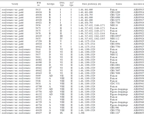
TABLE 1. C. neoformansstrains used in this study
Variety IFMno. Serotype DNAtype AAtypea Exon position(s) (nt) Source Accession no.
C. neoformansvar.gattii 5815 B I I 1–46, 461–800 Patient AB105913
C. neoformansvar.gattii 48634 B I I 1–46, 461–800 CBS 6289 AB105914
C. neoformansvar.gattii 48636 B I I 1–46, 454–793 CBS 7229 AB105915
C. neoformansvar.gattii 48819 B I I 1–46, 461–800 CBS 6998 AB105916
C. neoformansvar.gattii 48820 B I I 1–46, 461–800 CBS 8273 AB105917
C. neoformansvar.gattii 48821 B I I 1–46, 454–793 CBS 7749 AB105918
C. neoformansvar.gattii 5856 C II I 1–46, 317–632, 1148–1171 NIH 18 AB105919
C. neoformansvar.gattii 5873 C II I 1–46, 317–632, 1148–1171 Patient AB105920
C. neoformansvar.gattii 5875 C II I 1–46, 317–632, 1148–1171 Patient AB105921
C. neoformansvar.gattii 5878 B II I 1–46, 317–632, 1121–1144 Patient AB105922
C. neoformansvar.gattii 48635 C III I 1–46, 317–632, 1162–1185 CBS 6955T AB105923
C. neoformansvar.gattii 5855 B IV I 1–46, 317–632, 1162–1185 NIH 112 AB105924
C. neoformansvar.gattii 47258 B V I 1–46, 1175–1514 Patient AB105925
C. neoformansvar.gattii 48818 B V I 1–46, 1175–1514 CBS 6956 AB105926
C. neoformansvar.gattii 48822 B V I 1–46, 1175–1514 CBS 7750 AB105927
C. neoformansvar.neoformans 5844 D VI II 1–46, 1190–1529 Patient AB105928
C. neoformansvar.neoformans 5845 D VI II 1–46, 1190–1529 Patient AB105929
C. neoformansvar.neoformans 5857 D VI II 1–46, 1190–1529 NIH 52 AB105930
C. neoformansvar.neoformans 5881 D VI II 1–46, 1190–1529 Patient AB105931
C. neoformansvar.neoformans 46082 D VI II 1–46, 1190–1529 Patient AB105932
C. neoformansvar.neoformans 46090 D VI II 1–46, 1190–1529 Patient AB105933
C. neoformansvar.neoformans 48640 D VI II 1–46, 1190–1529 CBS 6901 AB105934
C. neoformansvar.neoformans 48641 Db VI II 1–46, 1190–1529 CBS 6995 AB105935
C. neoformansvar.neoformans 48642 D VI II 1–46, 1190–1529 CBS 7697 AB105936
C. neoformansvar.neoformans 48643 D VI II 1–46, 1190–1529 CBS 7698 AB105937
C. neoformansvar.neoformans 5889 AD VII II 1–46, 1190–1529 Patient AB105938
C. neoformansvar.neoformans 5505 A VIII II 1–46, 1189–1528 Patient AB105939
C. neoformansvar.neoformans 5506 A VIII II 1–46, 1189–1528 Patient AB105940
C. neoformansvar.neoformans 5854 A VIII II 1–46, 1189–1528 CDC 551 AB105941
C. neoformansvar.neoformans 45708 A VIII II 1–46, 1189–1528 Patient AB105942
C. neoformansvar.neoformans 45737 AD VIII II 1–46, 1189–1528 Pigeon droppings AB105943
C. neoformansvar.neoformans 45756 AD VIII II 1–46, 1189–1528 Pigeon droppings AB105944
C. neoformansvar.neoformans 46132 AD VIII II 1–46, 1189–1528 Pigeon droppings AB105945
C. neoformansvar.neoformans 46554 A VIII II 1–46, 1189–1528 Patient AB105946
C. neoformansvar.neoformans 46572 A VIII II 1–46, 1189–1528 Patient AB105947
C. neoformansvar.neoformans 46729 A VIII II 1–46, 1189–1528 Pigeon droppings AB105948
C. neoformansvar.neoformans 46734 A VIII II 1–46, 1189–1528 Pigeon droppings AB105949
C. neoformansvar.neoformans 48638 A VIII II 1–46, 1189–1528 CBS 996 AB105950
C. neoformansvar.neoformans 48639 A VIII II 1–46, 1189–1528 CBS 5756 AB105951
C. neoformansvar.neoformans 48637 D IX II 1–386 CBS 132T AB040655c
aAA, amino acid.
bIFM 48641 is regarded as a serotype A strain in the Centraalbureau voor Schimmelcultures (Baarn, The Netherlands), but repeated checking in our laboratory
showed serotype D.
cBiswas et al. (2).
on May 15, 2020 by guest
http://jcm.asm.org/
Nucleotide sequence accession numbers.Sequences of thecyt bgenes of theC. neoformansstrains sequenced in this study have been deposited in DDBJ/EMBL/ GenBank under the accession numbers listed in Table 1.
RESULTS
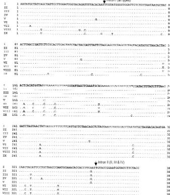
The 386-bp fragment corresponding to nucleotides (nt) 445
to 830 of theCandida glabrata cyt bcoding sequence (GenBank
accession no. X53862) was analyzed in this study. The type
strain of C. neoformansvar. neoformans, IFM 48637, had no
introns in this region ofcyt b. However, all other strains
inves-tigated contained one intron that started after nt 46 of the sequence (Fig. 1); the size of this intron varied from 270 to
1,143 bp. Some isolates ofC. neoformansvar.gattiicontained a
second intron that began at nt 633 of thecyt bsequence (Table
1) or nt 363 of the coding sequence (Fig. 1). The size of the second intron varied from 515 to 529 bp. The sizes and loca-tions of these introns were determined as described previously (2) through comparison of strains with and without introns to
maximize amino acid identities. Isolates ofC. neoformansvar.
neoformans contained the longest introns, which were 1,142
and 1,143 bp. Among isolates ofC. neoformansvar.gattii, IFM
47258, IFM 48818, and IFM 48822 contained introns similar in
size (1,128 bp) to that ofC. neoformansvar.neoformansand
with greater than 96% sequence identity. Although the intron
sizes were variable, they were fixed for specific cyt b types
(Table 1), with the exception of strains IFM 48636 and 48821 (cyt btype I) and IFM 5878 (cyt btype II).
Analysis of 386 bp of coding sequence ofcyt brevealed 27
(6.99%) variable nucleotide sites (Fig. 1). To ensure that these variations were not due to polymerase errors, we used the
TaKaRa ExTaqpolymerase, which has an approximately
four-fold-lower error rate than standard Taq DNA polymerase.
Moreover, we sequenced both strands of each PCR product and repeated each PCR and sequencing reaction. On the basis
of these differences in cyt b, the C. neoformans strains we
analyzed were divided into nine types:C. neoformansvar.gattii
comprising cyt btypes I to V andC. neoformansvar.
[image:3.603.122.459.76.462.2]neofor-manscomprisingcyt btypes VI to IX (Table 1 and Fig. 1). All
FIG. 1. Comparison of coding sequences of the mtcyt bgenes of variousC. neoformansisolates. Dots, nucleotides that are identical to those ofC. neoformans cyt btype I; arrows, positions of introns.Cyt btypes are in parentheses.
on May 15, 2020 by guest
http://jcm.asm.org/
strains withcyt btypes I, IV, and V were serotype B; all strains withcyt btypes II and III except IFM 5878 (serotype B) were
serotype C. Serotype D strains hadcyt btypes VI and IX, and
serotype A strains werecyt btype VIII. Of four serotype AD
strains, one wascyt btype VII and the remaining three werecyt
btype VIII. Although thecyt bsequences contained 27 variable
nucleotides (Fig. 1), the deduced amino acid sequences re-vealed that only one of these substitutions was
nonsynony-mous.C. neoformansvar.gattiiisolates, withcyt btypes I to V,
had identical amino acid sequences (type AA-I). Similarly,
isolates ofC. neoformansvar.neoformans, withcyt btypes VI
to IX, had identical amino acid sequences (type AA-II).
There-fore, isolates ofC. neoformansvar.neoformansdiffered from
those ofC. neoformansvar.gattiionly at amino acid position 55
(Thr instead of Ser) (Fig. 2).
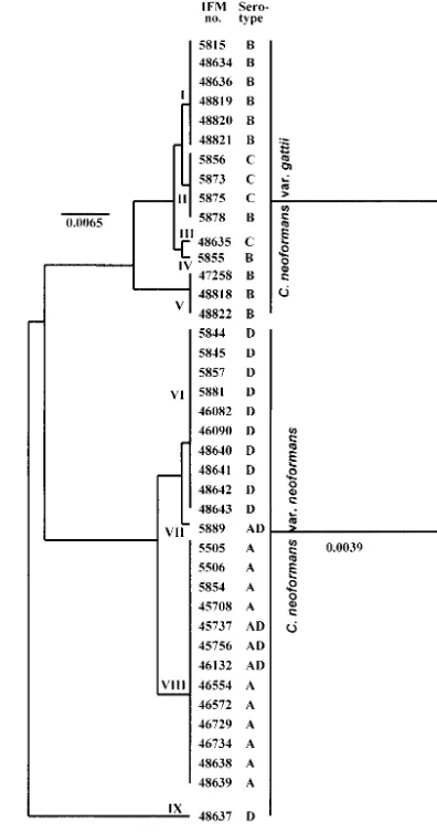
The phylogenetic trees (UPGMA) generated from the mtcyt
b DNA and amino acid sequences are shown in Fig. 3. The
strains of C. neoformans were distributed according to nine
DNA types. The type strain ofC. neoformansvar.neoformans
(IFM 48637) was the outgroup in the phylogenetic tree. IFM
48637 contained the most variable nucleotide sites incyt b(Fig.
1) and showed only a distant relation with other strains, even
those ofC. neoformansvar.neoformans. Strains withcyt btype
V (C. neoformansvar.gattiiIFM 47258, IFM 48818, and IFM 48822) contained introns similar in both length and content to
that ofC. neoformansvar. neoformans, and they were
phylo-genetically closer toC. neoformansvar.neoformans. Analysis of
mtcyt bsequences suggested the existence of nine DNA types ofC. neoformans; however, the phylogenetic tree based on the deduced amino acid sequences divided the strains into only
two varieties: C. neoformansvar. neoformans and C.
neofor-mansvar.gattii.
DISCUSSION
To our knowledge this is the first study where mt cyt b
sequences were used to analyze the genetic relations of strains of C. neoformans. Isolates of C. neoformans var. gattii, the
anamorphic state of F. neoformansvar. bacillispora, had five
types (I to V) for mtcyt b; however, the deduced amino acid
sequences were identical. Similarly, the amino acid sequences
encoded by mt cyt bgenes of isolates ofC. neoformans var.
neoformans, which is the anamorphic state ofF. neoformans
var.neoformans, were identical although there were fourcyt b
types (VI to IX). This result is consistent with the varieties of
C. neoformansvar.neoformansand var.gattiidistinguished by
D-prolineassimilationorreactiononL
-canavanine-glycine-brom-thymol blue agar (11, 14).
The present study also revealed that one serotype D strain (cyt btype IX) had no intron in the region sequenced; however,
all other strains ofC. neoformans contained one intron, and
some isolates ofC. neoformansvar.gattiicontained a second
intron (Table 1 and Fig. 2). A recent study of the mtCOX1
gene ofC. neoformansindicated that the presence or absence
of introns inCOX1is not serotype specific (22), which is similar
to the outcome of our study. The same group also reported
that serotype D strains contain more introns inCOX1than do
serotype A strains. However, in our study of thecyt bgene, all
serotype A and D strains except IFM 48637 contained a single intron and the introns from all strains were similar in size and
[image:4.603.320.518.72.449.2]FIG. 2. Comparison of the deduced amino acid sequences encoded by thecyt bgenes of variousC. neoformansisolates. Dots, amino acids that are identical to those of C. neoformans cyt b type I; arrows, inserted positions of introns.cyt btypes are in parentheses.
FIG. 3. UPGMA-based trees showing the relations of variousC. neoformansisolates generated from nucleotide sequences of thecyt b
gene (exon) (a) and deduced amino acid sequences (b). Bar, number of nucleotide and amino acid substitutions per nucleotide site and amino acid site.
on May 15, 2020 by guest
http://jcm.asm.org/
nucleotide sequence. The first intron incyt bofC. neoformans
started at nt 47 (Fig. 2), which is the same position as intron 2 ofNeurospora crassa cyt bgene.Rhodotorula acheniorumand
Rhodotorula ferulica, two other basidiomycetous yeasts, have
introns in the same location (2). Some isolates of C.
neofor-mansvar.gattii(cyt btypes II, III, and IV) contained a second intron that began at nt 633 (Table 1), which is similar to the
location of intron 5 of theSaccharomyces cerevisiae cyt bgene.
These findings suggest two possible evolutionary events. The first possibility is that these introns appeared in these locations prior to the separation of these species and that some species lost introns over time. The second is that these introns ap-peared in these locations after separation of these species.
Analysis of the mt large rRNA gene and NADH
dehydro-genase subunit 2 ofC. neoformansrevealed that serotype AD
strains had either the serotype A or serotype D mtDNA ge-notype (29). In the present study, we obtained almost similar
results for the distribution ofcyt bgene types in serotype AD
strains. Of four serotype AD strains, three had serotype
A-spe-cificcyt b(type VIII) and one had a unique specific cytb(type
VII) that was nearly identical to serotype D-specificcyt b(type
VI), with only 1 nt difference.
In conclusion, we have shown that isolates ofC. neoformans
represent ninecyt b types; however, the deduced amino acid
sequences indicate that there are only two varieties,C.
neofor-mansvar.neoformansandC. neoformansvar.gattii.
ACKNOWLEDGMENT
This study was performed as part of the program Frontier Studies and International Networking of Genetic Resources in Pathogenic Fungi and Actinomycetes (FN-GRPF) through the Special Coordina-tion Funds for Promoting Science and Technology from the Ministry of Education, Culture, Sports, Science and Technology of Japan (2001).
REFERENCES
1. Biswas, S. K., K. Yokoyama, L. Wang, K. Nishimura, and M. Miyaji.2001. Typing ofCandida albicansisolates by sequence analysis of the cytochrome bgene and differentiation fromCandida stellatoidea. J. Clin. Microbiol. 39:1600–1603.
2. Biswas, S. K., K. Yokoyama, K. Nishimura, and M. Miyaji.2001. Molecular phylogenetics of the genusRhodotorulaand related basidiomycetous yeasts inferred from the mitochondrial cytochromebgene. Int. J. Syst. Evol. Mi-crobiol.51:1191–1199.
3. Chaturvedi, S., B. Rodeghier, J. Fan, C. M. McClelland, B. L. Wickes, and V. Chaturvedi.2000. Direct PCR ofCryptococcus neoformans MAT␣and MATapheromones to determine mating type, ploidy, and variety: a tool for epidemiological and molecular pathogenesis studies. J. Clin. Microbiol.38: 2007–2009.
4. Diaz, M. R., T. Boekhout, B. Theelen, and J. W. Fell.2000. Molecular sequence analyses of the intergenic spacer (IGS) associated with rDNA of the two varieties of the pathogenic yeast,Cryptococcus neoformans. Syst. Appl. Microbiol.23:535–545.
5. Fell, J. W., T. Boekhout, A. Fonseca, G. Scorzetti, and A. Statzell-Tallman. 2000. Biodiversity and systematics of basidiomycetous yeasts as determined by large-subunit rDNA D1/D2 domain sequence analysis. Int. J. Syst. Evol. Microbiol.50:1351–1371.
6. Franzot, S. P., J. S. Hamdan, B. P. Currie, and A. Casadevall.1997. Mo-lecular epidemiology ofCryptococcus neoformansin Brazil and the United States: evidence for both local genetic differences and a global clonal pop-ulation structure. J. Clin. Microbiol.35:2243–2251.
7. Franzot, S. P., I. F. Salkin, and A. Casadevall.1999.Cryptococcus neofor-mansvar.grubii: separate varietal status forCryptococcus neoformans sero-type A isolates. J. Clin. Microbiol.37:838–840.
8. Hamari, Z., F. Kevei, E. Kovacs, J. Varga, Z. Kozakiewicz, and J. H. Croft.
1997. Molecular and phenotypic characterization ofAspergillus japonicusand Aspergillus aculeatusstrains with special regard to their mitochondrial DNA polymorphisms. Antonie Leeuwenhoek72:337–347.
9. Ikeda, R., T. Shinoda, Y. Fukazawa, and L. Kaufman.1982. Antigenic characterization ofCryptococcus neoformansserotypes and its application to serotyping of clinical isolates. J. Clin. Microbiol.16:22–29.
10. Kozlowski, M., and P. P. Stepien.1982. Restriction enzyme analysis of mitochondrial DNA of members of the genusAspergillusas an aid in taxon-omy. J. Gen. Microbiol.128:471–476.
11. Kwon-Chung, K. J., I. Polacheck, and J. E. Bennett.1982. Improved diag-nostic medium for separation ofCryptococcus neoformansvar.neoformans (serotypes A and D) andCryptococcus neoformansvar.gattii(serotypes B and C). J. Clin. Microbiol.15:535–537.
12. Kwon-Chung, K. J., and J. E. Bennett.1984. Epidemiologic differences between the two varieties ofCryptococcus neoformans. Am. J. Epidemiol. 120:123–130.
13. Kwon-Chung, K. J., B. L. Wickes, L. Stockman, G. D. Roberts, D. Ellis, and D. H. Howard.1992. Virulence, serotype, and molecular characteristics of environmental strains ofCryptococcus neoformansvar.gattii. Infect. Immun. 60:1869–1874.
14. Kwon-Chung, K. J.1998.FilobasidiellaKwon-Chung, p. 656–662,InC. P. Kurtzman and J. W. Fell (ed.), The yeasts, a taxonomic study, 4th ed. Elsevier, Amsterdam, The Netherlands.
15. Levitz, S. M.1991. The ecology ofCryptococcus neoformansand the epide-miology of cryptococcosis. Rev. Infect. Dis.13:1163–1169.
16. Li, A., K. Nishimura, H. Taguchi, R. Tanaka, S. Wu, and M. Miyaji.1993. The isolation ofCryptococcus neoformansfrom pigeon droppings and sero-typing of naturally and clinically sourced isolates in China. Mycopathologia 124:1–5.
17. Manceau, V., L. Despres, J. Bouvet, and P. Taberlet.1999. Systematics of the genusCaprainferred from mitochondrial DNA sequence data. Mol. Phylo-genet. Evol.13:504–510.
18. Mitchell, T. G., and J. R. Perfect.1995. Cryptococcosis in the era of AIDS— 100 years after the discovery ofCryptococcus neoformans. Clin. Microbiol. Rev.8:515–548.
19. Nakamura, Y., R. Kano, S. Watanabe, and A. Hasegawa.2000. Molecular analysis ofCAP59gene sequences from five serotypes ofCryptococcus neo-formans. J. Clin. Microbiol.38:992–995.
20. Nishikawa, M. M., M. S. Lazera, G. G. Barbosa, L. Trilles, B. R. Balassiano, R. C. Macedo, C. C. Bezerra, M. A. Perez, P. Cardarelli, and B. Wanke.2003. Serotyping of 467Cryptococcus neoformansisolates from clinical and envi-ronmental sources in Brazil: analysis of host and regional patterns. J. Clin. Microbiol.41:73–77.
21. Perfect, J. R., and A. Casadevall.2002. Cryptococcosis. Infect. Dis. Clin. N. Am.16:837–874.
22. Toffaletti, D. L., M. Del Poeta, T. H. Rude, F. Dietrich, and J. R. Perfect. 2003. Regulation of cytochromecoxidase subunit 1 (COX1) expression in Cryptococcus neoformansby temperature and host environment. Microbiol-ogy149:1041–1049.
23. Vanbreuseghem, R., and M. Takashio.1970. An atypical strain of Crypto-coccus neoformans(San Felice) Vuillemin 1894. II.Cryptococcus neoformans var.gattiivar. nov. Ann. Soc. Belg. Med. Trop.50:695–702.
24. Varga, J., F. Kevei, A. Vriesema, F. Debets, Z. Kozakiewicz, and J. H. Croft. 1994. Mitochondrial DNA restriction fragment length polymorphisms in field isolates of theAspergillus nigeraggregate. Can. J. Microbiol.40:612–621. 25. Wang, L., K. Yokoyama, M. Miyaji, and K. Nishimura.1998. The identifi-cation and phylogenetic relationship of pathogenic species ofAspergillus based on the mitochondrial cytochromebgene. Med. Mycol.36:153–164. 26. Wang, L., K. Yokoyama, M. Miyaji, and K. Nishimura.2000. Mitochondrial
cytochromeb gene analysis ofAspergillus fumigatusand related species. J. Clin. Microbiol.38:1352–1358.
27. Wang, L., K. Yokoyama, M. Miyaji, and K. Nishimura.2001. Identification, classification, and phylogeny of the pathogenic speciesExophiala jeanselmei and related species by mitochondrial cytochromebgene analysis. J. Clin. Microbiol.39:4462–4467.
28. Xu, J.2002. Mitochondrial DNA polymorphisms in the human pathogenic fungusCryptococcus neoformans. Curr. Genet.41:43–47.
29. Xu, J., R. Y. Ali, D. A. Gregory, D. Amick, S. E. Lambert, H. J. Yoell, R. J. Vilgalys, and T. G. Mitchell.2000. Uniparental mitochondrial transmission in sexual crosses inCryptococcus neoformans. Curr. Microbiol.40:269–273. 30. Yokoyama, K., S. K. Biswas, M. Miyaji, and K. Nishimura.2000.
Identifi-cation and phylogenetic relationship of the most common pathogenic Can-didaspecies inferred from mitochondrial cytochromeb gene sequences. J. Clin. Microbiol.38:4503–4510.
31. Yokoyama, K., L. Wang, M. Miyaji, and K. Nishimura.2001. Identification, classification and phylogeny of theAspergillussectionNigriinferred from mitochondrial cytochromebgene. FEMS Microbiol. Lett.200:241–246.