Preliminary Experience with Selective Laser Sintering Models of the
Human Temporal Bone
Richard A. Levy, Sashidhar Guduri, and Richard H. Crawford
PURPOSE: To assess the accuracy of three-dimensional models of the human temporal bone generated from CT data. METHODS: Thin-section CT of a left human cadaveric temporal bone was performed using multiple-scan planes (axial, coronal, and sagittal) at 1.5-mm section thickness and 0.25-mm pixel size with an edge-enhancement two-dimensional algorithm. CT data were converted to toggle point format based upon a threshold value of 200 (approximately -830 HU)
obtained from prior experimentation with a CT phantom. Selective laser sintering of polycarbonate powder was performed at a beam diameter of 0.060 inches (1.5 mm), 100 scan lines per inch,
layer thickness of 0.010 inches (0.25 mm), and layer repeat factor of 4. The polycarbonate models were then scanned in the axial, coronal, and sagittal planes and compared with the original CT data. Anatomic dissection of the models was performed for further verification of the imaging findings. RESULTS: Models of high anatomic accuracy were generated. Shortening by a factor of 0.67 along the Z axis secondary to the layer repeat factor of 4 resulted in distortion of the models. Distortion in the XY plane ranged from 0% to 20%. Differences in model accuracy based on the initial CT scan plane were observed. A significant amount of nonsintered or partially sintered polycarbonate resulted in intermediate density on the CT images. CONCLUSIONS: Selective laser sintering can result in accurate modeling of detailed anatomic structures in the human temporal bone. Further investigation of materials and factors contributing to the accuracy of selective laser sintering in the manufacturing of high-resolution anatomic models is warranted.
Index terms: Temporal bone, anatomy; Temporal bone, computed tomography; Models, anatomic; Computed tomography, technique; Computed tomography, experimental
AJNR Am J Neuroradio/15:473-477, Mar 1994
Computer-generated anatomic modeling using radiologic data is a well-known entity. Various technologies have been used in the development of anatomic models and prostheses. These tech-nologies include multiaxis milling, stereo lithog-raphy, and selective laser sintering (1). Our pur-pose in this research is to demonstrate the ac-curacy of selective laser sintered models of the human temporal bone. A qualitative comparison with other anatomic modeling technologies will be discussed.
Received January 13, 1993; accepted pending revision March 12;
revision received March 22.
From the Department of Radiology (R.A.L.), Neuroradiology Section,
University of Michigan Hospitals, Ann Arbor; and Department of Mechan-ical Engineering (S.G., R.H.C.), University of Texas at Austin.
Address reprint requests to Richard A. Levy, MD, Department of
Radiology, Neuroradiology Section, University of Michigan Hospitals, Ann Arbor, Ml 48109-0030.
AJNR 15:473-477, Mar 1994 0195-6108/94/1503-0473 © American Society of Neuroradiology
473
Methods
Thin-section computed tomography (CT) of a left hu
-man cadaveric temporal bone was performed using multi-ple scan planes (axial, coronal, and sagittal) at a 1.5-mm
section thickness and 0.25-mm pixel size with an
edge-enhancement two-dimensional algorithm on a General Electric (Milwaukee, Wis) 9800 CT scanner. CT data were networked from the Department of Radiology at the Uni-versity of Michigan to the Department of Mechanical En-gineering at the University of Texas at Austin via an Internet file transfer protocol file. CT data were converted to toggle point format (linear scalar transformation) resulting in a
threshold value of 200 (approximately -830 HU) obtained
from prior experimentation with a CT phantom. Selective
laser sintering of polycarbonate powder was performed with a 25 W carbon dioxide laser with power centered at 14 W at a beam diameter of 0.060 inches (1.5 mm), 100 scan lines per inch, polycarbonate layer thickness of 0.010
inches (0.25 mm), and layer repeat factor of 4. The poly-carbonate models were then rescanned, respectively, in the
474 LEVY AJNR: 15, March 1994
A
Bc
D
E
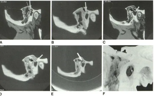
FFig. 1. A, Sagittal CT image of a left human cadaveric temporal bone. The thick arrow indicates the head of malleus. The thin arrow indicates the body of incus.
B, Sagittal CT image of polycarbonate laser-sintered model generated from sagittal CT data and corresponding to CT image in A.
The white arrow indicates the head of malleus. Black arrow indicates the body of incus. Ossicles appear less distinct than on CT
imaging.
C, Sagittal CT image medial to plane in A and B. The white arrow indicates the manubrium of malleus. Black arrow indicates the long process of incus.
D, Sagittal CT image of polycarbonate temporal bone model through imaging plane in C. The white arrow indicates the manubrium
of malleus. The black arrow indicates the long process of incus.
£, Sagittal CT image of polycarbonate temporal bone model as in Dafter simulated mastoidectomy. The white arrow indicates the resected area of loose polycarbonate powder in middle ear cavity, now with normal air density. Ossicles are better modeled than in Figures 2 and 3.
F, Postdissection polycarbonate model of the human cadaveric temporal bone specimen in A to E. Mirror-image modeling occurs by selecting the last CT section as the starting point for toggle point conversion before laser sintering. The white arrow indicates the manubrium of malleus.
original CT data. Anatomic dissection of the models was performed for further verification of the imaging findings.
Results
Models of high anatomic accuracy were gen-erated as verified by measurement of the margins of the models. Measurements from images of scanned models were compared with similar measurements from original CT data (Figs 1, 2, and 3). Shortening along the Z axis (defined as the craniocaudal axis for axial data, the antero-posterior axis for coronal data, and the mediola-teral axis for sagittal data) averaged 0.67 of the
[image:2.612.54.559.78.393.2]AJNR: 15, March 1994
A
8
significant amount of nonsintered or partially sin-tered polycarbonate resulted in intermediate den-sity on the CT images of the models.
Discussion
The utility of 3-D CT reconstructions in head and neck imaging remains controversial, having gained a number of proponents as well as those who find no significant contribution from 3-D imaging over conventional 2-D CT analysis. It may be that the ultimate usefulness of this tech-nology lies in its ability to generate anatomic models and prostheses. Based upon prior inves-tigations into the optimization of clinical variables that contribute to the accuracy of 3-D CT images (2, 3), a new technology, selective laser sintering, was adopted to test the relevance of these clinical variables to the model-generating process.
The term sintering refers to any process whereby particulates are caused to adhere into a solid mass by means of an externally applied energy. The selective laser sintering process be-gins with the deposition of a very thin layer of heat-fusible powder into a work-space container and heating to just below its melting point. An initial cross-section of the object under fabrication is traced on the layer of powder by a laser. In this experiment, the pattern traced by the laser is determined by a binary threshold value corre-sponding to a preselected Hounsfield value. Thus, the laser beam is on when the Hounsfield densities in the corresponding 2-D CT section are equal to or greater than the threshold value, and off when the Hounsfield values are less than the chosen threshold value.
The temperature of the powder impacted by the laser beam is raised to the point of sintering,
TEMPORAL BONE MODELS 475
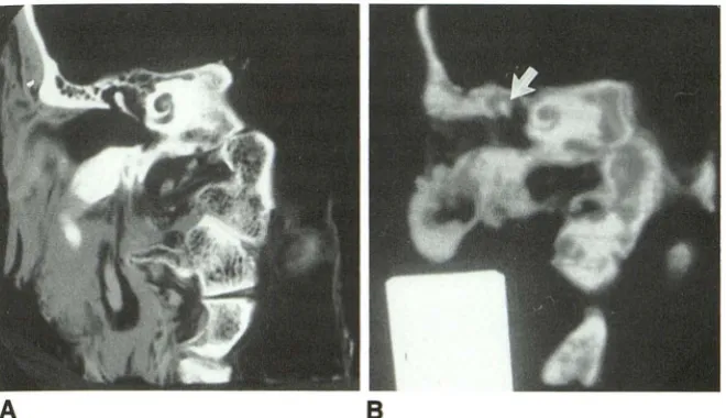
Fig. 2. A, Coronal CT image of left hu-man cadaveric temporal bone specimen used in Figure 1. The head, neck, and ma-nubrium of malleus are visualized. The CT image has been photographed in reverse to correspond to the orientation of the ana-tomic model.
8, Coronal CT image of a polycarbonate laser-sintered model generated from coronal CT data corresponding to the imaging plane in A. Only the head of the malleus is modeled
(white arrow).
forming a solid mass. As the process is repeated, each layer fuses to the underlying layer, and successive layers of powder are deposited and sintered until the object is complete. Thus, a three-dimensional mathematical model of the sur-face of interest is not required for solid model generation. The laser beam intensity is modulated to sinter the powder only in areas defined by the object's design geometry. In areas not sintered, powder remains loose and serves as a natural support for the next layer of powder and the object under fabrication (4).
It is obvious that the laser beam diameter (a function of beam intensity) is greater than the individual scan line, resulting in overlap sintering of contiguous scan lines. This may be the reason fine-detail modeled structures such as the audi-tory ossicles appear more accurate with CT im-aging than when the model is visually inspected; that is, there is a more densely sintered "core" creating greater x-ray beam attenuation than the outer, more loosely sintered coating (Fig 1).
One advantage of the selective laser sintering process is the ability to reproduce fine-detail anatomic structures within cavities (ie, the middle ear) as well as within anatomic recesses. Herein lies a major advantage over 3-D model-generating technologies, which rely upon multiaxis milling machines driven by computer-generated 3-D mathematical surface maps and which are unable to replicate enclosed cavities and are subject to tool path limitations (5).
[image:3.615.56.386.80.270.2]476 LEVY
Fig. 3. A, Axial CT image of left cadav-eric temporal bone as in Figures 1 and 2,
reversed to correspond to laser-sintered po-lycarbonate model in 8 and D. The black
arrow indicates the head of malleus. The
black arrowhead indicates the body of incus.
8, Laser-sintered polycarbonate model generated from axial CT data. Axial CT section through the model at the same plane as in A. The black arrow indicates the head of malleus. The white arrow indicates the body of incus.
C, Axial CT section caudal to that in A.
The white arrow indicates the manubrium of
malleus. The black arrow indicates the long process of incus.
D, Axial CT section through model cau-dal to the plane in B corresponding to C. The ossicles are not modeled.
A
c
may be used (6). At present our investigations are focused on the feasibility of hydroxyapatite, a form of synthetic bone, as a substrate for the selective laser sintering process. CT /selective laser sintering-generated hydroxyapatite grafts have potential usefulness in craniofacial recon-structive surgical procedures in which experimen-tation with hydroxyapatite grafts is already oc-curring (7).
One factor in the 3-D model-generating proc-ess previously investigated in 3-D imaging is the effect of partial volume averaging via changes in CT scan plane upon the accuracy of the 3-D images (2, 3). In this research, it is assumed that with faithful transformation of 2-D CT data to toggle point format by binary thresholding, partial volume averaging in the original 2-D CT data will be manifest in the 3-D models. This may be true in this preliminary research, as evidenced by more accurate modeling of the auditory ossicles
AJNR: 15, March 1994
8
D
using the sagittal CT scan plane (Fig 1). Because partial volume-averaging effects are difficult to predict (8, 9), a multiplicity of scan planes may be required to optimize the initial 2-D data set for 3-D model generation (US patent pending, serial number 07/861,947).
It must be emphasized that the potential for distortion in the models occurs not only when the
[image:4.613.228.561.78.484.2]2-AJNR: 15, March 1994
D data set occupying an entire voxel on 3-D CT reconstruction secondary to a combination of partial volume averaging, selection of a suffi-ciently low binary threshold before 3-D image generation, and interpolation throughout the en-tire voxel volume on 3-D reconstruction.
Another artifact, analogous to aliasing in 3-D
CT imaging, is the steplike borders created by successive sections in the laser sintigraphic models. Although this type of artifact can be reduced on 3-D images with the appropriate ap-plication of filtering (10), no corresponding mod-ifications have been devised for 3-D laser sinti-graphic modeling at present.
We conclude that selective laser sintering can result in accurate modeling of detailed anatomic structures in the human temporal bone. Further investigation of materials and factors contributing to t e accuracy of selective laser sintering in the manufacturing of high-resolution anatomic models is justified.
Acknowledgments ~
We thank Jane B. Mitchell for preparing the manuscript,
Mike Disher, MD, for dissection of the anatomic models, and Bob Combs for preparing the photographs.
TEMPORAL BONE MODELS 477
References
1. Lambrecht JT, Brix F. Individual skull model fabrication for cranio
-facial surgery. Cleft Palate Cranlofac J 1990;27:382-387
2. Levy RA, Edwards WT, Meyer JR, Rosenbaum AE. Facial trauma and 3-D reconstructive imaging: insufficiencies and correctives. AJNR
Am J Neuroradio/1992;13:885-892
3. Levy RA, Rosenbaum AE, Kellman RM, Bailey GL, Aravapalll SR.
Assessing whether the plane of section on CT affects accuracy in
demonstrating facial fractures in three-dimensional reconstruction
using the dried skull. AJNR Am J Neuroradio/1991;12:861-866
4. Nutt K. The selective laser sintering process, new dimensions in rapid prototyping and manufacturing technologies (monogr). DTM Corp,
Austin
5. Mankovich NJ, Cheeseman AM, Stoker NG. The display of three-dimensional anatomy with stereollthographic models. J Digit Imaging
1990;3:200-203
6. Stoker NG, Mankovich NJ, Valentino D. Stereolithographic models for surgical planning: preliminary report. J Oral Maxillofac Surg 1992;50:466-471
7. Ripamonti U. Calvarial reconstruction in baboons with porous hy-droxyapatite. J Craniofac Surg 1992;3:149-159
8. Chakeres DW. Clinical significance of partial volume averaging of the temporal.bone. AJNR Am J Neuroradio/1984;5:279-302
9. Goodenough D, Weaver K, Davis D, LaFalce S. Volume averaging limitations of computed tomography. AJR Am J Roentgenol 1982;138:313-316
10. Vannier M, Hlldebolt C, Gayou D, Marsh J. Introduction to 3D Imaging.