Animal Models for Panic Disorder
Sueslene Prado Rocha1, Rhayra Xavier do Carmo Silva2,3,4, Anderson Manoel Herculano3,
Monica Gomes Lima-Maximino1, Caio Maximino4
1 – Laboratório de Neurofarmacologia e Biofísica, Universidade do Estado do Pará, Campus
VIII – Marabá/PA, Brazil
2 – Programa de Pós-graduação em Neurociências e Biologia Celular, Universidade Federal do
Pará – Marabá/PA, Brazil
3 – Laboratório de Neurofarmacologia Experimental, Universidade Federal do Pará –
Marabá/PA, Brazil
4 – Laboratório de Neurociências e Comportamento “Frederico Guilherme Graeff”,
Universidade Federal do Sul e Sudeste do Pará – Marabá/PA, Brazil
Abstract
Panic disorder (PD) is characterized by recurrent and unexpected panic attacks associated with
behavioral changes and/or persistent anxiety due to the attacks. The development of behavioral
models in animals is important for the understanding of the psychobiological and behavioral
bases of PD. The present article reviews the main models used in the current literature. The
elevated T-maze, used in rats, presents good predictive validity, but its face validity has been
questioned. Models using electrical stimulation of the periaqueductal gray present good face
validity, but lesser construct validity. Models relying on predator exposure present good
predictive and construct validity. These three approaches seek coherence with theories on PD
as a way to increase its translational potential; thus, while the elevated T-maze is supported by
distance, and periaqueductal gray stimulation is based on the functional neuroanatomy of PD.
Moreover, to higher or lower degree the three models are supported by an “etho-experimental”
approach, with careful observation of animal behavior as a way of discriminating different
defensive strategies that model different aspects of anxiety, fear, and panic. Finally, an
alternative/complementary model is proposed that uses zebrafish alarm reaction to study this
disorder.
Keywords: Panic disorder; Animal experimentation; Defensive behavior (Animal); Escape
behavior (Animal)
1 Introduction
Panic disorder (PD) is characterized by recurrent and unexpected panic attacks that are
associated with behavioral changes and/or persistent anticipatory anxiety due to these attacks
(American Psychiatric Association, 2013). Panic attacks occur spontaneously, but there is
evidence that, for the majority of patients, moderate phobic or hypochondriac symptoms
precede the attacks (Craske, Miller, Rotunda, & Barlow, 1990; Fava, Grandi, & Canestrari,
1988); it was proposed, then, that panic attacks are “unpredictable” rather than “unexpected”
(Barlow, 2002). Besides symptom phenomenology, PD differs from other anxiety disorders due
to its particular pharmacological response (Donald F Klein, 1964, 1981) and, partially, due to
the independent structure of genetic and environmental risk factors (Gratacòs et al., 2007).
From a neuroanatomical point of view, PD presents important relationships with circuits
underlying fear, including different amygdala nuclei, insular cortex, anterior cingulate cortex,
and periaqueductal gray area (PAG) (Graeff & Del-Ben, 2008; Lovick, 2000). The mechanisms
associated with unconditional and conditional responses compose a fight/flight/freeze or
subnuclei, and the dorsal periaqueductal gray area (dPAG) (Brandão, Anseloni, Pandóssio, De
Araújo, & Castilho, 1999; Canteras, 2002; Cezario, Ribeiro-Barbosa, Baldo, & Canteras, 2008;
Gross & Canteras, 2012; LeDoux, 2003a; LeDoux & Pine, 2016; Sukikara, Mota-ortiz, Baldo,
Felício, & Canteras, 2006). The electrical stimulation of these areas elicits a response pattern
that characterize what is usually termed a defense reaction, and usually accompanies aversive
subjective states characteristic of fear and anxiety (Brandão, Troncoso, Silva, & Huston, 2003).
The functioning of this aversive brain system can also be related to panic attacks. The electrical
stimulation of dPAG elicits behaviours similar to the defense reaction as well as ubjective and
neurovegetative phenomena that resemble a panic attack not only in experimental animals, but
also in humans (Graeff & Del-Ben, 2008; Graeff, Silveira, Nogueira, Audi, & Oliveira, 1993;
Jenck, Moreau, & Martin, 1995; Lovick, 2000).
An important concept that organizes theories derived from animal models is that of
defensive direction and defensive distance (or predatory imminence continuum) (Deakin &
Graeff, 1991; Fanselow & Lester, 1988; McNaughton & Corr, 2004; Perusini & Fanselow,
2015). In general, it can be said that the more caudal portions of the system (especially the
PAG) directly and immediately control defensive responses when the defensive distance is very
small (proximal threat). As the distance increases, more complex defensive strategies emerge,
and those responses are controlled by progressively more rostral structures, with the cingulate
cortex representing the most distal threat. Defensive avoidance (“fear”) is controlled by the
amygdala and the anterior cingulate (Dresler et al., 2013; McNaughton & Corr, 2004).
Defensive approach (“anxiety”) occurs when a strong drive conflicts with avoidance, triggering
risk assessment behavior; these patterns are controlled by septo-hippocampal system and
posterior cingulate (R. J. Blanchard & Blanchard, 1988; Deakin & Graeff, 1991; Gray &
2. Animal models for panic disorder
The development of animal models for panic disorder is needed to establish the
psychological and biological bases of this disorder, as well as to allow the development of novel
therapeutics. Differently from models for generalized anxiety – that present a myriad of formats
and exploit a diversity of different behaviors (Belzung & Lemoine, 2011; Griebel & Holmes,
2013) –, few paradigms explicitly model PD. The distinction between models for PD and
models for generalized anxiety is based mainly on reference to the theoretical constructs that
support the models (McNaughton & Zangrossi Jr, 2008): while tests for generalized anxiety use
situations that impose approach/avoidance conflicts to animals – generating behavioral
inhibition and risk assessment responses –, models that are relevant for PD are based on
defensive responses elicited by aversive stimuli or by the stimulation of specific brain regions
(Moreira et al., 2013). In rodents, four main strategies are used to model panic attacks and/or
PD: conditional fear responses (fear conditioning), mouse defense test battery, and electrical
stimulation of the periaqueductal gray and/or medial hypothalamus (Moreira et al., 2013).
2.1. Fear conditioning
Conditional fear responses have been widely used to establish fear-like responses and
to elucidate the neural bases of these responses (LeDoux, 1998b, 2003b; Schafe, Nader, Blair,
& LeDoux, 2001). In fear conditioning, animals learn to associate a previously neutral stimulus
(CS) with an aversive unconditioned stimulus (US, such as an electrical shock (Pavlov, 1927).
In cued fear conditioning, the CS is explicit and reliably predicts the US, acting as a cue or
signal; in contextual fear conditioning, animals display a defensive reaction (usually freezing)
exposed to the US, and therefore the context represents a complex CS (Davis, Walker, Miles,
& Grillon, 2009). Typically, rats and mice display a initial activity burst to the footshock
followed by freezing (LeDoux, 1998b); reexposure to the CS in the absence of the US triggers
memory retrieval, which initiates reconsolidation and extinction processes.
While fear conditioning is adaptive, and although conditioning accounts of panic
disorder have been widely criticized, this process is an integral part of contemporary learning
explanations of PD that attempt to incorporate some complexities of contemporary learning
theory . Moreover, these learning accounts of PD provide a compelling explanation for the
persistence of PD, in spite of not always being able to explain the etiology of these disorders.
Bouton et al. (2001) have argued that panic attacks are salient aversive USs during which feeligs
of anxiety and fear can be associated with external and/or interoceptive cues that are present
during the attacks; subsequent exposure to these cues can trigger feelings of anticipatory
anxiety, which in its turn can exacerbate the next panic attack.
Enhanced fear conditioning has been associated with gene variants in rodent orthologues
of human PD risk genes. For example, Tmem132d has been recently found to be associated in
murine fear conditioning in a quantitative trait loci panel (Knoll, Halladay, Holmes, & Levitt,
2016), and female mice with humanized COMT Val158Met polymorphism show reduced
contextual fear, increased cued fear, and reduced extinction recall (Risbrough, Ji, Hauger, &
Zhou, 2014). Both genes have variants which increase the risk for PD in humans (Howe et al.,
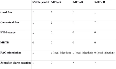
2015). A role for serotonin (5-HT) has also been suggested in conditional fear processes
(Homberg, 2012): acute treatment with selective 5-HT reuptake inhibitors (SSRIs) facilitates
fear conditioning, reduces contextual fear, and increases cued fear; 5-HT1A receptors inhibit the
acquisition and expression of contextual fear; 5-HT2A receptors facilitate the consolidation of
5-HT appears to exert opposite effects on contextual and cued fear (Homberg, 2012). While
consistent with the idea that contextual fear is more related to anxiety than fear, these results
are actually opposed to what is observed in other models (Table 1).
SSRIs (acute) 5-HT1AR 5-HT2AR 5-HT2CR
Cued fear ↑ ? ↑ ↓
Contextual fear ↓ ↓ ↑ ?
ETM escape ↓ 0 0 0
MDTB 0 0 0 0
PAG stimulation ↓ ↓ (local injection) ↓ (local injection) 0 (local injection)
Zebrafish alarm reaction ↓ 0 ? ?
Table 1 – Effect of serotonergic drugs on putative models of fear and panic-like behavior.
Abbreviations: ETM: Elevated T-mazed; MDTB: mouse defense test battery; PAG:
periaqueductal gray area; SSRIs: selective serotonin reuptake inhibitors.
2.2. Elevated T-maze
The elevated T-maze (ETM) is derived from the behavioral model for anxiety that is
most widely used contemporaneously – the elevated plus-maze –, and is explicitly based on the
Deakin/Greaff hypothesis on the role of 5-HT on defensive behavior (Graeff, Ferreira Netto, &
ETM attempts to explicitly separate defensive behaviors that are evoked by motivational
conflict (i.e., behavioral inhibition and risk assessment) from behaviors associated with fear
states (i.e., escape). The apparatus consists in two open arms and one closed arm in a
perpendicular configuration, elevated 50 cm from the floor (Viana, Tomaz, & Graeff, 1994). In
a ETM session, the rat executes two tasks: inhibitory avoidance and escape. First, the animal is
transferred to the end of the closed arm, and the latency to leave this arm is measured; after 30
s intervals, the task is repeated twice, and increased latency to leave the closed arm is interpreted
as the acquisition of inhibitory avoidance (Graeff et al., 1998). After this IA task, the animal is
positioned at the end of one of the open arms to measure the escape response (Viana et al.,
1994). These two responses – IA and escape – recruit different neuronal circuits (Leite Silveira,
Zangrossi, De Barros Viana, Silveira, & Graeff, 2001) and present opposite 5-HTergic
modulation (Graeff, Viana, & Mora, 1996; Sena et al., 2003; Zangrossi Jr & Graeff, 2014;
Zangrossi Jr et al., 2001; Table 1). IA is interpreted as modeling anxiety-like responses, while
escape is interpreted as modeling panic attacks (Graeff et al., 1998). Moreover, the model
appears to present good predictive validity for anxiolytic and panicolytic drugs (Graeff et al.,
1998). An important critique to the model, however, is its low face validity: the association of
escape from the open arm to panic attacks is tenuous, given that the escape observed in the
ETM is slow, directed, and controlled – very different from escape responses which are
observed in other models (Moreira et al., 2013).
2.3. Mouse defense test battery
The mouse defense test battery (MDTB) evaluates the behavior of mice exposed to a
natural predator, the rat, at different defensive distances (D. C. Blanchard, Griebel, &
anesthetized rat is manipulated by the experimenter so that its approaches the mouse at a fixed
velocity; when the rat approaches the mouse by about 1 m, the mouse usually starts running,
attempting to escape the aversive stimulus This model explicitly assumes defensive distance
(or predatory imminence continuum) – the psychological distance from the threat (R. J.
Blanchard & Blanchard, 1988; Fanselow & Lester, 1988) – as a central variable in decision
making by the mouse. The model presents excellent predictive validity, since acute treatment
with triazolobenzodiazepines and chronic treatment with imipramine, fluoxetine, and
moclobemide decrease escape responses, while acute treatment with imipramine and fluoxetine
or with panicogenic agents increase escape (D. C. Blanchard et al., 2001). Moreover, facilitating
5-HTergic neurotransmission in the periaqueductal gray inhibits escape in the MDTB (Pobbe,
Zangrossi Jr, Blanchard, & Blanchard, 2011). The model presents as advantages the high
ethological relevance, because it uses escape responses in an ecologically valid context and
avoids using artificial stimuli such as electric shocks; moreover, the model allows the evaluation
of two distinct responses – approach or escape the stimulus – that are predictive of anxiolytic
or panicolytic effects, respectively (Moreira et al., 2013).
2.4. Electrical stimulation of the dlPAG
The electrical stimulation of the dorsolateral periaqueductal grey (dPAG) evokes abrupt
escape and/or freezing behavior and cardiovascular responses that is reminiscent of a panic
attack (Beckett & Marsden, 1995; Brandão, Zanoveli, Ruiz-Martinez, Oliveira, &
Landeira-Fernandez, 2008; Schenberg, 2010; Schenberg et al., 2014). Currents with different amplitude,
or different concentrations of glutamatergic or nitrergic drugs, are needed to induce freezing
and escape (running and jumping) responses (de Oliveira, Del Bel, & Guimarães, 2001;
Landeira-Fernandez, 2001). Usually, lower concentrations or currents elicit alertness and escape, while
higher concentrations or currents elicit a sequence that is characterized by alertness, freezing,
and escape behavior interspersed with periods of tense immobility (Brandão et al., 2005, 2003,
2008). Similarly, disinhibiting the medial hypothalamus – a diencephalic structure that
composes, along with the periaqueductal gray, the encephalic circuits which mediate fear
responses and panic attacks (Canteras, 2002) – with GABAergic antagonists promote escape
responses, hypertension, and tachycardia (DiMicco, Samuels, Zaretskaia, & Zaretsky, 2002;
Johnson, Lowry, Truitt, & Shekhar, 2008; Johnson, Truitt, Fitz, Lowry, & Shekhar, 2008). Both
models are based on observations on the effects of electrically stimulating these regions in
humans, placing its construct validity in the similarity of neuronal circuits (Moreira et al.,
2013); these models also appear to present a good predictive validity, since panicolytic, but not
anxiolytic drugs, alter behavior after stimulation (Johnson, Lowry, et al., 2008; Schenberg et
al., 2002). However, from an etiological point of view, the induction of responses by direct
stimulation of brain regions has a tenuous relation to spontaneous panic attacks observed in
humans.
In addition to freezing that is observed during dPAG stimulation, post-stimulation
freezing is also observed (Brandão et al., 2008). Interrupting the electrical stimulation of the
dPAG at the escape threshold elicits this freezing behavior, that is accompanied by activation
of the laterodorsal nucleus of the thalamus, suggesting transfer of information to more rostral
structures (Brandão et al., 2008). This led Brandão et al. (2008) to speculate that
post-stimulation freezing is more related to risk assessment, as the animal freezes in order to better
accumulate information on threat levels (Hagenaars, Oitzl, & Roelofs, 2014); as a result, while
stimulation-elicited freezing should be treated as a model for a panic attack, post-stimulation
2.5. Summary
All four approaches are heavily based on construct validity, looking for coherence with
theories on PD as a way to elevate the translational potential of the model; as such, while the
ETM is grounded on the Deakin/Graeff theory, the MDTB is grounded mainly on the concept
of defensive distance. Moreover, to a greater or lesser degree, the four models are sustained by
an “etho-experimental” approach (R. J. Blanchard & Blanchard, 1988), based on the careful
observation of animal behavior as a way to discriminate between different defensive strategies
that model different aspects of anxiety, fear, and panic. The four approaches present important
advantages and disadvantages in modeling PD. Given the complexity of PD, it is to be expected
that the use of a single model will not mimic all its aspects, withal considering the limitations
related to the subjective aspects of the disorder (LeDoux & Pine, 2016; Pine & LeDoux, 2017).
Using different animal models can mimic different aspects of the disorder, allowing better
elucidation of the behavioral and neuropathological underpinnings of the disease. Ideally,
various animal models should be used to study the same disorder, including the use of different
species (de Mooij-van Malsen, Vinkers, Peterse, Olivier, & Kas, 2011; Kalueff, Ren-Patterson,
LaPorte, & Murphy, 2008; Kas, Gelegen, Schalkwyk, & Collier, 2009; van der Staay, 2006).
3. Complementary models: The alarm response of zebrafish
Aiming to enrich the breadth of etho-experimental approaches traditionally made with
rodents, the use of non-mammalian species has been proposed (Maximino et al., 2015). Among
these, zebrafish (Danio rerio Hamilton 1822), a cyprinid fish widely used in genetics and
developmental biology, has gained traction as model in behavioral research (Gerlai, 2010;
Rihel, Ortiz, & Gerlai, 2018). Importantly for the study of fear and panic is the range of
defensive responses that are observed in this species, among which the alarm reaction is
relatively well-characterized and relevant for modeling PD (Gerlai, 2010, 2013; Jesuthasan &
Mathuru, 2008; Maximino et al., 2018; Stewart et al., 2015). This is a response that is initiated
in the olfactory system by substances that are released by the damaged skin of conspecifics, and
is characterized by dramatic and measurable changes in swimming patterns, as well as in
well-defined physiological responses. The “alarm substance” produced by specialized skin cells
(“club cells”) and released after these cells are damaged is of unknown composition (Døving
& Lastein, 2009; Jesuthasan & Mathuru, 2008), but it presents hypoxanthine 3-N-oxide and
chondroitin fragments – substances which mimic part of the behavioral and physiological
reactions (Mathuru et al., 2012; Parra, Adrian Jr, & Gerlai, 2009). During exposure to this
substance, an increase in shoal cohesion (Speedie & Gerlai, 2008) and the initiation of patterns
of erratic swimming followed by freezing (Mathuru et al., 2012; Speedie & Gerlai, 2008) is
observed.
Differently from the behavior that is observed during exposure, behavior observed after
exposure usually involves increased bottom dwelling associated with erratic swimming and
freezing (Cachat et al., 2011; Egan et al., 2009), analgesia (Maximino, 2011), and increased
dark preference associated with erratic swimming, freezing, and thigmotaxis (Maximino, Lima,
Costa, Guedes, & Herculano, 2014). Moreover, the alarm substance also produces intense
autonomic resposnes, with increased plasma levels of glucose, haemoglobin, norepinephrine,
and epinephrine (Maximino et al., 2014), and a neuroendocrine stress response, with increased
whole-body cortisol levels (Abreu, Giacomini, Koakoski, Piato, & Barcellos, 2017; Schirmer,
Jesuthasan, & Mathuru, 2013). This array of behavioral and physiological adjustments simulate
significant face validity to the model. Moreover, the dissociation between responses produced
during alarm substance exposure (erratic swimming and freezing, without increased bottom
dwelling) and after exposure (increased bottom dwelling, erratic swimming, and freezing) is
reminiscent of the distinction between freezing responses during and after the electrical
stimulation of the dorsolateral column of the PAG in rats (Brandão et al., 2008), and could be
further exploited as dependent variables in models for panic attacks and PD, respectively.
4. Conclusions
The animal models reviewed here present different advantages that can be exploited to
discover the neurobiological and behavioral mechanisms associated with PD, as well as the
specific disadvantages that suggest the use of complementary approaches. These models vary
in terms of validity – for example, the face validity of the elevated T maze is lower than the
other presented models, but its construct validity is higher than the models involving electrical
or chemical stimulation.
Finally, the introduction of new models – including models with non-mammalian
species, such as the use of alarm substance in Danio rerio – is an approach with great potential
to amplify the field of investigation, testing novel psychobiological, behavioral, and
developmental hypotheses on this disorder and associated factors. The field of animal modeling
can only profit from the inclusion of these approaches in the roster of investigation strategies.
Acknowledgments
Part of the research reported was funded by Conselho Nacional de Desenvolvimento Científico
References
Abreu, M. S., Giacomini, A. C. V. V., Koakoski, G., Piato, A. L. S., & Barcellos, L. J. G.
(2017). Divergent effect of fluoxetine on the response to physical or chemical stressors
in zebrafish. PeerJ, 5, e3330. https://doi.org/10.7717/peerj.3330
American Psychiatric Association. (2013). Diagnostic and Statistical Manual of Mental
Disorders - Fifth Edition - DSM-5 (5th ed.). Washington: American Psychiatric
Publishing.
Barlow, D. H. (2002). Anxiety and its disorders. The nature and treatment of anxiety and
panic (2nd ed.). New York, NY: The Guilford Press.
Beckett, S., & Marsden, C. A. (1995). Computer analysis and quantification of periaqueductal
grey-induced defence behaviour. Journal of Neuroscience Methods, 58, 157–161.
Belzung, C., & Lemoine, M. (2011). Criteria of validity for animal models of psychiatric
disorders: Focus on anxiety disorders and depression. Biology of Mood & Anxiety
Disorders, 1, 9. https://doi.org/10.1186/2045-5380-1-9
Blanchard, D. C., Griebel, G., & Blanchard, R. J. (2001). Mouse defensive behaviors:
Pharmacological and behavioral assays for anxiety and panic. Neuroscience &
Biobehavioral Reviews, 25, 205–218.
Blanchard, R. J., & Blanchard, D. C. (1988). Ethoexperimental approaches to the biology of
emotion. Annual Review of Psychology, 39, 43–68.
Bouton, M. E., Mineka, S., & Barlow, D. H. (2001). A modern learning theory on the etiology
Brandão, M. L., Anseloni, V. Z., Pandóssio, J. E., De Araújo, J. E., & Castilho, V. M. (1999).
Neurochemical mechanisms of the defensive behavior in the dorsal midbrain.
Neuroscience & Biobehavioral Reviews, 23, 863–875.
Brandão, M. L., Borelli, K. G., Nobre, M. J., Santos, J. M., Albrechet-Souza, L., Oliveira, A.
R., & Martinez, R. C. (2005). Gabaergic regulation of the neural organization of fear in
the midbrain tectum. Neuroscience & Biobehavioral Reviews, 29, 1299–1311.
https://doi.org/10.1016/j.neubiorev.2005.04.013
Brandão, M. L., Troncoso, A. C., Silva, M. A. de S., & Huston, J. P. (2003). The relevance of
neuronal substrates of defense in the midbrain tectum to anxiety and stress: Empirical
and conceptual considerations. European Journal of Pharmacology, 463, 225–233.
https://doi.org/10.1016/S0014-2999(03)01284-6
Brandão, M. L., Zanoveli, J. M., Ruiz-Martinez, R. C., Oliveira, L. C., & Landeira-Fernandez,
J. (2008). Different patterns of freezing behavior organized in the periaqueductal gray of
rats: Association with different types of anxiety. Behavioural Brain Research, 188, 1–13.
https://doi.org/10.1016/j.bbr.2007.10.018
Cachat, J., Stewart, A., Utterback, E., Hart, P., Gaikwad, S., Wong, K., … Kalueff, A. V.
(2011). Three-dimensional neurophenotyping of adult zebrafish behavior. PLoS ONE, 6,
e17597. https://doi.org/10.1371/journal.pone.0017597
Canteras, N. S. (2002). The medial hypothalamic defensive system: Hodological organization
and functional implications. Pharmacology, Biochemistry and Behavior, 71, 481–491.
Cezario, A. F., Ribeiro-Barbosa, É. R., Baldo, M. V. C., & Canteras, N. S. (2008).
Hypothalamic sites responding to predator threats - The role of the dorsal premammillary
Journal of Neuroscience, 28, 1003–1015.
https://doi.org/10.1111/j.1460-9568.2008.06392.x
Craske, M. G., Miller, P. P., Rotunda, R., & Barlow, D. H. (1990). A descriptive report of
features of initial unexpected panic attacks in minimal and extensive avoiders. Behaviour
Research and Therapy, 28, 395–400. https://doi.org/10.1016/0005-7967(90)90158-F
Davis, M., Walker, D. L., Miles, L., & Grillon, C. (2009). Phasic vs sustained fear in rats and
humans: Role of the extended amygdala in fear vs anxiety. Neuropsychopharmacology,
35, 105–135. https://doi.org/10.1038/npp.2009.109
de Mooij-van Malsen, A. J. G., Vinkers, C. H., Peterse, D. P., Olivier, B., & Kas, M. J. H.
(2011). Cross-species behavioural genetics: A starting point for unravelling the
neurobiology of human psychiatric disorders. Progress in Neuro-Psychopharmacology
& Biological Psychiatry, 35, 1383–1390. https://doi.org/10.1016/j.pnpbp.2010.10.003
de Oliveira, R. M. W., Del Bel, E. A., & Guimarães, F. S. (2001). Effects of excitatory amino
acids and nitric oxide on flight behavior elicited from the dorsolateral periaqueductal
gray. Neuroscience & Biobehavioral Reviews, 25, 679–685.
https://doi.org/S0149763401000501 [pii]
Deakin, J. F. W., & Graeff, F. G. (1991). 5-HT and mechanisms of defense. Journal of
Psychopharmacology, 5, 305–315.
DiMicco, J. A., Samuels, B. C., Zaretskaia, M. V, & Zaretsky, D. V. (2002). The dorsomedial
hypothalamus and the response to stress. Part renaissance, part revolution.
Døving, K. B., & Lastein, S. (2009). The alarm reaction in fishes - Odorants, modulations of
responses, neural pathways. Annals of the New York Academy of Sciences, 1170, 413–
423. https://doi.org/10.1111/j.1749-6632.2009.04111.x
Dresler, T., Guhn, A., Tupak, S. V., Ehlis, A. C., Herrmann, M. J., Fallgatter, A. J., …
Domschke, K. (2013). Revise the revised? New dimensions of the neuroanatomical
hypothesis of panic disorder. Journal of Neural Transmission, 120(1), 3–29.
https://doi.org/10.1007/s00702-012-0811-1
Egan, R. J., Bergner, C. L., Hart, P. C., Cachat, J. M., Canavello, P. R., Elegante, M. F., …
Kalueff, A. V. (2009). Understanding behavioral and physiological phenotypes of stress
and anxiety in zebrafish. Behavioural Brain Research, 205, 38–44.
https://doi.org/10.1016/j.bbr.2009.06.022
Fanselow, M. S., & Lester, L. (1988). A functional behavioristic approach to aversively
motivated behavior: Predatory imminence as a determinant of the topography of
defensive behavior. In R. C. Bolles & M. D. Beecher (Eds.), Evolution and Learning (pp.
185–211). Hillsdale: Erlbaum.
Fava, G. A., Grandi, S., & Canestrari, R. (1988). Prodromal symptoms in panic disorder with
agoraphobia. American Journal of Psychiatry, 145, 1564–1567.
Gerlai, R. (2010). Zebrafish antipredatory responses: A future for translational research?
Behavioural Brain Research, 207, 223–231. https://doi.org/10.1016/j.bbr.2009.10.008
Gerlai, R. (2013). Antipredatory behavior of zebrafish: Adaptive function and a tool for
Graeff, F. G., & Del-Ben, C. M. (2008). Neurobiology of panic disorder: From animal models
to brain neuroimaging. Neuroscience & Biobehavioral Reviews, 32, 1326–1335.
https://doi.org/10.1016/j.neubiorev.2008.05.017
Graeff, F. G., Ferreira Netto, C., & Zangrossi, H. (1998). The elevated T-maze as an
experimental model of anxiety. Neuroscience & Biobehavioral Reviews, 23, 237–246.
https://doi.org/10.1016/S0149-7634(98)00024-4
Graeff, F. G., Silveira, M. C., Nogueira, R. L., Audi, E. A., & Oliveira, R. M. (1993). Role of
the amygdala and periaqueductal gray in anxiety and panic. Behavioural Brain Research,
58, 123–131.
Graeff, F. G., Viana, M. de B., & Mora, P. O. (1996). Opposed regulation by dorsal raphe
nucleus 5-HT pathways of two types of fear in the elevated T-maze. Pharmacology,
Biochemistry and Behavior, 53, 171–177.
Gratacòs, M., Sahún, I., Gallego, X., Amador-Arjona, a, Estivill, X., & Dierssen, M. (2007).
Candidate genes for panic disorder: insight from human and mouse genetic studies.
Genes, Brain, and Behavior, 6 Suppl 1, 2–23.
https://doi.org/10.1111/j.1601-183X.2007.00318.x
Gray, J. A., & McNaughton, N. (2000). The Neuropsychology of Anxiety: An Enquiry into the
Functions of the Septo-Hippocampal System (2a ed.). Oxford: Oxford University Press.
Griebel, G., & Holmes, A. (2013). 50 years of hurdles and hope in anxiolytic drug discovery.
Nature Reviews Drug Discovery, 12, 667–687. https://doi.org/10.1038/nrd4075
Gross, C. T., & Canteras, N. S. (2012). The many paths to fear. Nature Publishing Group, 13,
Guimarães, F. S., Carobrez, A. P., & Graeff, F. G. (2008). Modulation of anxiety behaviors
by 5-HT-interacting drugs. In R. J. Blanchard, D. C. Blanchard, G. Griebel, & D. J. Nutt
(Eds.), Handbook of Anxiety and Fear (pp. 241–268). Amsterdam: Elsevier B. V.
Hagenaars, M. A., Oitzl, M., & Roelofs, K. (2014). Updating freeze: Aligning animal and
human research. Neuroscience & Biobehavioral Reviews, 47, 165–176.
https://doi.org/10.1016/j.neubiorev.2014.07.021
Homberg, J. R. (2012). Serotonergic modulation of conditioned fear. Scientifica, 2012, Article
ID 821549. https://doi.org/10.6064/2012/821549
Howe, A., Buttenschøn, H., Bani-Fatemi, A., Maron, E., Otowa, T., Erhardt, A., … Mors, O.
(2015). Candidate genes in panic disorder: Meta-analyses of 23 common variants in
major anxiogenic pathways. Molecular Psychiatry, 21, 665–679.
https://doi.org/10.1038/mp.2015.138
Jenck, F., Moreau, J.-L., & Martin, J. R. (1995). Dorsal periaqueductal gray-induced aversion
as a simulation of panic anxiety: Elements of face and predictive validity. Psychological
Research, 57, 181–191.
Jesuthasan, S. J., & Mathuru, A. S. (2008). The alarm response in zebrafish: Innate fear in a
vertebrate genetic model. Journal of Neurogenetics, 22, 211–229.
https://doi.org/10.1080/01677060802298475
Johnson, P. L., Lowry, C. A., Truitt, W., & Shekhar, A. (2008). Disruption of GABAergic
tone in the dorsomedial hypothalamus attenuates responses in a subset of serotonergic
neurons in the dorsal raphe nucleus following lactate-induced panic. Journal of
Johnson, P. L., Truitt, W. A., Fitz, S. D., Lowry, C. A., & Shekhar, A. (2008). Neural
pathways underlying lactate-induced panic. Neuropsychopharmacology, 33(9), 2093–
2107. https://doi.org/10.1038/sj.npp.1301621
Kalueff, A. V, Ren-Patterson, R. F., LaPorte, J. L., & Murphy, D. L. (2008). Domain
interplay concept in animal models of neuropsychiatric disorders: A new strategy for
high-throughput neurophenotyping research. Behavioural Brain Research, 188, 243–
249. https://doi.org/10.1016/j.bbr.2007.11.011
Kalueff, A. V, Stewart, A. M., Kyzar, E. J., Cachat, J., Gebhardt, M., Landsman, S., …
Zebrafish Neuroscience Research Consortium. (2012). Time to recognize zebrafish
“affective” behavior. Behaviour, 149, 1019–1036.
https://doi.org/10.1163/1568539X-00003030
Kas, M. J. H., Gelegen, C., Schalkwyk, L. C., & Collier, D. A. (2009). Interspecies
comparisons of functional genetic variations and their implications in neuropsychiatry.
American Journal of Medical Genetics, Part B: Neuropsychiatric Genetics, 150, 309–
317. https://doi.org/10.1002/ajmg.b.30815
Klein, D. F. (1964). Delineation of two drug-responsive anxiety syndromes.
Psychopharmacologia, 5, 397–408.
Klein, D. F. (1981). Anxiety reconceptualized. In D. F. Klein & J. G. Rabkin (Eds.), Anxiety:
New Research and Changing Concepts (pp. 235–263). Nova Iorque: Raven Press.
Knoll, A. T., Halladay, L. R., Holmes, A., & Levitt, P. (2016). Quantitative trait loci and a
novel genetic candidate for fear learning. Journal of Neuroscience, 36, 6258–6268.
LeDoux, J. E. (1998a). Fear and the brain: Where have we been, and where are we going?
Biological Psychiatry, 44, 1229–1238.
LeDoux, J. E. (1998b). Fear and the brain: Where have we been, and where are we going?
Biological Psychiatry, 44, 1229–1238.
LeDoux, J. E. (2003a). The emotional brain, fear, and the amygdala. Cellular and Molecular
Neurobiology, 23, 727–738.
LeDoux, J. E. (2003b). The emotional brain, fear, and the amygdala. Cellular and Molecular
Neurobiology, 23, 727–738.
LeDoux, J. E., & Pine, D. S. (2016). Using neuroscience to help understand fear and anxiety:
A two-system framework. American Journal of Psychiatry, 173, 1083–1093.
https://doi.org/10.1176/appi.ajp.2016.16030353
Leite Silveira, M. C., Zangrossi, H., De Barros Viana, M., Silveira, R., & Graeff, F. G.
(2001). Differential expression of Fos protein in the rat brain induced by performance of
avoidance or escape in the elevated T-maze. Behavioural Brain Research, 126(1–2), 13–
21. https://doi.org/10.1016/S0166-4328(01)00233-9
Lovick, T. A. (2000). Panic disorder—A malfunction of multiple transmitter control systems
within the midbrain periaqueductal gray matter? Neuroscientist, 6, 48–59.
Mathuru, A. S., Kibat, C., Cheong, W. F., Shui, G., Wenk, M. R., Friedrich, R. W., &
Jesuthasan, S. (2012). Chondroitin fragments are odorants that trigger fear behavior in
Maximino, C. (2011). Modulation of nociceptive-like behavior in zebrafish (Danio rerio) by
environmental stressors. Psychology & Neuroscience, 4, 149–155.
https://doi.org/10.3922/j.psns.2011.1.017
Maximino, C., Lima, M. G., Costa, C. C., Guedes, I. M. L., & Herculano, A. M. (2014).
Fluoxetine and WAY 100,635 dissociate increases in scototaxis and analgesia induced
by conspecific alarm substance in zebrafish (Danio rerio Hamilton 1822).
Pharmacology, Biochemistry, and Behavior, 124C, 425–433.
https://doi.org/10.1016/j.pbb.2014.07.003
Maximino, C., Silva, R. X. do C., Campos, K. dos S., Oliveira, J. S. de, Rocha, S. P.,
Pyterson, M. P., … Maximino, M. L. (2018). Sensory ecology of Ostariophysan alarm
substances. Preprints.Org. https://doi.org/10.20944/preprints201803.0279.v1
Maximino, C., Silva, R. X. do C., da Silva, S. de N. S., Rodrigues, L. do S. D. S., Barbosa, H.,
de Carvalho, T. S., … Herculano, A. M. (2015). Non-mammalian models in behavioral
neuroscience: Consequences for biological psychiatry. Frontiers in Behavioral
Neuroscience, 9, Article 233. https://doi.org/10.3389/fnbeh.2015.00233
McNaughton, N., & Corr, P. J. (2004). A two-dimensional neuropsychology of defense:
Fear/anxiety and defensive distance. Neuroscience & Biobehavioral Reviews, 28, 285–
305. https://doi.org/10.1016/j.neubiorev.2004.03.005
McNaughton, N., & Zangrossi Jr, H. (2008). Theoretical approaches to the modeling of
anxiety in animals. In R. J. Blanchard, D. C. Blanchard, G. Griebel, & D. J. Nutt (Eds.),
Moreira, F. A., Gobira, P. H., Viana, T. G., Vicente, M. A., Zangrossi Jr, H., & Graeff, F. G.
(2013). Modeling panic disorder in rodents. Cell & Tissue Research.
https://doi.org/10.1007/s00441-013-1610-1
Norton, W., & Bally-Cuif, L. (2010). Adult zebrafish as a model organism for behavioural
genetics. BMC Neuroscience, 11, 90. https://doi.org/10.1186/1471-2202-11-90
Parra, K. V, Adrian Jr, J. C., & Gerlai, R. (2009). The synthetic substance hypoxanthine
3-N-oxide elicits alarm reactions in zebrafish (Danio rerio). Behavioural Brain Research,
205, 336–341. https://doi.org/10.1016/j.bbr.2009.06.037
Pavlov, I. P. (1927). Conditioned reflexes. Londres: Oxford University Press.
Perusini, J. N., & Fanselow, M. S. (2015). Neurobehavioral perspectives on the distinction
between fear and anxiety. Learning & Memory, 22, 417–425.
https://doi.org/10.1101/lm.039180.115
Pine, D. S., & LeDoux, J. E. (2017). Elevating the role of subjective experience in the clinic:
Response to Fanselow and Pennington. American Journal of Psychiatry, 174, 1121–
1122. https://doi.org/10.1176/appi.ajp.2017.17070818r
Pobbe, R. L. H., Zangrossi Jr, H., Blanchard, D. C., & Blanchard, R. J. (2011). Involvement
of dorsal raphe nucleus and dorsal periaqueductal gray 5-HT receptors in the modulation
of mouse defensive behaviors. European Neuropsychopharmacology, 21, 306–315.
https://doi.org/10.1016/j.euroneuro.2010.05.004
Rinkwitz, S., Mourrain, P., & Becker, T. S. (2011). Zebrafish: An integrative system for
neurogenomics and neurosciences. Progress in Neurobiology, 93, 231–243.
Risbrough, V., Ji, B., Hauger, R., & Zhou, X. (2014). Generation and characterization of
humanized mice carrying COMT158 Met/Val Alleles. Neuropsychopharmacology, 39,
1823–1832. https://doi.org/10.1038/npp.2014.29
Schafe, G. E., Nader, K., Blair, H. T., & LeDoux, J. E. (2001). Memory consolidation of
Pavlovian fear conditioning: A cellular and molecular perspective. Trends in
Neurosciences, 24, 540–546. https://doi.org/10.1016/S0166-2236(00)01969-X
Schenberg, L. C. (2010). Towards a translational model of panic attack. Psychology &
Neuroscience, 9–37.
Schenberg, L. C., Bittencourt, A. S., Sudré, E. C. M., & Vargas, L. C. (2002). Modeling panic
attacks. Neuroscience & Biobehavioral Reviews, 25, 647–659.
https://doi.org/10.1016/S0149-7634(01)00060-4
Schenberg, L. C., Schimitel, F. G., Armini, R. D. S., Bernabé, C. S., Rosa, C. A., Tufik, S., …
Quintino-dos-Santos, J. W. (2014). Translational approach to studying panic disorder in
rats: Hits and misses. Neuroscience & Biobehavioral Reviews, 46, 472–496.
https://doi.org/10.1016/j.neubiorev.2014.10.002
Schirmer, A., Jesuthasan, S., & Mathuru, A. S. (2013). Tactile stimulation reduces fear in
fish. Frontiers in Behavioral Neuroscience, 7, Article 167.
https://doi.org/10.3389/fnbeh.2013.00167
Sena, L. M., Bueno, C., Pobbe, R. L. H., de Andrade, T. G. C. S., Zangrossi Jr, H., & Viana,
M. de B. (2003). The dorsal raphe nucleus exerts opposed control on generalized anxiety
and panic-related defensive responses in rats. Behavioural Brain Research, 142, 125–
Shams, S., Rihel, J., Ortiz, J. G., & Gerlai, R. T. (2018). The zebrafish as a promising tool for
modeling human brain disorders: A review based upon an IBNS Symposium.
Neuroscience & Biobehavioral Reviews, 75, 176–190.
Speedie, N., & Gerlai, R. (2008). Alarm substance induced behavioral responses in zebrafish
(Danio rerio). Behavioural Brain Research, 188, 168–177.
https://doi.org/10.1016/j.bbr.2007.10.031
Stewart, A. M., Ullmann, J. F. P., Norton, W. H. J., Parker, M. O., Brennan, C. H., Gerlai, R.,
& Kalueff, A. V. (2015). Molecular psychiatry of zebrafish. Molecular Psychiatry, 20,
2–17. https://doi.org/10.1038/mp.2014.128
Sukikara, M. H., Mota-ortiz, S. R., Baldo, M. V. C., Felício, L. F., & Canteras, N. S. (2006).
A role for the periaqueductal gray in switching adaptive behavioral responses. Journal of
Neuroscience, 26, 2583–2589. https://doi.org/10.1523/JNEUROSCI.4279-05.2006
van der Staay, F. J. (2006). Animal models of behavioral dysfunctions: Basic concepts and
classifications, and an evaluation strategy. Brain Research Reviews, 52, 131–159.
https://doi.org/10.1016/j.brainresrev.2006.01.006
Viana, M. de B., Tomaz, C., & Graeff, F. G. (1994). The elevated T-maze: A new animal
model of anxiety and memory. Pharmacology, Biochemistry & Behavior, 49, 549–554.
https://doi.org/10.1016/0091-3057(94)90067-1
Vianna, D. M. L., Graeff, F. G., Brandão, M. L., & Landeira-Fernandez, J. (2001). Defensive
freezing evoked by electrical stimulation of the periaqueductal gray: Comparison
Yang, M., Augustsson, H., Markham, C. M., Hubbard, D. T., Webster, D., Wall, P. M., …
Blanchard, D. C. (2004). The rat exposure test: A model of mouse defensive behaviors.
Physiology & Behavior, 81, 465–473. https://doi.org/10.1016/j.physbeh.2004.02.010
Zangrossi Jr, H., & Graeff, F. G. (2014). Serotonin in anxiety and panic: Contributions of the
elevated T-maze. Neuroscience & Biobehavioral Reviews, 46, 397–406.
https://doi.org/10.1016/j.neubiorev.2014.03.007
Zangrossi Jr, H., Viana, M. de B., Zanoveli, J., Bueno, C., Nogueira, R. L., & Graeff, F. G.
(2001). Serotonergic regulation of inhibitory avoidance and one-way escape in the rat
elevated T-maze. Neuroscience & Biobehavioral Reviews, 25, 637–645.