An Adaptive MRI Tumor Detection Using Neural
Network Based Adaboost Algorithm
V.Trinadh babu Sk.Salma begum
M.Tech STudent Assistant Professor
Gudlavalleru Engineering College Gudlavalleru Engineering College
ABSTRACT- A stochastic approach for tumor texture in human brain magnetic resonance images is suggested. The efficiency of the approach is proved in patient self-reliant tumor surface characteristic segmentation and extraction in MRIs. As a consequence of complex visual appearance in magnetic resonance images, brain tumor texture is taken up by using a multi-resolution-fractal approach generally known as multifractional Brownian motion. Comprehensive mathematical derivating factor Bm form and appropriate unique procedure to obtain spatial varying multi-fractal characteristics are suggested. A multi-fractal attribute dependent brain tumor clustering technique is improved. To assess, tumor separation performance using suggested multi-fractal characteristic is compared with that using Gabor like multi-scale texture characteristic. Additionally, an improved patient self-reliant tumor clustering scheme is proposed by improving the well known AdaBoost procedure. The improvement of AdaBoost procedure includes allocating weights to element classification algorithms dependent on their capability to classify challenging analytical samples and assurance in such classifier. Experimental outcome exhibit over 10 MRIs efficiency of the proposed procedure in automatic separation of tumors in brain MRIs. Additionally, evaluation with other state-of- the-art brain tumor separation along with separation operates with openly accessible low grade BRATS2012 data and shows that our segmented outcomes are more persistent and on the average performs for the patients.
I INTRODUCTION
Image clustering operates an essential role in lots
of health-related imaging usage by automating
or assisting the differentiation of anatomical structures. Amongst the human brain diagnosis and imaging, MRI can offer volumetric graphics of a given brain using the soft tissue distinct separation is a post processing procedure
in which quantitative explanation of anatomically
applicable buildings[1, 2]. The aim of segmenting different kinds of soft-tissue in MRI mind graphics could be to mark challenging constructs by using tricky lights, as light colored subject, gray topic, CSF in addition to other different varieties of cells in neurological issues. This prospects strait onto producing of quantitative techniques to assess the neuro anatomical constructions. Furthermore, the messages between problem condition and measure of form deformations in scientific neurology necessitate the applying of computational methods that you can greatly enhance their techniques. Within this particular thesis, techniques for two-dimensional and three-dimensional separation and home improvement of anatomical subjects of MR mind photographs are featured [3]. Separation is naturally a significant treatment procedure to effectively
remove important information from intricate health care illustrations. Segmented has vast use in health-related discipline. The very best unbiased of graphic subdivision is often to actually separate a picture into joined special and worn-out areas in a manner that the each maturing liquid delivering location fascination is spatial immediate along with enjoying pixels around the zone are homogeneous in the context of a put criterion. Made use of extensively homogeneity guidelines normally include beliefs of passion, feel, and shade, vary, and carpet surface ordinary and carpet surface back curves. Before couple of years, many methodologies carries through formed to put together the guidelines acquired from a variety of records methods, with the intention to boost classification end result, usually titled records mixture. One precise method hard statistics recuperation blend is data-level mixture, especially the mixture regarding a large spatial quality panchromatic photo besides lessened spatial outcome hyper spectral picture, as a means acquire a sole picture by using much higher ghostly and spatial quality resolution, respectively. Mathematical morphology was also primarily formed for twofold graphics. We've now different steps specifically for your delay of double morphology to really morphology for grayscale photographs.
steady, and demands no situation of characteristics.
Graphic denoising is more often than not utilized on the thrill of photographic or advertising by which a photo appeared to be in some way lowered but is flawless to remain better before it very well could even be printed out. For that often company of usage it can be needed to know something useful in regards to the destruction treatment to help pick a product by it. Because we have a very superb incredibly product for your chosen own private destruction procedure, the inverse method may be considered placed on photography to effectively restage it back directly into preliminary kind. Similar to the ones of graphic revitalization can be work in place investigation for supporting terminate artifacts presented by instrument jitter throughout the spacecraft in order to entire crookedness among the list of many visible technique associated with a scopes. Graphic denoising realizes apps in professions to be quite clearly pointing out astronomy where the quality resolution limitation are intense, in health imaging where the wellness visit specifications with reference to quality top quality imaging are required for interpreting photographs of exceptional activities, and additionally to in forensic knowledge at which likely valuable optical solid facts is all too often of tremendously negative top notch quality [1-5].
Double illustrations could well be the only real route to pictures and might consider only number of discrete beliefs, created arrangement. A twofold graphic realizes functions in desktop eyesight parts and the final contour or describe important information of one's graphic is vital. These are also referred to as a singular piece/pixel graphics. Gray-scale graphics are labeled as black white or one hue photographs. The images administered for comparison intentions in such a thesis are gray-scale graphics. They will contain no hue data. They actually characterize the sunlight of one's graphic. This graphic contains 6 chunks/pixel facts, which actually leads to refer it thoroughly would have about 256 (0-255) different daylight degrees. If they save the passion data, also they are definitely often known as magnitude graphics. Hue photographs commit as a few ensemble black white photographs, at which each ring is of resulting in different shade. Each ring allows the sunlight data of one's corresponding unearthly ring. Stereotypical hue graphics are ruddy, canary and canary photographs and also an potential to have titled Backslide illustrations. This can be commonly a 24yrs pieces/pixel image [8-10].
The image s(x, y) is fuzzy by a linear procedure and noise n(x, y) is integrated to form the reduced image w(x, y). This is often convolved using the improved procedure g(x,y) to provide the restored image z(x,y).
II.LITERATURE SURVEY
With regards to revealing this process, we're considering a pair of dimensional vector place as indicated in Fig.1. This is often gathered by considering two different sequential pixel ideals as by and eliminates co-ordinates ensuring that each set is displayed by aspect with smooth. In this particular system centroid is analyzed just like the first system vector C1 when it comes to the courses establish. In Fig. a singular a pair of characteristics v1 & v2 are produced by including consistent fault into the program vector. Euclidean kilometers of every the workout capabilities are examined with the use of capabilities v1 & v2 and a couple of groupings are fashioned dependent on adjacent of v1 or v2. Processes are done over and over again these a pair of groups to build four new normally. This method gets repeated almost every new lot until such time as the essential volume of codebook has been reached or specific MSE has been reached.
Fig.1. LBG for 2 dimensional cases
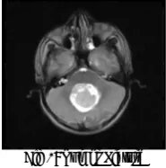
[2] Stationary comes with a computerized image segmented procedure utilizing persistence tactic. This can be driven by judgment that often adjoining pixels whom benefit (gray point, shade advantages, surface, etc) resides within one certain array were created by precisely the same lesson and following that, wonderful segmented of images including only couple opposite elements can be achieved as shown in Fig 2.
Fig 2: Tumor Picture
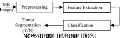
Fig 3: Tumor detection process
[5-6] planned region-based approaches through use of an judgment which typically adjacent pixels inside the same areas have related viewable capabilities namely gray stage, shade benefit, or texture. Cut up and fuse specialist techniques were utilized & its efficiency basically depends upon the chosen homogeneity criterion.
[7] Paper identifies the several segmentation techniques applied to the concept of diagnostic medical monographers and SAR Image Processing. Initially this paper takes a look at and compiles a few of the innovations utilized for image segmentation. Afterwards a bibliographical review of present segmentation approaches obtains with this paper and eventually universal habits in image segmentation are shown. This paper applies to the prevalence of straightforward Cluster Approach [4] for innovation of array and formation of tumor in person human brain MR images. This employs desktop assisted way of segmentation (innovation) of human brain tumor according to the mixing of a couple of approaches. Towards the end of building a song the tumor is taken from that are caused by the MR image together with its accurate pose plus the formation also established & the tumors platform is exhibited driven by number of territory smart that are caused by the cluster.
III. PROPOSED WORK
Applying Adaptive Median Filter Noise Removal Technique on the Noised Image:
Adaptive Median Filtering
Therefore the adaptive median filtering has been applied widely as an advanced method compared with standard median filtering. The Adaptive Median Filter performs spatial processing to determine which pixels in an image have been affected by impulse noise. The Adaptive Median Filter classifies pixels as noise by comparing each pixel in the image to its surrounding neighbor pixels. The size of the neighborhood is adjustable, as well as the threshold for the comparison. A pixel that is different from a majority of its neighbors, as well as being not structurally aligned with those pixels to which it is similar, is labeled as impulse noise. These noise pixels are then replaced by the median pixel value of the pixels in the neighborhood that have passed the noise labeling test.
VARYING intensity of tumors in brain magnetic resonance images (MRIs) makes the automatic segmentation of such tumors extremely challenging. Brain tumor segmentation using MRI has been an intense research area as shown in Fig 4.
Fig 4: Simplified overall flow diagram
Neural network classifier
Neural network continues to be extensively utilized for
classification of varying tissue regions in medical
images. In this particular work, we utilize multilayer back
propagation neural network to be classifier
to distinguish the tumor regions from nontumor regions. The most basic form of back-propagation procedure which generally studies the network biases and weights in the negative direction of the condition.
PROPOSED APPROACH MODEL
5. For any pixel in the above angx and below angy mask or clear the regions.
6. Extract the tumor bounded region. The area calculation formula is:
Size_of_tumor, S= [(√P)*0.264] mm2 P = no of white pixels; W = width; H = height.
IV.
EXPERIMENTAL RESULTSAll experiments are performed with the configurations Intel(R) Core(TM) 2 CPU 2.13GHz, 2 GB RAM, and the operating system platform is Microsoft Windows XP Professional (SP2).
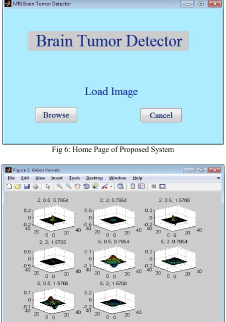
Fig 6: Home Page of Proposed System
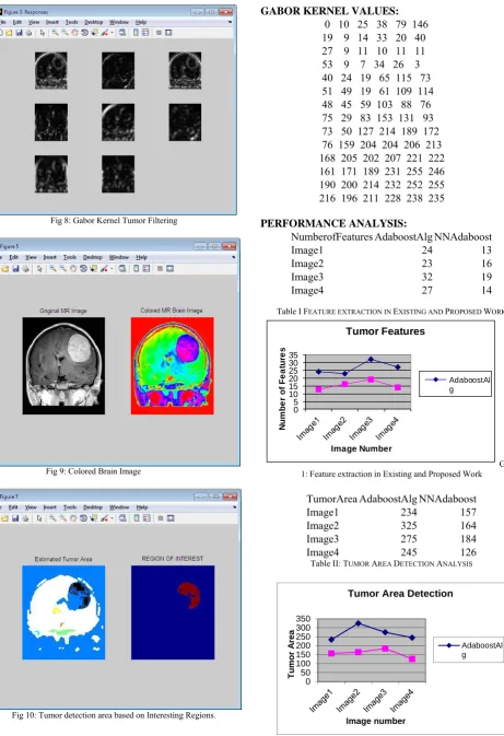
Fig 8: Gabor Kernel Tumor Filtering
Fig 9: Colored Brain Image
Fig 10: Tumor detection area based on Interesting Regions.
GABOR KERNEL VALUES: 0 10 25 38 79 146 19 9 14 33 20 40 27 9 11 10 11 11 53 9 7 34 26 3 40 24 19 65 115 73 51 49 19 61 109 114 48 45 59 103 88 76 75 29 83 153 131 93 73 50 127 214 189 172 76 159 204 204 206 213 168 205 202 207 221 222 161 171 189 231 255 246 190 200 214 232 252 255 216 196 211 228 238 235
PERFORMANCE ANALYSIS:
NumberofFeatures AdaboostAlg NNAdaboost
Image1 24 13
Image2 23 16
Image3 32 19
Image4 27 14
Table I FEATURE EXTRACTION IN EXISTING AND PROPOSED WORK
0 5 10 15 20 25 30 35
N
u
m
b
er
o
f F
eat
u
res
Image Number
Tumor Features
AdaboostAl g
Graph 1: Feature extraction in Existing and Proposed Work
TumorArea AdaboostAlg NNAdaboost
Image1 234 157
Image2 325 164
Image3 275 184
Image4 245 126
Table II: TUMOR AREA DETECTION ANALYSIS
0 50 100 150 200 250 300 350
Tumor A
rea
Image number
Tumor Area Detection
AdaboostAl g
V. CONCLUSION
Image segmentation is a major issue in image processing and realizes comprehensive application in many sectors. The improvement of AdaBoost procedure includes allocating weights to element classification algorithms dependent on their capability to classify challenging analytical samples and assurance in such classifier. Experimental outcome exhibit over 10 MRIs efficiency of the proposed procedure in automatic separation of tumors in brain MRIs. Additionally, evaluation with other state-of- the-art brain tumor separation along with separation operates with openly accessible low grade BRATS2012 data and shows that our segmented outcomes are more persistent and on the average performs for the patients. Evaluation of the segmentation results was performed through quantitative comparisons with manual segmentations, using tumor area and olume and surface measures.
ACKNOWELDGMENT
The authors would like to express appreciation to Children Hospital of Philadelphia for providing brain MR images for this paper.The paper also uses brain tumor image data obtained from MICCAI 2012 challenge on Multimodal Brain Tumor Segmentation.Atiq Islam,Syed M.S Reza and Khan M.The challenge database contains fully anonymized images from the following institutions:ETH Zurich, University of Bern and University of Utah.
R
EFERENCES[1] “Study on the Edge Detection Algorithms”, Chun-ling
FanYuan-yuan Ren, ISIP ‘10 Pages 217-220 IEEE Computer Society Washington, DC, USA ©2010.
[2] . H. B. Kekre, Tanuja K. Sarode, Saylee Gharge, “Image Segmentation of MRI Images using Vector Quantization Techniques” March 2010.
[3]. Donald Mcrobbie. “Medical Image Segmentation”
[4]. Dr. H. B. Kekre , Saylee Gharge , “Image Segmentation of MRI using Texture Features,” 05-06 December, 2008.
[5]. “Vector quantization”, R. M. Gray, IEEE ASSP Mag., pp.: 4-29,
Apr. 1984
[6]. “An algorithm for vector quantizer design”, Y. Linde, A. Buzo, and R. M. Gray, IEEE Trans.Commun., vol. COM- 28, no. 1, pp.: 84- 95, 1980
[7]. H.B.Kekre, Tanuja K. Sarode, “New Fast Improved Clustering Algorithm for Codebook Generation for Vector Quantization”,
13th –14th January 2008.
[8] Deorah S, Charles L, Zita A, Timothy R. “Trends in brain cancer incidence and survival in the United States: Surveillance, Epidemiology and End results program”. Neurosurgery Focus
2006; 20:1-7.
[9] "A Weighted K-means Algorithm applied to Brain Tissue Classification", Guillermo N. Abras and Virginia L. Ballarin,; JCS&T Vol. 5 No. 3, October 2005.