O R I G I N A L R E S E A R C H
Long noncoding RNA SNHG16 silencing inhibits the
aggressiveness of gastric cancer via upregulation of
microRNA-628-3p and consequent decrease of
NRP1
This article was published in the following Dove Press journal: Cancer Management and Research
Weifeng Pang1
Mingcui Zhai2
Yue Wang3
Zhiqiang Li4
1Department of Internal Oncology, The Fourth Affiliated Hospital of Harbin Medical University, Harbin, People’s Republic of China;2Department of Burn, Heilongjiang Province Hospital, Harbin, People’s Republic of China;3Department of Pharmacology and Toxicology, Wright State University, Fairborn, OH, USA; 4Department of General Surgery, Suihua First Hospital in Heilongjiang Province, Suihua, People’s Republic of China
Background:MicroRNA-628-3p (miR-628) has been reported to play important roles in the
progression of multiple human cancer types. Nonetheless, whether the expression profile of miR-628 is altered in gastric cancer remains unclear and whether its aberrant expression plays a crucial part in the aggressiveness of gastric cancer is yet to be determined. Therefore, in this study, we systematically investigated the involvement of miR-628 in gastric cancer progression.
Materials and methods:MiR-628 expression in gastric cancer tissues and cell lines were
determined via reverse transcription-quantitative polymerase chain reaction (RT-qPCR). A CCK-8 assay,flow-cytometric analysis, Transwell assays, and a xenograft model experiment were performed to evaluate the influence of miR-628 overexpression on gastric cancer cells. Notably, the mechanisms underlying the tumor-suppressive activity of miR-628 in gastric cancer cells were explored by bioinformatics analysis, a luciferase reporter assay, RT-qPCR, and Western blotting.
Results:MiR-628 expression was low in gastric cancer tissue samples and cell lines. The
low expression of miR-628 was closely associated with the lymph node metastasis, invasive depth and TNM stage among patients with gastric cancer. Further clinical analysis indicated that patients with gastric cancer underexpressing miR-628 had a worse prognosis than did the patients with high miR-628 expression in the tumor. Overexpressed miR-628 restrained proliferation, migration, and invasion; induced apoptosis; and impaired tumor growth of gastric cancer cells. In addition, neuropilin 1 (NRP1) mRNA was validated as the direct target of miR-628 in gastric cancer. Long noncoding RNA small nucleolar RNA host gene 16 (SNHG16) was demonstrated to sponge miR-628 in gastric cancer. Moreover, miR-628 knockdown abrogated the influence of SNHG16 silencing on gastric cancer cells.
Conclusion:Ourfindings elucidate how the SNHG16–miR-628–NRP1 pathway serves as a
regulatory network playing crucial roles in gastric cancer progression, suggesting that this pathway may be a novel target of anticancer therapy.
Keywords: gastric cancer, microRNA-628-3p, long noncoding RNA, neuropilin 1, small
nucleolar RNA host gene 16
Introduction
Gastric cancer is the fifth most prevalent type of malignant tumor and the third leading cause of cancer-associated deaths globally.1The morbidity of gastric cancer and resulting deaths decreased in the past decade owing to notable progress in the
Correspondence: Zhiqiang Li
Department of General Surgery, Suihua First Hospital in Heilongjiang Province, 80 Beilin Road, Suihua 152000, People’s Republic of China
Tel +86 1 384 558 5516 Email [email protected]
Cancer Management and Research
Dovepress
open access to scientific and medical research
Open Access Full Text Article
Cancer Management and Research downloaded from https://www.dovepress.com/ by 118.70.13.36 on 20-Aug-2020
diagnostic and therapeutic methods.2 Unfortunately, there are still many thousands of patients with gastric cancer who have received the diagnosis at late stages, thus, result-ing in unsatisfactory clinical outcomes.3 The recurrence and metastasis after surgical management are the major causes of the poor prognosis of patients with gastric cancer.4 Various factors, such as heredity, Helicobacter pylori infection, dietary habits, smoking, and drinking, are known to be implicated in gastric carcinogenesis and progression;5,6however, the mechanisms are still incom-pletely understood, and this situation is another barrier to successful therapy for gastric cancer. Therefore, studying the molecular alterations that occur during the initiation and progression of gastric cancer may facilitate the
identi-fication of promising therapeutic strategies and should improve the prognosis.
MicroRNAs (miRNAs) are a family of evolutionarily conserved and noncoding short RNAs with a length of ~23 nucleotides.7MiRNAs can recognize and directly bind to complementary sequences in the 3′-untranslated regions (3′-UTRs) of their target mRNAs, thereby causing transla-tion inhibitransla-tion and/or mRNA degradatransla-tion.8Thus far, over 1800 miRNAs have been confirmed in the human genome, and their aberrant expression has been observed in nearly all human cancer types.9Some studies have indicated that a number of miRNAs are dysregulated in gastric cancer, and the dysregulation of miRNAs is involved in gastric cancer progression by affecting numerous tumor behaviors.10–12 These findings have collectively proved the regulatory importance of miRNAs in the progression of gastric cancer, suggesting that miRNAs may be effec-tive therapeutic targets in gastric cancer.
Long noncoding RNAs (lncRNAs) are a novel group of noncoding RNAs of >200 nucleotides.13 Thousands of lncRNA genes have been identified in the human genome, many of which perform regulatory functions in the aggres-sive behaviors of cancer cells during tumorigenesis including tumor progression.14LncRNAs are reported to be regulators of gene expression through a variety of mechanisms, includ-ing genomic interactions, protein amounts, miRNA competi-tion, and chromatin modifications.15,16Various lncRNAs are abnormally expressed in gastric cancer and regulate multiple pathological processes, including cell proliferation, cell cycle, apoptosis, metastasis, and angiogenesis.17–19 Hence, lncRNAs might be potential molecular targets for the diag-nosis and treatment of gastric cancer.
In recent years, miR-628-3p (miR-628) was reported to substantially participate in the progression of several types
of human cancer, including colorectal cancer,20acute mye-loid leukemia,21 pancreatic cancer,22 and non-small-cell lung cancer.23Nevertheless, whether the expression profile of miR-628 is altered in gastric cancer remains unclear and whether its aberrant expression is important for the aggres-siveness of gastric cancer is yet to be studied. Therefore, the aim of our current study was to evaluate miR-628 expression in gastric cancer and explore its specific roles in the regulation of the malignant characteristics of gastric cancer.
Materials and methods
Patient samples
In total, 54 pairs of gastric cancer tissue samples and matched adjacent normal tissue samples were collected at Suihua First Hospital in Heilongjiang Province. None of the patients had received preoperative radiotherapy, che-motherapy, or other anticancer interventions. All tissue specimens were obtained after surgical resection, immedi-ately frozen in liquid nitrogen, and then stored at−80 °C. All the participants agreed to take part in this study and provided written informed consent prior to the surgical operation. The study protocol was approved by the Ethics Committee of Suihua First Hospital in Heilongjiang Province and was carried out in compliance with the ethical standards of the Declaration of Helsinki.
Cell culture
Human gastric cancer cell lines (BGC-823, SGC-7901, MKN-45, and AGS) and immortalized human gastric epithelial cells (GES-1) were bought from the American Type Culture Collection (Manassas, VA, USA). All the above cells were maintained at 37 °C in a humidified 95% air and 5% CO2 atmosphere and cultured in Dulbecco’s
Modified Eagle’s Medium (DMEM) supplemented with 10% of fetal bovine serum (FBS),100 U/mL penicillin, and 100 μg/mL streptomycin (All from Gibco; Thermo Fisher Scientific, Inc., Waltham, MA, USA).
Oligonucleotides, small interfering RNAs
(siRNAs), plasmids, and cell transfection
MiR-628 agomir (agomir-628), negative control (NC) ago-mir (agoago-mir-NC), miR-628 antagoago-mir (antagoago-mir-628), and NC antagomir (antagomir-NC) were purchased from Shanghai GenePharma Co., Ltd. (Shanghai, China). The siRNA that was used to silence SNHG16 (si-SNHG16) and negative control siRNA (si-NC) were generated by
Cancer Management and Research downloaded from https://www.dovepress.com/ by 118.70.13.36 on 20-Aug-2020
Guangzhou RiboBio Co., Ltd. (Guangzhou, China). The plasmid expressing NRP1 (pcDNA3.1-NRP1) was con-structed by GenScript Biotech Corp. (Nanjing, China). Cells were seeded in 6-well plates 24 h before transfection. The above-mentioned oligonucleotides and plasmid were transfected into cells by means of Lipofectamine 2000 (Invitrogen; Thermo Fisher Scientific, Inc.).
RNA isolation and reverse
transcription-quantitative polymerase chain reaction
(RT-qPCR)
Total RNA was isolated from tissue samples or cells using the TRIzol reagent (Invitrogen; Thermo Fisher Scientific, Inc.) and was reverse-transcribed into complementary DNA (cDNA) with the miScript Reverse Transcription Kit (Qiagen GmbH, Hilden, Germany). MiR-628 expression was detected via qPCR with the miScript SYBR Green PCR kit (Qiagen GmbH, Hilden, Germany) and normalized to U6 small nuclear RNA. To quantify NRP1 and SNHG16 expression, the synthesis of cDNA was conducted using the PrimeScript RT Reagent Kit, followed by qPCR with SYBR® Premix Ex Taq™ (both from Takara Biotechnology Co., Ltd., Dalian, China). The expression levels of NRP1 and SNHG16 were normalized to GAPDH. The 2−ΔΔCqmethod was employed for quantification.
Cell Counting Kit-8 (CCK-8) assay
Transfected cells were seeded in 24-well plates at a density of 2×10³cells/well. After 0–72 h of incubation, the CCK-8 assay was carried out every 24 h to determine cell prolif-eration. Briefly, 10 μL of the CCK-8 solution (Dojindo Molecular Technologies, Inc., Kumamoto, Japan) was added into each well, and the transfected cells were incu-bated at 37 °C for additional 2 h. The absorbance was measured at a 450 nm wavelength on a microplate reader (Bio-Rad Laboratories, Inc., Hercules, CA, USA).
Detection of cell apoptosis by
fl
ow-cytometric analysis
Transfected cells were treated with trypsin (Gibco; Thermo Fisher Scientific), harvested by centrifugation, and subjected to cell apoptosis detection by means of the Annexin V Fluorescein Isothiocyanate (FITC) Apoptosis Detection Kit (Biolegend, San Diego, CA, USA). In particular, transfected cells were resuspended in 100 µL of 1× binding buffer, double-stained with 5 µL of the Annexin V solution and 5 µL of the propidium iodide solution, and incubated at
room temperature in the dark for 15 min. Finally, the apop-totic rate (early stage + late stage) was determined on aflow cytometer (FACScan; BD Biosciences, Bedford, MA, USA).
Transwell assay
The migratory and invasive abilities were assessed using noncoated or Matrigel-coated Transwell chambers (BD Biosciences), respectively. Transfected cells were col-lected after 48 h incubation, centrifuged, and resuspended in FBS-free DMEM. The cell concentration was adjusted to 1×10⁵cells/mL. A total of 200μL of the cell suspension was added into the upper chambers, while the bottom chambers were covered with 500 μL of DMEM supple-mented with 10% of FBS. The chambers were next kept at 37 °C and 5% CO2 for 24 h, following by cell fixation
with 4% paraformaldehyde, staining with 0.5% crystal violet, and washing with phosphate-buffered saline (PBS; Gibco; Thermo Fisher Scientific, Inc.). After that, the images of the migratory and invading cells were captured under an inverted microscope (×200 magnification; CKX41SF; Olympus Corporation, Tokyo, Japan). The numbers of migratory and invading cells in five random visualfields per group were determined, and the mean and standard deviation (SD) were calculated to describe the migratory and invasive abilities.
Xenograft model experiment
Agomir-628–transfected or agomir-NC–transfected cells in the logarithmic growth phase were collected and injected subcutaneously into female 4–6-week-old BALB/c nude mice (n=4 for each group; the Laboratory Animal Center of Yangzhou University; Yangzhou, China). Two weeks after the injection, the tumor volume was measured using the formula: volume (mm3) = 0.5×length×width2. All the nude mice were then euthanized by dislocation of cervical vertebrae at 4 weeks after the inoculation for excision of the tumor xenografts. The tumor xenografts were stored for the isolation of total RNA and protein. The study protocol was approved by the Animal Care and Use Committee of Suihua First Hospital in Heilongjiang Province and was performed in compliance with the Animal Protection Law of the People’s Republic of China-2009 for experimental animals.
Bioinformatics analysis
Bioinformatics tools, namely, miRDB (http://mirdb.org/) and TargetScan (http://www.targetscan.org), were utilized to search for the putative target of miR-628. DIANA tools -LncBase Experimental v2 (http://carolina.imis.athena-inno
Cancer Management and Research downloaded from https://www.dovepress.com/ by 118.70.13.36 on 20-Aug-2020
vation.gr/diana_tools/web/index.php?r=lncbasev2%2fi ndex-experimental) was applied to analyze the binding site for miR-628 in SNHG16.
Luciferase reporter assay
The 3′-UTR of NRP1, which contains the predicted wild-type (wt) miR-628–binding site, and the mutant (mut) NRP1 3′-UTR, were amplified by Shanghai GenePharma Co., Ltd. The synthesized DNA fragments were cloned into the pmirGLO vector (Promega Corporation, Madison, WI, USA) to generate the wt-NRP1 and NRP1 reporter plasmids. The wt-SNHG16 and mut-SNHG16 reporter plasmids were chemically produced in the same way. For the reporter assay, cells were seeded in 24-well plates, followed by cotransfection with agomir-628 or agomir-NC and either the wt or mut reporter plasmid. Following a 48 h transfection period, the lucifer-ase activity was determined using a dual-luciferlucifer-ase repor-ter assay system (Promega Corporation). The Renilla luciferase activity was assayed for normalization.
Protein extraction and Western blot
analysis
RIPA lysis buffer (Beyotime Institute of Biotechnology, Haimen, China) was utilized to isolate total protein from tissue samples, cells, or tumor xenografts. The isolated total protein was quantified with the Bradford Kit (Beyotime Institute of Biotechnology). Protein samples containing equal amounts of protein were separated by sodium dodecyl sulfate polyacrylamide gel electrophoresis in a 10% gel and transferred onto polyvinylidene difl uor-ide membranes. Blocking with 5% nonfat milk was per-formed at room temperature for 2 h to prevent nonspecific binding of antibodies. The membranes were next probed with primary antibodies against NRP1 (cat. # ab81321; 1:1000 dilution; Abcam) or GAPDH (ab181603; 1:1000 dilution; Abcam), followed by incubation with a goat anti-rabbit IgG horseradish peroxidase-conjugated antibody (ab6721; 1:5000 dilution; Abcam) (secondary antibody) at room temperature for 1 h and visualization with an enhanced chemiluminescence detection system (GE Healthcare, Chicago, IL, USA).
Statistical analysis
All data are presented as mean ± SD and were subjected to analysis in SPSS 13.0 software (IBM Corporation, Armonk, NY, USA). Pearson’sχ2test was conducted to investigate
the association between miR-628 expression and clinical parameters among the patients with gastric cancer. The expression correlation between miR-628 and NRP1 in gas-tric cancer tissue samples was evaluated by Spearman’s correlation analysis, which was also carried out to test the expression correlation between miR-628 and SNHG16. The Kaplan–Meier method was employed to build a survival curve, and the differences among groups were assessed by the logrank test. A comparison between two groups was performed with Student’st-test, whereas one-way analysis of variance along with Tukey’s post-hoc test was performed to assess differences among multiple groups. Data with P<0.05 were considered statistically significant.
Results
Downregulation of miR-628 is associated
with an unfavorable prognosis among
patients with gastric cancer
To determine the expression profile of miR-628 in gastric cancer, we measured miR-628 expression in 54 pairs of gastric cancer tissue samples and matched adjacent normal tissue samples through RT-qPCR. The results showed that expres-sion levels of miR-628 were lower in gastric cancer tissue samples relative to adjacent normal tissues (Figure 1A, P<0.05). In addition, miR-628 turned out to be underexpressed in the four tested gastric cancer cell lines (BGC-823, SGC-7901, MKN-45, and AGS) in comparison with the immorta-lized human gastric epithelial cells (GES-1; Figure 1B, P<0.05).
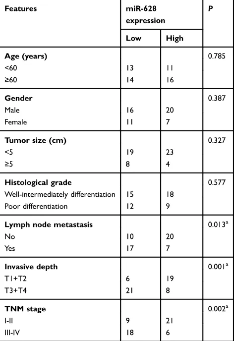
To uncover the clinical relevance and prognostic significance of miR-628 in gastric cancer, we subdi-vided all the 54 patients with gastric cancer into two groups: low-miR-628-expression group and high-miR-628-expression group, according to the median value of miR-628 among the gastric cancer tissue samples. Low miR-628 expression was significantly associated with lymph node metastasis (P=0.013), invasive depth (P=0.001) and TNM stage (P=0.002) among the patients with gastric cancer (Table 1). By contrast, no obvious correlation with age, gender, tumor size or histological grade was detected (all P>0.05). Moreover, patients with gastric cancer harboring low miR-628 expression had a lower probability of better overall survival than did patients in the high-miR-628-expression group (Figure 1C, P=0.0264). These results suggested that miR-628 may play a critical part in the aggressiveness of gastric cancer.
Cancer Management and Research downloaded from https://www.dovepress.com/ by 118.70.13.36 on 20-Aug-2020
miR-628 inhibits gastric cancer cell
proliferation, migration, and invasion in
vitro and promotes apoptosis
BGC-823 and SGC-7901 cell lines manifested relatively low miR-628 expression as compared with MKN-45 and AGS cells; therefore, thefirst two cell lines were chosen as the model to explore the detailed functions of miR-628 in the oncogenicity of gastric cancer. Agomir-628 was trans-fected into BGC-823 and SGC-7901 cells to increase endogenous miR-628 expression (Figure 2A, P<0.05). Data from the CCK-8 assays showed that transfection with agomir-628 greatly reduced the proliferative ability of BGC-823 and SGC-7901 cells (Figure 2B,P<0.05). In line with this finding, upregulation of miR-628 notably increased the apoptosis of BGC-823 and SGC-7901 cells, as revealed by flow-cytometric analysis (Figure 2C, P<0.05). After that, Transwell assays were carried out to test the effects of miR-628 upregulation on the migration and invasiveness of gastric cancer cells. The migratory (Figure 2D, P<0.05) and invasive (Figure 2E, P<0.05) abilities of BGC-823 and SGC-7901 cells were decreased
by miR-628 upregulation. The above results indicated that miR-628 may function as a tumor-suppressive modulator in gastric cancer.
NRP1 mRNA is directly targeted by
miR-628 in gastric cancer
To gain an in-depth understanding of the mechanisms behind the activity of miR-628 in gastric cancer, the putative targets of miR-628 were predicted via bioinfor-matics analysis. NRP1 was chosen for further verifi ca-tion because this gene has two major miR-628 binding sites in the 3′-UTR of its mRNA (Figure 3A) and sub-stantially participates in gastric carcinogenesis.24–26 To test our assumption, the wt-NRP1 (1 and 2) and mut-NRP1 (1 and 2) reporter plasmids were constructed based on the predicted binding site and were cotrans-fected with agomir-628 or agomir-NC into BGC-823 and SGC-7901 cells. The transfection with agomir-628 efficiently impaired the luciferase activity of the plasmid containing the wild-type NRP1-binding site (both 1 and 2; P<0.05). Conversely, no obvious alterations in the 2.5
A
B
C
*
*
*
*
*
1.5Relative
miR-628 expression
Relative
miR-628 expression
Overall survival (%)
1.0
0.5
0.0 2.0
1.5
1.0
0.5
0.0
100
80
60
40
20
0
0 20 40 60
Time (months)
Low miR-628 expression group
P=0.0264
High miR-628 expression group Normal Gastric
cancer
GES-1
BGC-823SGC-7901MKN-45 AGS
Figure 1The expression of miR-628 in gastric cancer and its correlation with patients’survival. (A) MiR-628 expression in 54 pairs of gastric cancer tissue samples and matched adjacent normal tissues was analyzed using RT-qPCR. All tissue samples were collected at Suihua First Hospital in Heilongjiang Province. *P<0.05 vs normal tissues. (B) MiR-628 levels in four gastric cancer cell lines (BGC-823, SGC-7901, MKN-45, and AGS) and immortalized human gastric epithelial cells (GES-1) were measured by RT-qPCR. *P<0.05 vs GES-1. (C) The correlation between miR-628 expression and overall survival among patients with gastric cancer.P=0.0264.
Cancer Management and Research downloaded from https://www.dovepress.com/ by 118.70.13.36 on 20-Aug-2020
luciferase activity were seen in the BGC-823 and SGC-7901 cells cotransfected with agomir-628 and the mut-NRP1 reporter plasmid (both 1 and 2; Figure 3B). To investigate whether NRP1 is scientifically and clinically relevant to the expression of miR-628, the expression profile of NRP1 was determined in the 54 pairs of gastric cancer tissue samples and matched adjacent nor-mal tissues. The mRNA level of NRP1 was higher in the gastric cancer tissue samples than in the adjacent normal tissues (Figure 3C, P<0.05). Additionally, Spearman’s correlation analysis of the 54 gastric cancer tissue samples confirmed that the expression of NRP1 inversely correlated with miR-628 expression (Figure 3D; R2=0.4138, P<0.0001). Furthermore, the mRNA (Figure 3E, P<0.05) and protein (Figure 3F, P<0.05) levels of NRP1 obviously diminished after overexpres-sion of miR-628 in BGC-823 and SGC-7901 cells. In summary, NRP1 is a direct target of miR-628 in gastric cancer.
Tumor-suppressive activities of miR-628
in gastric cancer cells are NRP1
dependent
MiR-628 inhibited the growth and metastasis of gastric cancer cells in vitro, and NRP1 mRNA was validated as a direct target of miR-628 in the experiments above. Hence, we assumed that the tumor-suppressive roles of miR-628 in gastric cancer are dependent on NRP1. We restored NRP1 expression in the agomir-628–transfected BGC-823 and SGC-7901 cells by cotransfecting the plas-mid expressing NRP1 pcDNA3.1-NRP1 (Figure 4A, P<0.05). Then, the results of CCK-8 andflow-cytometric assays revealed that ectopic miR-628 expression decreased the proliferation (Figure 4B, P<0.05) and increased the apoptosis rate (Figure 4C, P<0.05) of BGC-823 and SGC-7901 cells; these effects were attenu-ated by cotransfection with pcDNA3.1-NRP1. Furthermore, restoration of NRP1 expression weakened the miR-628–mediated inhibitory actions on the migra-tion (Figure 4D, P<0.05) and invasiveness (Figure 4E, P<0.05) of BGC-823 and SGC-7901 cells. Taken together, the above observations suggested that NRP1 is the functional target of miR-628 and that NRP1 down-regulation is essential for the tumor-suppressive activities of miR-628 in gastric cancer.
SNHG16 functions as a sponge for
miR-628 in gastric cancer
A plethora of studies indicate that lncRNAs can act as competing endogenous RNAs (ceRNAs) to sponge miRNAs. Therefore, we next attempted to test whether miR-628 can be sponged by a certain lncRNA in gastric cancer. Bioinformatics analysis was carried out and iden-tified two potential miR-628–binding sites in an lncRNA called SNHG16 (Figure 5A). The luciferase reporter assay was then conducted to confirm the prediction, and the results showed that restoration of miR-628 expression greatly decreased the luciferase activities of wt-SNHG16 (both 1 and 2; Figure 5B, P<0.05) but not mut-SNHG16 (both 1 and 2) in BGC-823 and SGC-7901 cells.
To further examine the interaction between miR-628 and SNHG16 in gastric cancer, we quantitated SNHG16 expression in the 54 pairs of gastric cancer tissue samples and the matched adjacent normal tissue samples. In line with other studies,27,28the expression of SNHG16 turned out to be higher in the gastric cancer tissue samples than in the adjacent normal tissues (Figure 5C, P<0.05).
Table 1The association between miR-628 expression and
clin-icopathological features in patients with gastric cancer
Features miR-628
expression
P
Low High
Age (years) 0.785
<60 13 11
≥60 14 16
Gender 0.387
Male 16 20
Female 11 7
Tumor size (cm) 0.327
<5 19 23
≥5 8 4
Histological grade 0.577 Well-intermediately differentiation 15 18
Poor differentiation 12 9
Lymph node metastasis 0.013a
No 10 20
Yes 17 7
Invasive depth 0.001a
T1+T2 6 19
T3+T4 21 8
TNM stage 0.002a
I-II 9 21
III-IV 18 6
Note:aP<0.05.
Cancer Management and Research downloaded from https://www.dovepress.com/ by 118.70.13.36 on 20-Aug-2020
Moreover, SNHG16 expression was inversely related with miR-628 expression among the gastric cancer tissue sam-ples, as revealed by Spearman’s correlation analysis (Figure 5D; R2=0.4296, P<0.0001). Lastly, si-SNHG16 silenced SNHG16 expression (Figure 5E, P<0.05),
increased miR-628 expression (Figure 5F, P<0.05), and reduced NRP1 protein expression (Figure 5G,P<0.05) in BGC-823 and SGC-7901 cells. Collectively, thesefindings confirmed that SNHG16 functions as a molecular sponge for miR-628 in gastric cancer.
60
A
B
C
D
E
*
*
* *
*
*
*
*
*
*
*
*
50
40
30 2.0 1.5 1.0 0.5 0.0
10
4
10
3
10
2
10
1
10
0
10
4
10
3
10
2
10
1
10
0
10
4
10
3
10
2
10
1
10
0
104 103 102 101 100
10
4
10
3
10
2
10
1
10
0
104 103 102 101
100 100 101 102 103 104 100 101 102 103 104 Agomir-NC
Agomir-NC Agomir-628 Agomir-NC Agomir-628 Agomir-NC
25
20
Cell apoptosis rate (%)
Migratory
cell number
Invasion
cell number
15
10
5
0
250
200
150
100
50
0
250
200
150
100
50
0
Agomir-628
Agomir-NC Agomir-628
Agomir-NC Agomir-628
Agomir-NC Agomir-628
Agomir-NC Agomir-628
Agomir-NC Agomir-628
Agomir-NC Agomir-628
Agomir-NC Agomir-628 Agomir-NC
Agomir-628
Agomir-NC Agomir-628 Agomir-628
BGC-823 SGC-7901
60
70 1.5
1.0
0.5
0.0
1.5
1.0
Absorbance
Absorbance
Relative
miR-628 expression
Relative
miR-628 expression
0.5
0.0 0 h 24 h 48 h
BGC-823
BGC-823 SGC-7901
SGC-7901
BGC-823 SGC-7901
BGC-823 SGC-7901
BGC-823 SGC-7901
BGC-823 Annexin V
PI PI
Annexin V SGC-7901
BGC-823 SGC-7901
72 h 0 h 24 h 48 h 72 h
50
40 2.0 1.5 1.0 0.5 0.0
Figure 2MiR-628 overexpression suppresses the proliferation, induces apoptosis, and decreases the migration and invasiveness of BGC-823 and SGC-7901 cells. (A) RT-qPCR was conducted to measure miR-628 expression in BGC-823 and SGC-7901 cells after transfected with agomir-628 or agomir-NC. *P<0.05 vs agomir-NC. (B) The proliferative ability of BGC-823 and SGC-7901 cells treated with agomir-628 or agomir-NC was examined using the CCK-8 assay. *P<0.05 vs agomir-NC. (C) Flow-cytometric analysis was carried out to determine the influence of agomir-628 transfection on the apoptosis of BGC-823 and SGC-7901 cells. *P<0.05 vs agomir-NC. (D, E) The migration and invasiveness of miR-628–overexpressing BGC-823 and SGC-7901 cells were assessed in Transwell assays. The migration and invasion abilities were quantified as cell numbers (× 200 magnification). *P<0.05 vs agomir-NC.
Cancer Management and Research downloaded from https://www.dovepress.com/ by 118.70.13.36 on 20-Aug-2020
Downregulation of SNHG16 inhibits the
proliferation, migration, and invasiveness
and induces apoptosis of gastric cancer
cells
To explore the roles of SNHG16 in the biological char-acteristics of gastric cancer, si-SNHG16 was used to silence endogenous SNHG16 expression in BGC-823 and SGC-7901 cells, and then a series of functional assays were conducted. The influence of SNHG16 downregulation on gastric cancer cell proliferation and apoptosis was investigated in the CCK-8 assay and
flow-cytometric experiment. The proliferative capacity (Figure 6A, P<0.05) of the BGC-823 and SGC-7901 cells transfected with si-SNHG16 diminished, whereas the apoptosis rate (Figure 6B, P<0.05) increased. We also performed the Transwell assay to determine the
actions of the SNHG16 knockdown on the migration and invasiveness of gastric cancer cells. BGC-823 and SGC-7901 cells transfected with si-SNHG16 had weaker migratory (Figure 6C,P<0.05) and invasive (Figure 6D, P<0.05) abilities. The results revealed that SNHG16 may have an oncogenic influence on the aggressive phenotypes of gastric cancer.
SNHG16 exerts its effects in gastric
cancer via the miR-628
–
NRP1 axis
Because the above results indicated that SNHG16 plays oncogenic roles in gastric cancer progression and could regulate NRP1 expression by sponging miR-628, we next conducted rescue experiments to determine whether silen-cing of SNHG16 expression inhibits the growth and metastasis of gastric cancer cells in vitro by releasing
1.5
A
B
C
D
E
F
Agomir-NC Agomir-628
*
*
*
*
*
*
Agomir-NCAgomir-628Agomir-NCAgomir-628
Agomir-NC Agomir-628
Agomir-NC 5
4
3
2
1
0
5 R2=0.4138 P<0.0001
4
3
2
1
0 Gastric
cancer
Normal 0.0 0.2 0.4 0.6
Relative miR-628 expression
0.8 1.0
Agomir-628 Agomir-NC
Agomir-628
BGC-823 SGC-7901 BGC-823 SGC-7901
BGC-823
NRP1
GAPDH
SGC-7901 1.0
0.5
0.0
1.5
1.0
0.5
0.0
1.5
1.0
0.5
0.0 1.5
Luciferase activity Luciferase activity
Relative
NRP1 mRNA
expression
Relative
NRP1 mRNA
expression
Relative
NRP1 mRNA
expression
Relative
NRP1 protein expression
1.0
0.5
0.0
wt 1 wt 2
BGC-823
mut 1 mut 2 wt 1 wt 2
SGC-7901
mut 1 mut 2
Figure 3Validation of NRP1 mRNA as a direct target of miR-628 in gastric cancer. (A) The putative miR-628 target sequences in the 3′-UTR of NRP1 revealed by bioinformatics software. The 3′-UTR regions of NRP1 containing the wt or mut binding sites as well as miR-628 sequences are presented. (B) BGC-823 and SGC-7901 cells were cotransfected with agomir-628 or agomir-NC and wt-NRP1 or mut-NRP1. Following transfection, a luciferase reporter assay was performed to assess the interaction between miR-628 and NRP1 mRNA in gastric cancer. *P<0.05 vs agomir-NC. (C) RT-qPCR was carried out to measure the expression levels of NRP1 mRNA in 54 pairs of gastric cancer tissue samples and matched adjacent normal tissue samples. *P<0.05 vs normal tissue samples. (D) A negative expression correlation between miR-628 and NRP1 in gastric cancer tissue samples was confirmed via Spearman’s correlation analysis. R2
=0.4138,P<0.0001. (E, F) NRP1 mRNA and protein expression levels in BGC-823 and SGC-7901 cells with restored miR-628 expression were detected through RT-qPCR and Western blotting. *P<0.05 vs agomir-NC.
Cancer Management and Research downloaded from https://www.dovepress.com/ by 118.70.13.36 on 20-Aug-2020
BGC-823 BGC-823 NRP1
A
B
D
E
C
GAPDH NRP1 GAPDH SGC-7901 SGC-7901 BGC-823 SGC-7901 BGC-823 SGC-7901 BGC-823 SGC-7901 BGC-823 BGC-823 SGC-7901 SGC-7901 SGC-7901 BGC-823 SGC-7901 BGC-823 1.5 30 20 10 0 1.0 AbsorbanceCell apoptosis rate (%)
0.5 0.0 Agomir-NC 1.5 1.0 Relative
NRP1 protein expression
0.5 0.0 1.5 Absorbance 1.0 0.5 0.0
0 h 24 h 48 h 72 h
0 h 24 h
250 200 150 100 50 0 200 150 Invasive cell number
Migratory cell number
100
50
0 48 h 72 h
Agomir-628+pcDNA3.1 Agomir-628+pcDNA3.1-NRP1 # # # # #
*
*
*
*
*
# #*
*
# #*
*
*
# Agomir-NC Agomir-628+pcDNA3.1 Agomir-628+pcDNA3.1-NRP1 Agomir-NC Agomir-628+pcDNA3.1Agomir-628+ pcDNA3.1-NRP1 Agomir-NC Agomir-628+pcDNA3.1Agomir-628+ pcDNA3.1-NRP1 Agomir-NC Agomir-628+pcDNA3.1 Agomir-628+pcDNA3.1-NRP1 Agomir-NC Agomir-628+pcDNA3.1 Agomir-628+pcDNA3.1-NRP1 Agomir-NC Agomir-628+pcDNA3.1 Agomir-628+ pcDNA3.1-NRP1 Agomir-NC Agomir-628+pcDNA3.1 Agomir-628+ pcDNA3.1-NRP1 Agomir-NC Agomir-628+pcDNA3.1 Agomir-628+pcDNA3.1-NRP1 Agomir-NC Agomir-628+pcDNA3.1 Agomir-628+pcDNA3.1-NRP1Agomir-NC Agomir-628+ pcDNA3.1 Agomir-628+ pcDNA3.1-NRP1 10 4 10 3 10 2 10 1 10 0 10 4 10 3 10 2 10 1 10 0 10 4 10 3 10 2 10 1 10 0 10 4 10 3 10 2 10 1 10 0 10 4 10 3 10 2 10 1 10 0 10 4 10 3 10 2 10 1 10 0 104 103 102 101 100 104 103 102 101 100 104 103 102 101 100 Annexin V 104 103 102 101
100 102 103 104 101
100 104
103
102
101
100
PI
Figure 4Restoring NRP1 expression neutralizes the influence of miR-628 overexpression on gastric cancer cells. (A) Western blot analysis of NRP1 protein expression in BGC-823 and SGC-7901 cells after cotransfection with agomir-628 and pcDNA3.1-NRP1 or pcDNA3.1. *P<0.05 vs group agomir-NC.#
P<0.05 vs group agomir-628+pcDNA3.1. (B, C) The proliferation and apoptosis of BGC-823 and SGC-7901 cells with restored NRP1 expression were quantified by the CCK-8 assay andflow-cytometric analysis. *P<0.05 vs group agomir-NC.#
P<0.05 vs group agomir-628+pcDNA3.1. (D, E) The migratory and invasive abilities of BGC-823 and SGC-7901 cells treated with the above-mentioned constructs were evaluated in Transwell assays. *P<0.05 vs group agomir-NC (× 200 magnification).#
P<0.05 vs group agomir-628+pcDNA3.1.
Cancer Management and Research downloaded from https://www.dovepress.com/ by 118.70.13.36 on 20-Aug-2020
sponged miR-628 and decreasing NRP1 expression. Hence, antagomir-628, which was used to knock down miR-628 expression (Figure 7A, P<0.05), was cotrans-fected with si-SNHG16 into BGC-823 and SGC-7901 cells, and the miR-628 amount and NRP1 protein levels were detected via RT-qPCR and Western blotting. After the transfection, the increased level of miR-628 (Figure 7B, P<0.05) and decreased level of the NRP1 protein (Figure 7C, P<0.05) in SNHG16 knockdown BGC-823 and SGC-7901 cells were almost reversed by cotransfec-tion with antagomir-628. Furthermore, cotransfeccotransfec-tion with antagomir-628 abrogated si-SNHG16–mediated effects on the proliferation (Figure 7D, P<0.05), apoptosis (Figure 7E,P<0.05), migration (Figure 8A,P<0.05), and invasive-ness (Figure 8B, P<0.05) of BGC-823 and SGC-7901 cells. These findings suggested that SNHG16 performs
its biological activities in gastric cancer cells at least in part via the miR-628–NRP1 axis.
miR-628 suppresses the growth of gastric
cancer in vivo
The xenograft model experiment was conducted to test whether miR-628 can hinder tumor growth of gastric cancer cells in vivo. Agomir-628–transfected BGC-823 cells were injected subcutaneously into nude mice, and cells treated with agomir-NC served as the control. Consistently with the in vitro results, the agomir-628 group showed an obvious decrease in tumor volume compared with that in the agomir-NC group (Figure 9A and B, P<0.05). Meanwhile, measurements of the tumor xenografts revealed that miR-876 overexpression markedly reduced tumor weight (Figure 9C, P<0.05).
1.5
*
*
*
*
*
*
*
*
*
*
*
Agomir-NC Agomir-628
Agomir-NC Agomir-628
1.0
0.5
Luciferase activity Luciferase activity
wt 1 wt 2
BGC-823
si-NC si-SNHG16
si-NC si-NC
si-SN HG16
si-SN HG16 si-NC
si-SNHG16 si-NC
si-SNHG16
mut 1 mut 2 wt 1 mut 1 wt 2 mut 2
0.0
1.5
1.0
0.5
0.0
SGC-7901
BGC-823 SGC-7901 BGC-823 SGC-7901
1.5 6
4
2
0 1.0
0.5
0.0
Relative
SNHG16 expression
Relative
miR-628 expression
NRP1
GAPDH
BGC-823 SGC-7901
4
3
2
1
0.0 0.2 0.4 0.6 Relative miR-628 expression
0.8 1.0
Relative
SNHG16 expression
Relative
SNHG16 expression
5
4
3
2
1
0
Gastric cancer Normal
Relative
NRP1 protein expression
1.5
1.0
0.5
0.0
BGC-823 SGC-7901
R2=0.4296 P<0.0001
A
B
C
D
E
F
G
Figure 5SNHG16 functions as a ceRNA and regulates NRP1 expression in gastric cancer by competitively binding 628. (A) The predicted two binding sites for miR-628 on SNHG16 as predicted by bioinformatics software. (B) Agomir-miR-628 or agomir-NC was cotransfected with wt-SNHG16 or mut-SNHG16 into BGC-823 and SGC-7901 cells, and sequentially the luciferase activity was quantified. *P<0.05 vs agomir-NC. (C) Expression of SNHG16 in 54 pairs of gastric cancer tissue samples and matched adjacent normal tissue samples was examined via RT-qPCR. *P<0.05 vs normal tissues. (D) Spearman’s correlation analysis uncovered an inverse association between miR-628 and SNHG16 in gastric cancer tissue samples. R2=0.4296,P<0.0001. (E) The expression levels of SNHG16 in BGC-823 and SGC-7901 cells when they were treated with si-SNHG16 or si-NC were determined by RT-qPCR. *P<0.05 vs si-NC. (F, G) RT-qPCR and Western blot analysis were performed to assess miR-628 and NRP1 protein expression, respectively, in SNHG16-depleted BGC-823 and SGC-7901 cells. *P<0.05 vs si-NC.
Cancer Management and Research downloaded from https://www.dovepress.com/ by 118.70.13.36 on 20-Aug-2020
After that, the expression levels of miR-876 and NRP1 protein in the tumor xenografts were determined. The results meant that in the agomir-628 group, the expres-sion of NRP1 protein (Figure 9D, P<0.05) was lower, whereas miR-628 (Figure 9E, P<0.05) was pressed. These results suggested that miR-628 overex-pression inhibited gastric cancer tumor growth in vivo by decreasing NRP1 expression.
Discussion
In recent decades, dysregulation of miRNAs has been reported to be involved in gastric cancer initiation and progression, and it has become clear that miRNAs may serve as oncogenic or tumor-suppressive factors.29–31 Hence, exploring the specific functions of cancer-asso-ciated miRNAs in gastric cancer should be useful for identifying promising targets for the diagnosis and
0 h 24 h 48 h
* *
* *
*
*
*
*
*
*
72 h 0 h 24 h 48 h 72 h
30
20
Cell apoptosis rate (%)
10
0
BGC-823 SGC-7901
BGC-823 SGC-7901
BGC-823 SGC-7901
BGC-823 SGC-7901
1.5 si-NC
si-SNHG16
si-NC si-SNHG16 si-NC si-SNHG16
si-NC si-SNHG16 si-NC si-SNHG16
si-NC si-SNHG16 si-NC si-SNHG16
si-NC si-SNHG16
si-NC si-SNHG16
si-NC si-SNHG16
si-NC si-SNHG16
Absorbance
1.0
0.5
0.0 1.5
A
B
C
D
Absorbance
1.0
0.5
0.0
Migratory cell number 250
200
150
100
50
0
Invasive cell
number
200
150
100
50
0
BGC-823 SGC-7901
BGC-823 SGC-7901
BGC-823 SGC-7901
10
4
10
3
10
2
10
1
10
0
10
4
10
3
10
2
10
1
10
0
10
4
10
3
10
2
10
1
10
0
10
4
10
3
10
2
10
1
10
0
104 103 102 101
100 100 101 102 103 104 100 101 102 103 104100 101 102 103 104 Annexin V
PI
Annexin V
Figure 6The knockdown of SNHG16 inhibits the proliferation, migration and invasion as well as promotes apoptosis of BGC-823 and SGC-7901 cells. (A, B) The CCK-8 assay andflow-cytometric analysis were carried out to evaluate the proliferation and apoptosis of BGC-823 and SGC-7901 cells after si-SNHG16 or si-NC transfection. *P<0.05 vs si-NC. (C, D) The influence of si-SNHG16-induced SNHG16 silencing on the migration and invasiveness of BGC-823 and SGC-7901 cells was tested in Transwell assays (× 200 magnification). *P<0.05 vs si-NC.
Cancer Management and Research downloaded from https://www.dovepress.com/ by 118.70.13.36 on 20-Aug-2020
treatment of gastric cancer. To the best of our knowl-edge, this study is the first to systematically investigate
the involvement of miR-628 in gastric cancer. The expression status and prognostic value of miR-628 in
1.5 1.0 0.5 0.0 1.5
A
B
C
D
E
1.0 6 si-NC si-SNHG16+antagomir-NC si-SNHG16+antagomir-628 si-NC si-SNHG16+antagomir-NC si-SNHG16+antagomir-628 si-NC si-SNHG16+antagomir-NC si-SNHG16+antagomir-628 si-NC si-SNHG16+antagomir-NC si-SNHG16+antagomir-628 si-NC si-SNHG16+antagomir-NC si-SNHG16+antagomir-628 si-NC si-SNHG16+ antagomir-NC si-SNHG16+ antagomir-628 4 2 0 Relative miR-628 expression Relative miR-628 expression 0.5 0.0 Antagomir-NC Antagomir-628 Antagomir-NC Antagomir-628 BGC-823*
* *
# #* *
# # #*
*
#*
*
*
# # #*
*
# SGC-7901 Relative miR-628 expression BGC-823 SGC-7901 NRP1 GAPDH NRP1 GAPDH BGC-823 BGC-823 BGC-823 SGC-7901 SGC-7901 SGC-7901 PI RelativeNRP1 protein expression
1.5 1.0 0.5 0.0 BGC-823 SGC-7901 BGC-823 SGC-7901 1.5 1.0 Absorbance Absorbance 0.5 0.0 1.5 30 20
Cell apoptosis rate (%)
10
0 1.0
0.5
0.0
0 h 24 h 48 h 72 h
0 h 24 h 48 h 72 h
10 4 10 3 10 2 10 1 10 0 10 4 10 3 10 2 10 1 10 0 10 4 10 3 10 2 10 1 10 0 10 4 10 3 10 2 10 1 10 0 10 4 10 3 10 2 10 1 10 0 10 4 10 3 10 2 10 1 10 0 104 103 102 101
100 100 101 102 103 104 100 101 102 103 104
104 103 102 101 100 104 103 102 101 100 104 103 102 101 100 Annexin V
Figure 7The miR-628 knockdown abrogates the effects of SNHG16 silencing on the proliferation and apoptosis of BGC-823 and SGC-7901 cells. (A) The transfection efficiency of antagomir-628 in BGC-823 and SGC-7901 cells was examined via RT-qPCR. *P<0.05 vs antagomir-NC. (B) RT-qPCR analysis was conducted to measure miR-628 expression in BGC-823 and SGC-7901 cells pretransfected with si-SNHG16 and transfected with antagomir-628 or antagomir-NC. *P<0.05 vs the si-NC group.#
P<0.05 vs the si-SNHG16+antagomir-NC group. (C) Total protein was extracted from the aforementioned cells and then subjected to Western blotting for the quantification of NRP1 protein expression. *P<0.05 vs group si-NC.#
P<0.05 vs group si-SNHG16+antagomir-NC. (D, E) The proliferation and apoptosis of BGC-823 and SGC-7901 cells treated as described above were assessed respectively by the CCK-8 assay andflow-cytometric analysis. *P<0.05 vs the si-NC group.#
P<0.05 vs the si-SNHG16+antagomir-NC group.
Cancer Management and Research downloaded from https://www.dovepress.com/ by 118.70.13.36 on 20-Aug-2020
gastric cancer were explored in detail. In particular, we examined the detailed actions of miR-628 on the malig-nant characteristics of gastric cancer cells and unraveled the mechanisms of its action.
MiR-628 is downregulated in colorectal cancer,20acute myeloid leukemia,21and pancreatic cancer.22On the con-trary, the expression of miR-628 is high in non-small-cell lung cancer.23 These conflicting observations piqued our si-NC
A
B
si-SNHG16+ antagomir-NC
si-SNHG16+ antagomir-628
si-NC
250
*
*
#
#
*
*
# #
200
150
Migratory cell number Invasive cell number
100
50
0
200
150
100
50
0 BGC-823 SGC-7901
BGC-823
SGC-7901
BGC-823
SGC-7901
BGC-823 SGC-7901 si-SNHG16+antagomir-NC
si-SNHG16+antagomir-628
si-NC
si-SNHG16+antagomir-NC si-SNHG16+antagomir-628 si-NC
si-SNHG16+ antagomir-NC
si-SNHG16+ antagomir-628
Figure 8The miR-628 knockdown abolishes the influence of SNHG16 silencing on the migration and invasion of BGC-823 and SGC-7901 cells. (A, B) Transwell assays were employed to determine the migration and invasion of BGC-823 and SGC-7901 cells that were cotransfected with si-SNHG16 and antagomir-628 or antagomir-NC. *P<0.05 vs the si-NC group.#
P<0.05 vs the si-SNHG16+antagomir-NC group (× 200 magnification).
2000
T
umor volume (mm
3) Agomir-NCAgomir-628
Agomir-NC Agomir-628Agomir-NCAgomir-628 Agomir-NC Agomir-628
Agomir-NC
A
B
C
D
E
Agomir-628 5
4
3
T
umor weight (g)
2
1
0
20
15
10
5
0 1.5
NRP1
GAPDH
1.0
0.5
0.0
Agomir-NC Agomir-628
*
*
*
* * * *
Agomir-NC
Relative
miR-628 expression
Relative
NRP1 protein expression
Agomir-628 1500
1000
500
0
14 1618 20 22 24 26 Time (days)
28
Figure 9MiR-628 decreases tumor growth in vivo by decreasing NRP1 expression. BGC-823 cells transfected with agomir-628 or agomir-NC were harvested and then injected subcutaneously into female 4- to 6-week-old BALB/c nude mice. (A) A representative image of the tumor xenografts obtained from the agomir-628 and agomir-NC groups. (B, C) The tumor growth and tumor weight were obviously lower in the agomir-628 group than in the agomir-NC group. *P<0.05 vs agomir-NC. (D) Western blotting was performed to detect NRP1 protein expression in the tumor xenografts obtained from the agomir-628 and agomir-NC groups. *P<0.05 vs agomir-NC. (E) The expression of miR-628 in the tumor xenografts was determined by RT-qPCR. *P<0.05 vs agomir-NC.
Cancer Management and Research downloaded from https://www.dovepress.com/ by 118.70.13.36 on 20-Aug-2020
interest in determining the expression profile of miR-628 in gastric cancer. Herein, we demonstrated aberrant down-regulation of miR-628 in gastric cancer tissues and cell lines. Decreased miR-628 expression was found to be closely related to lymph node metastasis, invasive depth and TNM stage among our patients with gastric cancer. Patients with gastric cancer that underexpressed miR-628 had a worse prognosis than did the patients with high miR-628 expression. These results suggest that miR-miR-628 might be an effective predictor of the clinical outcomes of patients with gastric cancer. However, in this study, we did not assess the correlation betwee nmiR-628 and dis-ease-free survival rate among patients with GC. It was a limitation of our study, and we will resolve it in the near further.
MiR-628 plays tumor-suppressive roles by regulating the progression of multiple human cancer types. For instance, miR-628 overexpression suppresses acute mye-loid leukemia cell proliferation, induces cell cycle arrest and promotes cell apoptosis in vitro, and decreases tumor growth in vivo.21 Resumption of miR-628 expression restricts epithelial–mesenchymal transition and metastasis in breast cancer.32 On the contrary, miR-628 performs oncogenic activities in non-small-cell lung cancer by pro-moting cell proliferation, motility, adhesion and decreasing apoptosis.23Nevertheless, the functions of miR-628 in the malignancy of gastric cancer remain poorly understood. In this study, we showed that restoration of miR-628 expres-sion suppressed gastric cancer cell proliferation, migration, and invasion as well as increased apoptosis. Additionally, ectopic miR-628 expression inhibited tumor growth in vivo. These findings suggest miR-628 may be a target for the anticancer therapy of patients with gastric cancer.
MiRNAs function by repressing the expression of their target protein and can be sponged by certain lncRNAs. In this study, we demonstrated that NRP1 mRNA is the direct target of miR-628 in gastric cancer. In addition, SNHG16 was found to act as a ceRNA to sponge miR-628, thereby regulating the expression of NRP1. NRP1, being a mem-ber of the neuropilin family, is a type I transmembrane glycoprotein expressed on the cell surface.33 NRP1 is upregulated in gastric cancer, and its overexpression is closely associated with a diffuse subtype, poor differentia-tion grade, tumor size, tumor stage, lymph node metasta-sis, and TNM stage.24,25 Patients with gastric cancer overexpressing NRP1 show shorter overall survival and median survival period than do the patients with low NRP1 expression in the tumor.24 NRP1 exerts a
tumorigenic effect on the malignant phenotype of gastric cancer and is implicated in the regulation of cell prolifera-tion, apoptosis, migraprolifera-tion, invasion, epithelial– mesenchy-mal transition, and chemotherapy responses.24–26Here, we report that miR-628 directly downregulates NRP1, thereby restraining the aggressive behaviors of gastric cancer.
SNHG16 is overexpressed in gastric cancer, and its high expression obviously correlates with invasion depth, lymph node metastasis, TNM stage, and histological differentiation.27 Functionally, silencing of SNHG16 reduces cell proliferation, colony formation, and metasta-sis; induces cell cycle arrest; increases apoptosis in vitro; and decreases tumor growth in vivo.27,28 The oncogenic activities of SNHG16 in gastric cancer are mediated by sponging of miR-135a and stimulation of the JAK2– STAT3 pathway.28 In the present study, we demonstrated a new mechanism underlying the tumor-promoting action of SNHG16 in gastric cancer. SNHG16, which contains miR-628–binding sites, can act as a ceRNA to reduce the effective miR-628 amount, thereby upregulating NRP1. Consequently, targeting the SNHG16–miR-628–NRP1 pathway might be an innovative modality for managing gastric cancer.
Conclusion
In summary, we revealed that miR-628 has a tumor-sup-pressive influence on the progression of gastric cancer. In addition, NRP1 mRNA was identified as a direct target of miR-628 in gastric cancer, and miR-628 was found to be sponged by SNHG16. Our results may be applicable to the treatment of patients with gastric cancer and could improve their prognosis.
Disclosure
The authors report no conflicts of interest in this work.
References
1. Kim H, Keum N, Giovannucci EL, Fuchs CS, Bao Y. Garlic intake and gastric cancer risk: results from two large prospective US cohort studies.Int J Cancer.2018;143(5):1047–1053. doi:10.1002/ijc.31396 2. Thiel A, Ristimaki A. Gastric cancer: basic aspects. Helicobacter.
2012;17(Suppl 1):26–29. doi:10.1111/j.1523-5378.2012.00979.x 3. Yang L. Incidence and mortality of gastric cancer in China.World J
Gastroenterol.2006;12(1):17–20. doi:10.3748/wjg.v12.i1.17
4. Lin XL, Xu Q, Tang L, et al. Regorafenib inhibited gastric cancer cells growth and invasion via CXCR4 activated Wnt pathway.PLoS One.
2017;12(5):e0177335. doi:10.1371/journal.pone.0177335
5. Kankeu Fonkoua L, Yee NS. Molecular characterization of gastric carcinoma: therapeutic implications for biomarkers and targets.
Biomedicines.2018;6:1. doi:10.3390/biomedicines6010032
Cancer Management and Research downloaded from https://www.dovepress.com/ by 118.70.13.36 on 20-Aug-2020
6. Cavatorta O, Scida S, Miraglia C, et al. Epidemiology of gastric cancer and risk factors. Acta Bio-medica. 2018;89(8–S):82–87. doi:10.23750/abm.v89i8-S.7966
7. Bartel DP. MicroRNAs: genomics, biogenesis, mechanism, and func-tion.Cell.2004;116(2):281–297. doi:10.1016/s0092-8674(04)00045-5 8. Bartel DP. MicroRNAs: target recognition and regulatory functions.
Cell.2009;136(2):215–233. doi:10.1016/j.cell.2009.01.002 9. Garzon R, Marcucci G. Potential of microRNAs for cancer
diagnos-tics, prognostication and therapy.Curr Opin Oncol.2012;24(6):655– 659. doi:10.1097/CCO.0b013e328358522c
10. Wu X, Shen J, Xiao Z, et al. An overview of the multifaceted roles of miRNAs in gastric cancer: spotlight on novel biomarkers and therapeu-tic targets. Biochem Pharmacol. 2019;163:425–439. doi:10.1016/j. bcp.2019.03.011
11. Kang W, Zheng Q, Lei J, Chen C, Yu C. Prognostic value of long noncoding RNAs in patients with gastrointestinal cancer: a systema-tic review and meta-analysis. Dis Markers. 2018;2018:5340894. doi:10.1155/2018/5340894
12. Link A, Kupcinskas J. MicroRNAs as non-invasive diagnostic biomar-kers for gastric cancer: current insights and future perspectives.World J
Gastroenterol.2018;24(30):3313–3329. doi:10.3748/wjg.v24.i30.3313
13. Kugel JF, Goodrich JA. Non-coding RNAs: key regulators of mam-malian transcription. Trends Biochem Sci. 2012;37(4):144–151. doi:10.1016/j.tibs.2011.12.003
14. Fanelli GN, Gasparini P, Coati I, et al. LONG-NONCODING RNAs in gastroesophageal cancers.Non-coding RNA Res.2018;3(4):195– 212. doi:10.1016/j.ncrna.2018.10.001
15. Khalil AM, Guttman M, Huarte M, et al. Many human large inter-genic noncoding RNAs associate with chromatin-modifying com-plexes and affect gene expression. Proc Natl Acad Sci U S A.
2009;106(28):11667–11672. doi:10.1073/pnas.0904715106 16. Guttman M, Rinn JL. Modular regulatory principles of large
non-coding RNAs. Nature. 2012;482(7385):339–346. doi:10.1038/ nature10887
17. Zhang S, Xu J, Wang H, Guo H. Downregulation of long noncoding RNA LINC00460 expression suppresses tumor growth in vitro and in vivo in gastric cancer.Cancer Biomarkers.2019. doi:10.3233/CBM-182177 18. Liu Y, Guo G, Zhong Z, et al. Long non-coding RNA FLVCR1-AS1
sponges miR-155 to promote the tumorigenesis of gastric cancer by targeting c-Myc.Am J Transl Res.2019;11(2):793–805.
19. Cen D, Huang H, Yang L, Guo K, Zhang J. Long noncoding RNA STXBP5-AS1 inhibits cell proliferation, migration, and invasion through inhibiting the PI3K/AKT signaling pathway in gastric cancer cells. Onco Targets Ther. 2019;12:1929–1936. doi:10.2147/OTT. S194463
20. Hamfjord J, Stangeland AM, Hughes T, et al. Differential expression of miRNAs in colorectal cancer: comparison of paired tumor tissue and adjacent normal mucosa using high-throughput sequencing.PLoS One.2012;7(4):e34150. doi:10.1371/journal.pone.0034150
21. Chen L, Jiang X, Chen H, Han Q, Liu C, Sun M. microRNA-628 inhibits the proliferation of acute myeloid leukemia cells by directly targeting IGF-1R.Onco Targets Ther.2019;12:907–919. doi:10.2147/ OTT.S192137
22. Qadir MI, Faheem A. miRNA: a diagnostic and therapeutic tool for pancreatic cancer.Crit Rev Eukaryot Gene Expr. 2017;27(3):197– 204. doi:10.1615/CritRevEukaryotGeneExpr.2017019494
23. Jiang M, Zhou LY, Xu N, An Q. Down-regulation of miR-500 and miR-628 suppress non-small cell lung cancer proliferation, migration and invasion by targeting ING1. Biomed Pharmacother.
2018;108:1628–1639. doi:10.1016/j.biopha.2018.09.145
24. Wang G, Shi B, Fu Y, et al. Hypomethylated gene NRP1 is co-expressed with PDGFRB and associated with poor overall survival in gastric cancer patients. Biomed Pharmacother. 2019;111:1334– 1341. doi:10.1016/j.biopha.2019.01.023
25. Zhang L, Xing Y, Gao Q, Sun X, Zhang D, Cao G. Combination of NRP1-mediated iRGD with 5-fluorouracil suppresses proliferation, migration and invasion of gastric cancer cells.Biomed Pharmacother.
2017;93:1136–1143. doi:10.1016/j.biopha.2017.06.103
26. Peng Y, Liu YM, Li LC, Wang LL, Wu XL. MicroRNA-338 inhibits growth, invasion and metastasis of gastric cancer by targeting NRP1 expression. PLoS One. 2014;9(4):e94422. doi:10.1371/journal. pone.0094422
27. Lian D, Amin B, Du D, Yan W. Enhanced expression of the long non-coding RNA SNHG16 contributes to gastric cancer progression and metastasis. Cancer Biomarkers. 2017;21(1):151–160. doi:10.3233/ CBM-170462
28. Wang X, Kan J, Han J, Zhang W, Bai L, Wu H. LncRNA SNHG16 functions as an oncogene by sponging MiR-135a and promotes JAK2/STAT3 signal pathway in gastric cancer.J Cancer.2019;10 (4):1013–1022. doi:10.7150/jca.29527
29. Merhautova J, Demlova R, Slaby O. MicroRNA-based therapy in animal models of selected gastrointestinal cancers.Front Pharmacol.
2016;7:329. doi:10.3389/fphar.2016.00323
30. Zhang M, Du X. Noncoding RNAs in gastric cancer: research pro-gress and prospects.World J Gastroenterol.2016;22(29):6610–6618. doi:10.3748/wjg.v22.i29.6610
31. Tsai MM, Wang CS, Tsai CY, et al. Potential diagnostic, prognostic and therapeutic targets of microRNAs in human gastric cancer.Int J
Mol Sci.2016;17:6. doi:10.3390/ijms17060945
32. Lin C, Gao B, Yan X, et al. MicroRNA 628 suppresses migration and invasion of breast cancer stem cells through targeting SOS1.Onco
Targets Ther.2018;11:5419–5428. doi:10.2147/OTT.S164575
33. Fantin A, Vieira JM, Plein A, et al. NRP1 acts cell autonomously in endothelium to promote tip cell function during sprouting angiogen-esis. Blood. 2013;121(12):2352–2362. doi:10.1182/blood-2012-05-424713
Cancer Management and Research
Dovepress
Publish your work in this journal
Cancer Management and Research is an international, peer-reviewed open access journal focusing on cancer research and the optimal use of preventative and integrated treatment interventions to achieve improved outcomes, enhanced survival and quality of life for the cancer patient.
The manuscript management system is completely online and includes a very quick and fair peer-review system, which is all easy to use. Visit http://www.dovepress.com/testimonials.php to read real quotes from published authors.
Submit your manuscript here:https://www.dovepress.com/cancer-management-and-research-journal
Cancer Management and Research downloaded from https://www.dovepress.com/ by 118.70.13.36 on 20-Aug-2020