© 2016, IJCSMC All Rights Reserved
738
Available Online at www.ijcsmc.comInternational Journal of Computer Science and Mobile Computing
A Monthly Journal of Computer Science and Information Technology
ISSN 2320–088X
IMPACT FACTOR: 5.258
IJCSMC, Vol. 5, Issue. 5, May 2016, pg.738 – 742
A Comparative Study on Brain Tumor
Analysis Using Image Mining Techniques
Jokin Arul Raj.A
1, Sathees Kumar.B
2¹Computer Science & Bishop Heber College, India ²Computer Science & Bishop Heber College, India
1
[email protected]; 2 [email protected]
Abstract— The growth of abnormal cells in the brain is called as Brain Tumor which affects the normal functionalities of the brain. It is one among the major leading cause for the human death. The causes for the brain tumors are unpredictable, because there are more than 120 types of brain tumors. Earlier diagnosis can save the life of a person. National Brain Tumor Society has given a survey that 31.7% in male and 34.4% in female are affected by brain tumor. American brain tumor association has given a survey that, more or less 70,000 people will be diagnosed in this year. Therefore, many researchers have proposed new system for the diagnosis of the brain tumor. We analyzed those research and finally summarized this paper with some conclusion about the efficiently and accuracy of those proposed systems.
Keywords— ―Image mining, Preprocessing, Feature extraction, Image classification, Segmentation‖
I. INTRODUCTION
Image mining is used to analyze and reveal many useful information to various Sectors. It is all about recovering relevant images and discovering image patterns that are noteworthy from the collection of images. It involves few steps like preprocessing of image, retrieval of image, matching, data mining, pattern recognition, machine learning and also the most recent trends of artificial intelligence. There are basically two methods in image mining. First method is, to extracts images from collection of images or image databases. Second is, mining the gold like alphanumeric data from those collection of images. It is always a complex process to establish the image mining system. Hence image mining is in extending process.
The tasks involved in image mining are performed using suitable framework. As the data present in image database are raw data, it cannot be used directly. Ji Zhang [11] explained about function driven framework and information driven framework. Function driven framework involves acquisition of data, preprocessing, storage and retrieval system. Information driven framework delivers four levels of information. They are pixel level, object level, Semantic concept level and finally, Pattern and Knowledge level. Image mining deals with Preprocessing, Feature Extraction, Classification and Segmentation.
II. TECHNIQUES INVOLVED IN IMAGE MINING
© 2016, IJCSMC All Rights Reserved
739
Feature extraction is followed by preprocessing. Color, shape, texture etc. are some of the features. It also includes pixel level feature which is calculated at each pixel, Global feature which is calculated with entire image, and local feature which is calculated in the subdivision of the image. It is the process that analyze the objects and images in order to extract the most relevant features that are correspondence of various classes of objects [5]. It is difficult to classify the tissues of brain using shape and or intensity level of information. So the texture feature extraction is very important for further classification.
The main objective of image classification is to categorize and portray, as a unique grey level or color. This helps to understand the anatomy of the parts that is affected by the disease for early diagnosis and to learn the progress of a disease. In classification process, individual items are clustered based on the similarities of data present in the image. It determines the part of the image that belongs to the object of interest. It is a supervised method as the training data are partitioned manually and also used as reference for new test data. It is based on probability distribution models. It could be parametric or non-parametric. Few classification algorithms are nearest-neighbor, error back propagation, rule-based learning, decision tree induction, and lazy learning [5].
Finding regions in the image which will share some of the common characteristics and division of an image into meaningful structure is Segmentation. It labels each voxel in a medical image. It may be 2D or 3D images also. It segments the regions of the medical image. Brain tissues of MRI are visualized through the region classification. Two widely used segmentation algorithms are Normalized Cuts algorithm, Spectral Segmentation with multiscale graph decomposition. Edge based, Clustering based and Region based are the three major categories in image segmentation algorithms.
III.BRAIN TUMOR TYPES
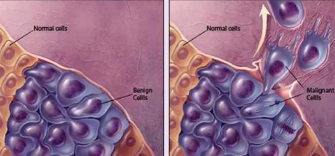
Brain tumors are basically classified into benign brain tumors and malignant brain tumors. Benign tumors are not risky and noncancerous. They do not spread to the near tissues, and are not aggressive. The exact cause for the benign tumor is unpredictable. However genetic history, exposure to radiations, continuous contact with hazardous chemicals like vinyl chloride or formaldehyde are few reasons for the cause.
Fig. 1 Example of the benign tumor cells and malignant tumor cells
Malignant brain tumors are highly risky, cancerous and aggressive. They spread to the nearby tissues and affects the central nervous system very fast. It is complicated to identify the brain tumor. Medical imaging techniques are used to determine whether the tumor is benign, or malignant. Computed tomography (CT), Magnetic Resonance Imaging (MRI) and Positron Emission Tomography (PET) are the most commonly used imaging tests to diagnose brain tumors.
IV.LITERATURE REVIEW
Quratul Ain [1] has proposed a system with multiple phases which classifies and segments the brain tumor. In this Genetic algorithm(GA) is used for weights optimization. Based on natural selection, it solves the optimization problems. Initially, preprocessing of images involves noise removal technique for removing noise. After this, Features are extracted and they are classified. Classification is the phase in which normal and tumorous images are classified. Finally segmentation is involved to extract the tumor portion separately.
© 2016, IJCSMC All Rights Reserved
740
using features which differentiate the subclass. Similar images are identified by its texture, shape, location and the grey level features of the images. An indexing technique is used which is developed from PCA, KD-tree and modified K-means clustering.
P. Rajendran [9] used pruned association rule with MARI algorithm for classification of brain tumor images into normal, benign and malignant tumor. He describes the importance of data cleaning phase for image classification that deals with the accurate data mining architecture. He extracts the features from the transactional database and finds the association between the items using Association Rule Mining.
Saima Anwar Lashari [5] says that the main objective of the image classification is to attain good accuracy as well as to find the parts that are affected by the disease to help in diagnosis. Coincidence matrix or contingency table is the major source of classification to validate the performance of the classification methods. Texture classification is used to obtain the spectral properties of an image, k-NN pattern classification can handle binary and continuous attributes.
TABLE 1
Comparison Between Image Mining Techniques Used for Brain Tumor Analysis
No. Title of Paper Year Author(s) Datasets Name of Algorithm
Overview Limitation Advantages
1 Fuzzy anisotropic diffusion based segmentation and texture based ensemble
classification of brain tumor
2014 Quratul Ain, M. Arfan Jaffar, Tae-Sun Choi
Holy Family hospital and abrar MRI &
CT Scan
center Rawalpindi Genetic Algorithm Phase1: Classifying MR Images into normal and tumorous Image Phase2: Feature Extraction Phase3: Image segmentation
Works perfectly with local dataset. But it is questionabl e with new datasets
Specificity and sensitivity is very accurate and good
2 An intelligent content based image retrieval system for clinical decision support in
brain tumor
diagnosis
2013 Megha P. Arakeri, G. Ram Mohana Reddy
1.5-T MRI clinical scanner at Shridi Cancer Hospital, Manipal, India K-means clustering algorithm, PCA, KD-tree. 1.Database building (offline phase) 2.Query processing (online phase) Inbound classificatio n is not clear
The semantic gap between high level feature and low-level feature is reduced
3 Study of
techniques used for medical image segmentation and computation of statistical test for region
classification of Brain MRI
2013 Anamika Ahirwar
Keith databse Neuro Fuzzy algorithm
Classification of brain MRI through Self Organizing map (SOM) and neuro fuzzy scheme
Only axial view images of MRI is tested
Proposed system automatically classifies the WM, GM, CSF
and tumor
© 2016, IJCSMC All Rights Reserved
741
4 Medical Image
Feature Extraction, Selection And Classification
2010 M. Vasantha, Dr. V. Subbiah Bharathi, R. Dhamodh aran BRATS Dataset Greedy stepwise method & Genetic Algorithm Features Extracted: 1.Intensity Histogram Features 2.Gray Level Co-occurrence Matrix(GLCM)
Compared only with few classifiers
Hybrid approach helps to reduce features
5 An Improved
Image Mining Technique for
Brain Tumor
Classification Using Efficient classifier
2009 P.Rajendr an, M. Madhesw aran
Pandima CT scan centre, Dindigul MARI Algorithm Phase1: Training Phase-Data Cleaning & Feature Extraction Phase2: Test Phase-Images are tested for diagnosis
Consistent result is under question for several combined collection of images
Selects most relevant features during mining process
V. CONCLUSION
In this paper, comparison between many proposed techniques in image mining which is used to diagnose brain tumor has been presented. This survey has recorded the advantages, and limitations of few techniques and still few more systems are needed to identify.
New techniques are still emerging and many areas are left for the future enhancement. Image mining techniques for the tensor based images which describes the linear relations and perfusion based images which shows the blood flow in vessels are yet to be developed.
R
EFERENCES[1] Quratul Ain, M. Arfan Jaffar, Tae-Sun Choi, “Fuzzy anisotropic diffusion based segmentation and texture based ensemble
classification of brain tumor”, Elsevier Science, (2014).
[2] Gaurav Mandloi, “A Survey on Feature Extraction Techniques for Color Images”, International Journal of Computer Science and Information Techniques, Vol 5 (3), 2014, 4615-4620.
[3] Megha P. Arakeri, G. Ram Mohana Reddy, “An intelligent content based image retrieval system for clinical decision
support in brain tumor diagnosis”, Springer (2013).
[4] Anamika Ahirwar, “Study of techniques used for medical image segmentation and computation of statistical test for region
classification of Brain MRI”, I. J Information Technology and Computer Science, MECS (2013)
[5] Saima Anwar Lashari, Rosziati Ibrahim,”A framework for medical image classification using soft set” Elsevier, SciVerse ScienceDirect, 2013.
[6] A. Hema, E.Annasaro, “A Survey in need of Image Mining Techniques”, Imternational Journal of Advanced Research in Computer and Communication Engineering, Vol. 2, Issue 2, Feb 2013.
[7] Dong ping Tian, “A Review on Image Feature Extraction and Representation Techniques”, International Journal of Multimedia and Ubiquitous Engineering, Vol. 8, No 4, July, 2013
© 2016, IJCSMC All Rights Reserved
742
[9] P.Rajendran, M. Madheswaran, “An Improved Image Mining Technique for Brain Tumor Classification Using Efficient
classifier”, (IJCSIS) International Journal of computer and Information Security, Vol 6 No.3, 2009.
[10] Ryszard S. Choras, “Image Feature Extraction Techniques and Their Application for CBIR and Biometrics System”, International Journal of Biology And Biomedical Engineering, Issue 1, Vol 1, 2007.