Brain Tumor Segmentation Using K-Means
Clustering and Fuzzy C-Means Algorithms
and Its Area Calculation and Disease
Prediction Using Naive-Bayes Algorithm
Sonali Mukund Chore, Kalpana Malpe
M. E Student, Department of Computer Science Engineering, Guru Nanak Institute of Engineering and Technology,
Nagpur, India
Professor, Department of Computer Science Engineering, Guru Nanak Institute of Engineering and Technology,
Nagpur, India
ABSTRACT: This paper shows the implementation of Simple Algorithm for detection of range and shape of tumor in
brain MR images and predicts the disease risk details from the given area of tumor. Tumor is an uncontrolled growth of tissues in any part of the human body. Tumors are of various types and they have various characteristics and different treatment. A brain tumor is inherently serious and life-threatening because of its character in the limited space of the intracranial cavity (space formed inside the skull). In developed countries most researchshows that the numbers of peoples who have brain tumors were died due to the fact of inaccurate detection. Generally, a CT scan or MRI that is conducted into intracranial cavity creates a complete image of brain. Researching a lot statistical search which is based on those people whose are affected in brain tumor some general Risk factors and Symptoms have been found. The development of technology in science day night tries to develop new techniques of treatment. This image is visually checked by the physician for detection & diagnosis of brain tumor. However this technique accurate shows the accurate of stage & size of tumor and also predicts the disease details from the area of tumor. This work uses segmentation of brain tumor based on the k-means and fuzzy c-means algorithms. This method allows the segmentation of tumor tissue with accuracy comparable to manual segmentation. In addition, it also decrease the time for analysis and predicts the disease details from the given area of tumor.
Finally implement a system using java to predict Brain tumor risk level which is easier, cost reducible and time savable.
KEYWORDS: Abnormalities, Magnetic Resonance Imaging (MRI), Brain tumor, Pre-processing, K-means, fuzzy
c-means, Thresholding, Naive-Bayesclassification.
I. INTRODUCTION
This paper shows the concept for brain tumor segmentation and finally the detection of brain tumor and risk of disease. The anatomy of the Brain can be explored by the MRI scan or CT scan. In this paper the MRI scanned image is taken for the whole process. The MRI scan is more convenient than CT scan for diagnosis. It is not affect the human body because it doesn't use any dissemination. It is located on the magnetic field and radio waves.
it is detected at current stage. That will increase the lifetime about 1 to 2 years of person. Normally tumor cells are of two types. Those are Mass and Malignant. The detection of the malignant tumor is difficult to mass tumor. In this work we focused on detection of brain tumor with the help of Brain MRI images and predict the disease details from the given area of tumor.
Treatment for brain tumor is depends on the type and stage of the disease, the size and place of the tumor, and your general medical history and health history. In most cases, the aim of treatment is to remove or destroy the tumor completely. Most brain tumor can be cured if found and treated early.
A person who was affected by any kind of tumor has an increased risk of developing another brain tumor of any type. A person who has two or more close relatives (mother, father, sister, brother, or child) who are responsible for developing brain tumor has a risk factor of developing brain tumor for his own. Rarely, members of a family will have an inherited disorder that makes the brain more sensitive and increases the risk of brain tumor. About 5% of brain tumors may be linked to hereditary (genetic) factors or conditions.
Another risk factor of Brain tumor as well as other diseases is taking any camo therapy. A person who has taken any therapy is responsible for occurring different kinds of disease compare to other people who don’t take any therapy. Day by day the number of brain tumor person is increasing rapidly because of unconsciousness. The Objective of this work is to contract such a tool which can tell people about his/her approximate condition about brain tumor ,that is he or she in risk or not and how much?
The developing platform for the detection is java. At the end, we are providing systems that detect the tumor and its shape and disease details from the given area of tumor.
II. LITERATURE SURVEY
1. Author and Title: Samir Kumar Bandhyopadhyay, Tuhin Utsab Paul, “Automatic Segmentation of Brain Tumor
fromMultiple Images of Brain MRI”.
ImplementedConcept: This paper has proposed a system of image registration and data fusion theory suitable for the
segmentation of MR images. This system provides an efficient and fast way for diagnosis of the brain tumor called K-means technique.
2. Author and Title: A. Meena, K. Raja,” Spatial Fuzzy C-Means PET Image Segmentation of Neurodegenerative
Disorder”
ImplementedConcept: This paperproposed an approach of Spatial Fuzzy C means (PET-SFCM) clustering technique
on Positron Emission Tomography (PET) scan image datasets.
3. Author and Title: Suman Tatiraju, Avi Mehta, “Image Segmentation using k-means clustering, EM and
Normalized Cuts”
ImplementedConcept: In this system, we look at three techniques namely K Means clustering, Expectation
Maximization and the Normalized cuts and compare them for image segmentation.
4. Author and Title: Ajala Funmilola,” Fuzzy k-c-means Clustering Algorithm for Medical ImageSegmentation”
ImplementedConcept:This workproposed the Fuzzy K-C-means technique, which carries more of Fuzzy C-means
properties than that of K-means.
5. Author and Title:Beshiba Wilson, Julia Punitha Malar Dhas,”An Experimental Analysis of Fuzzy C-Means and
K-Means Segmentation Algorithm for Iron Detection in Brain SWI using Matlab”
ImplementedConcept:Wilson and Dhas used K-means and Fuzzy C-means techniques respectively to detect the iron
6. Author and Title:M.H. Fazel Zarandia, “Systematic image processing for diagnosing brain tumors: A Type-II fuzzy expert system”
ImplementedConcept:This paper proposed a dip work of brain tumor. It shows different type of diagnosis approaches.
7. Author and Title:Samarjit Das,” Systematic image processing for diagnosing brain tumors: A Type-II fuzzy
expert system approach”
ImplementedConcept:In pattern recognition due to the fundamental involvement of human perception and inadequacy
of standard Mathematics to deal with its complex and incoherently defined system; different fuzzy techniques have been applied as an appropriate alternative.
8. Author and Title:Vignesh Rajesh,” brain tumor segmentation and its area calculation in brain MRI images
using k-mean clustering and fuzzy c mean algorithm”
ImplementedConcept:This paper suggested a synergistic and an effective technique for the detection of brain tumors
based on Median filtering, K Means Segmentation, FCM Segmentation, and finally, threshold segmentation.
9. Author and Title:Krishna Kant Singh,” A Study of Image Segmentation Algorithms ForDifferent Types of
Images”
ImplementedConcept:In this paper the author gives a study of the various techniques that are available for color
images, text and gray scale images.
10. Author and Title:payal mistry, shagun akhauri, sayali patil, s.p.tondare, ”Segmentation of brain tumor and its
area calculation in brain MRI images using k-mean clustering and fuzzy c- mean algorithm”
ImplementedConcept:In this paper proposed k-means and C-mean techniques to extract the features from the images.
III. PROPOSED SYSTEM ARCHITECTURE
1. Pre-processing
According to the need of the next level in pre-processing step, performs filtering of noise and other artifacts in the image and sharpening the edges in the image. RGB image to grey image conversion and Reshaping of image also takes place here. It includes median filter for removing noise of image. The possibilities of arrival of noise in modern MRI scan imageare very less. Noise may arrive due to the thermal effect. The main goal of this paper is to detect and segment the tumor cells. But for the complete system it needs the process of noise removal of image.
2. Segmentation using K-means
Steps:
1. Give the no of cluster value as k. 2. Randomly choose the k cluster centers 3. Calculate mean or center of the cluster
4. Calculate the distance b/w each pixel to each cluster center
5. If the distance is near to the cluster center then move to that cluster. 6. Otherwise move pixel to next cluster.
7. Re-estimate the cluster center.
8. Repeat the process until the center doesn't move.
3. Segmentation using Fuzzy C means
The fuzzy logic is a way to processing the data by giving the fractional membership value to each pixel in the image.
The membership value of the fuzzy set is ranges from 0 to 1.
Fuzzy clustering is basically a multi valued logic that allows intermediate values i.e., member of one fuzzy set can also be member of other fuzzy sets in the same image.There is no direct transition between full membership and non-membership.
The membership function defines the fuzziness of an image and also to define the information contained in the image.
4. Approximate reasoning
In the approximate reasoning step the tumor area is calculated using the binarization/thresholding method. And also identify the stage of tumor. That is the output image having only two values either black or white (0 or 1).
So,
Calculate the area of tumor=
Area= √P * 0.264
Where, p= total no of white pixels of threshold image.
And 0.264 is the 1 pixel size.
5. Naive-BayesClassification:
IV. MATHEMATICAL MODEL
Mathematical equation in K-means clustering
1. =∑: ( )
, k=1, 2,……, K.
2. D (i) = arg min||Xi- Mk||2, i=1, 2,….., N.
Mathematical equation in Fuzzy-C means clustering
Ym =∑ ∑ ijm||Xi – Cj ||2
Where,
m= any real number greater than 1,
Mij= degree of membership of X; in the cluster j,
Xi= data measured in d-dimensional,
Rj= d-dimension center of the cluster,
The update of membership Mij and the cluster centers R are given by:
Mij =
∑ || ||
|| ||
Rj=
∑ .
∑
Let consider S is a system for Brain Tumor Segmentation Using K-Means clustering and Fuzzy C-Means techniques and Its Area Calculation and Disease Prediction Using Naive-Bayestechnique.
S= {….}
INPUT:
Identify the inputs
F= {f1, f2, f3 ..., fn| ‘F’ as set of functions to execute commands.}
I= {i1, i2, i3…|’I’ sets of inputs to the function set}
O= {o1, o2, o3….|’O’ Set of outputs from the function sets}
S= {I, F, O}
I = {Query submitted by the user}
O = {Output of desired query}
F = {Functions implemented to get the output,
Fuzzy C –means clustering, Naive-BayesAlgorithm}
SPACE COMPLEXITY
The space complexity depends on Presentation and visualization of discovered patterns. More the storage of data more is the space complexity.
TIME COMPLEXITY
Check No. of patterns available in the datasets= n
If (n>1) then retrieving of information can be time consuming.
So the time complexity of this algorithm is O( ).
= Failures and Success conditions. Failures:
Huge database can lead to more time consumption to get the information.
Hardware failure.
Software failure.
Success:
Search the required information from available in Datasets. User gets result very fast according to their needs.
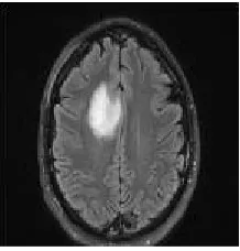
V. RESULT
Let us consider the brain tumor image procured from MRI, containing the tumor in figure 1. Median filtering is implemented on the acquired images to get rid of the unwanted noises. The outcomes are displayed in the figure 2.
Fig. 2 Median Filtering Outcome
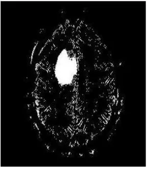
K means algorithm is implemented on such noise filtered images containing brain tumors. In figure 5, a white spot is seen in image, which isfinal output of application of threshold segmentation on the input image. This region is the area having higher intensity values compared to the defined threshold. Areas with higher intensity values mostly contains ulcer. The outcomes of thresholding segmentation are shown below in figure 5.
Fig. 3 K Means Clustering
Fig. 4 FCM Segmentation
Eventually, the thresholding segmentation is implemented, once the FCM segmentation is completed. The outcomes are spectacular and proposed approach is efficacious in nature to an extent. Figure 5 shows the resultant image obtainsafter the implementation of thresholding segmentation.
Fig. 5 Thresholding Segmentation
Fig. 6 Output Images Of Tumor Area Estimation
The proposed work is also very sensitive to the errors, because the small error will take the situation in ambiguous state which is not good for diagnosis of tumor. Again same FCM mean and k means techniques are use to compare individual performance with the proposed system and the result of all are compare and we find that the proposed system having less errors in the system.
VI. CONCLUSION
There are various types of tumors are available.They may be as mass or malignant over thebrain. Suppose if it is a mass then K- means technique is enough to extract it from the brain cells.If there is any noise are present in the MR scan image it is removed before the K-means process. The noise freeimage is given as an input to the k-means technique and tumoris extracted from the MRI scan image. Thenusing Fuzzy C means techniquesegmentation for accurate tumor shape extraction froman image. Finallyapproximate reasoning step for calculating tumor shape and position calculation and finally using the navy bias classification technique to classify the disease risk from resultant area of tumor. I.e. predict Brain tumor risk level which is easier, cost reducible and time savable.
The experimental results arecompared with other techniques so the proposedmethod gives more accurate result.
REFERENCES
[1] J.selvakumar, A.Lakshmi and T.Arivoli, “Brain Tumor Segmentation and Its Area
Calculation in Brain MR Images using K-Mean Clustering and Fuzzy C-Mean Algorithm”, IEEE-International Conference On Advances In Engineering, Science And Management (ICAESM -2012) March 30, 31, 2012.
[2] Samir Kumar Bandhyopadhyay and Tuhin Utsab Paul, “Automatic Segmentation of Brain Tumor from Multiple Images of Brain MRI” International Journal of Application or Innovation in Engineering & Management (IJAIEM),Volume 2, Issue 1, January 2013.
[3] A. Meena, “Spatial Fuzzy C-Means PET Image Segmentation of Neurodegenerative Disorder” , A. Meena et.al / Indian Journal of Computer Science and Engineering (IJCSE).
[4] Suman Tatirajua and Avi Mehta, “Image Segmentation using k-means clustering,EM and Normalized Cuts” IEEE Trans. Parallel Diatribe. Syst., vol. 19, no. 5, pp. 710–720, May 2008.
[5] Ajala Funmilola A*, Oke O.A, Adedeji T.O and Alade O.M, Adewusi E.A, “Fuzzy k-c-means Clustering Algorithm for Medical Image Segmentation”, Journal of Information Engineering and Applications ISSN 2224-5782 (print) ISSN 2225-0506 (online)Vol 2, No.6, 2012.
[6] M.H. Fazel Zarandia, M. Zarinbal and M. Izadi, “Systematic image processing for diagnosing brain tumors”, Department of Industrial
Engineering, Amirkabir University of Technology, P.O. Box 15875-4413, Tehran, Iran , journal homepage:www.elsevier.com/locate/asoc
[7] Samarjit Das, “Pattern Recognition using the Fuzzy c-means Technique” International Journal of Energy, Information and Communications Vol. 4, Issue 1, February, 2013.
[8] Vignesh Rajesh, Bharathan Venkat, Vikesh Karan and M. Poonkodi, “Brain Tumor Segmentation and its Area Calculation in Brain MR Images Using K-Mean Clustering andFuzzy C-Mean Algorithm”, Department of Computer Science and Engineering, SRM University.
[9] Krishna Kant Singh1 and Akansha Singh, “A Study of Image Segmentation Algorithms forDifferent Types of Images”, IJCSI International Journal of Computer Science Issues, Vol. 7, Issue 5, September 2010.