ABSTRACT
JACOBSEN, THOMAS. Developing Tools and Techniques for Expanding the Synthetic Biology Toolkit (Under the direction of Dr. Chase L. Beisel and Dr. Gregory T. Reeves).
The field of synthetic biology has revolutionized biological research by making biology more predictable to engineer. By combining biology with fundamental engineering principles, synthetic biology has provided a platform to solve various challenges faced in society today. Synthetic biology has advanced due to the availability of gene regulatory tools. While these tools have been developed for eukaryotic systems, their development has been outpaced compared to unicellular systems. This thesis helps expand upon the tools and techniques available for synthetic biology, with a focus on eukaryotic systems.
First, a set of self-cleaving ribozymes, tools previously used to silence gene expression, was engineered to modulate gene expression. These ribozymes were flanked with upstream competing sequences designed to alter the ribozyme’s secondary structure, thus potentially resulting in complete or partial inability of the ribozyme to self-cleave. As a proof-of-concept, these tools were implemented in two eukaryotic systems: HEK293T cells and Drosophila embryos. In both systems, these ribozymes modulated gene expression, with each competing sequence associated with varying extents of gene repression. Additionally, we correlated the empirical data with RNA folding algorithms and observed a lack of correlation. While this tool is currently not predictive, a set of ribozymes to modulate gene expression in various model systems are now available.
Using this TXTL PAM screen, we discovered unreported non-canonical PAMs of the previously characterized Acidaminococcus sp. Cas12a (AsCas12a, also known as AsCpf1). While AsCas12a is associated with the canonical TTTV (V = A/C/G) PAM, this nuclease also recognizes CTTV, TCTV, and TTCV as non-canonical PAMs. The PAM screen revealed AsCas12a’s ability to recognize GTTV and GCTV as non-canonical PAMs, while plasmid clearance in E. coli, DNA cleavage in TXTL, and indel formation in mammalian cells validated recognition of these PAMs. While this finding increases the targeting range of AsCas12a, it also increases the number of potential off-target sites within a genome.
In a separate study, the TXTL PAM screen was used to characterize the PAMs of previously unreported Cas12a nucleases, with Fn3Cas12a and PiCas12a sharing high sequence homology with the previously characterized FnCas12a and PdCas12a, respectively. While the characterized nucleases recognized T-rich PAMs commonly associated with Cas12a nucleases, PiCas12a recognized G-rich motifs. Mutating residues within PiCas12a transitioning PiCas12a to PdCas12a altered its PAM profile, thus increasing the targeting range of PiCas12a. These findings were validated using DNA cleavage in TXTL and indel formation in mammalian cells. Due to sequence homology between PiCas12a and PdCas12a, as well as the PAM flexibility of PiCas12a and its variants, PiCas12a provides a platform for CRISPR evolutionary studies and expands upon the CRISPR toolkit.
Developing Tools and Techniques for Expanding the Synthetic Biology Toolkit
by
Thomas Jacobsen
A thesis submitted to the Graduate Faculty of North Carolina State University
in partial fulfillment of the requirements for the degree of
Doctor of Philosophy
Chemical and Biomolecular Engineering
Raleigh, North Carolina
2019
APPROVED BY:
Dr. Balaji Rao Dr. Jeffrey Yoder
Dr. Chase Beisel Dr. Gregory Reeves
DEDICATION
BIOGRAPHY
ACKNOWLEDGEMENTS
First, I would like to thank my PhD advisors, Dr. Chase Beisel and Dr. Greg Reeves, for all of their support throughout my graduate career. Their patience and encouragement throughout the years have been invaluable during my time here.
Also, I thank two former graduate students, Dr. Ashley Jermusyk and Dr. Michelle Luo, that I had the honor of learning under during the early years of my graduate career. Thank you for helping me acquire the skills and techniques I needed to become successful.
Moreover, I thank all of my colleagues, collaborators, and fellow Beisel and Reeves lab members for their technical guidance and expertise. I would especially like to thank Dr. Colin Maxwell, Dr. Chunyu Liao, Hadel Al Asafen, Gloria Yi, and collaborators from Benson Hill Biosystems for their significant contributions to my projects.
Furthermore, I would like to thank my family and close friends. They have loved me, supported me, and have always lent an ear whenever I needed it.
TABLE OF CONTENTS
List of Tables ... x
List of Figures ... xi
List of Documents ... xiii
CHAPTER 1: An overview of gene regulatory tools in Drosophila ... 1
1.1 - INTRODUCTION ... 2
1.2 - GENE REGULATORY TOOLS DEVELOPED IN DROSOPHILA ... 2
1.1.1 - Promoter swapping ... 2
1.1.2 - Orthogonal transcription factors ... 4
1.1.3 - Tetracycline-inducible systems ... 5
1.1.4 - RNAi ... 5
1.1.5 - CRISPR-Cas9/12a ... 6
1.3 - POTENTIAL TOOLS TO REGULATE GENE EXPRESSION IN DROSOPHILA ... 8
1.2.1 - Short upstream open reading frames (uORFs) ... 8
1.2.2 - Self-cleaving ribozymes ... 8
1.2.3 - CRISPR-Cas13a ... 8
1.4 - CONCLUSIONS ... 9
1.5 - REFERENCES ... 10
CHAPTER 2: Tunable self-cleaving ribozymes for modulating gene expression in eukaryotic systems ... 17
ABSTRACT ... 18
2.1 - INTRODUCTION ... 19
2.2 - MATERIALS AND METHODS ... 21
2.2.1 - Strains, plasmids, oligonucleotides, and fly lines. ... 21
2.2.3 - Transient transfections of pcDNA3.1(+)-ribozyme constructs. ... 22
2.2.4 - Flow cytometry analysis of transfected HEK293T cells. ... 23
2.2.5 - Fluorescent in situ hybridization of Drosophila embryos. ... 23
2.2.6 - Imaging and analysis of embryos. ... 23
2.3 - RESULTS ... 24
2.3.1 - Designing self-cleaving ribozymes containing tunable upstream competing sequences. ... 24
2.3.2 - Self-cleaving ribozymes combined with upstream competing sequences can tune gene expression in mammalian cells. ... 26
2.3.3 - Self-cleaving ribozymes/upstream competing sequences can tune gene expression in Drosophila. ... 27
2.4 - DISCUSSION ... 30
2.5 - CONCLUSIONS ... 32
2.6 - ACKNOWLEDGMENTS ... 32
2.7 - REFERENCES ... 33
CHAPTER 3: A detailed cell-free transcription-translation (TXTL)-based assay to decipher CRISPR protospacer-adjacent motifs ... 38
ABSTRACT ... 39
3.1 - INTRODUCTION ... 40
3.1.1 - Previous methods to characterize PAMs of CRISPR-Cas systems ... 41
3.1.2 - TXTL-based PAM determination ... 43
3.2 - MATERIALS AND METHODS ... 45
3.2.1 - Creating DNA required for PAM assay ... 45
3.2.2 - PAM library cleavage in TXTL ... 52
3.2.3 - Assessing cleavage of the PAM library ... 55
3.2.4 - Troubleshooting Cas nuclease cleavage in TXTL ... 56
3.2.5 - NGS library preparation ... 57
3.3 - RESULTS AND DISCUSSION ... 61
3.4 - CONCLUSIONS ... 63
3.4 - ACKNOWLEDGMENTS ... 63
3.5 - REFERENCES ... 64
CHAPTER 4: The Acidaminococcus sp. Cas12a nuclease recognizes GTTV and GCTV as non-canonical PAMs ... 70
ABSTRACT ... 71
4.1 - INTRODUCTION ... 72
4.2 - MATERIALS AND METHODS ... 74
4.2.1 - Strains, plasmids, and oligonucleotides ... 74
4.2.2 - TXTL-based PAM screen and DNA cleavage assay ... 74
4.2.3 - Plasmid clearance assay in E. coli ... 75
4.2.4 - Indel formation in DNMT1 ... 75
4.2.5 - Tracking of Indels by Decomposition (TIDE) analysis ... 76
4.3 - RESULTS ... 77
4.3.1 - PAM screen of AsCas12a reveals non-canonical motifs ... 77
4.3.2 - AsCas12a can recognize the GYTV motif in vitro ... 78
4.3.3 - The -5 PAM position influences target recognition in E coli ... 80
4.3.4 - Indel formation can be achieved with AsCas12a using GYTV PAMs ... 81
4.4 - DISCUSSION ... 82
4.5 - ACKNOWLEDGEMENTS ... 83
4.6 - REFERENCES ... 84
CHAPTER 5: Characterization of Cas12a nucleases reveals diverse PAM profiles between closely-related orthologs ... 89
ABSTRACT ... 90
5.1 - INTRODUCTION ... 91
5.2.1 - Strains, plasmids, and oligonucleotides ... 93
5.2.2 - DNA cleavage assay using a cell-free transcription-translation (TXTL) system ... 93
5.2.3 - TXTL-based PAM screen ... 94
5.2.4 - Indel formation in HEK293T cells ... 94
5.2.5 - Tracking of Indels by Decomposition (TIDE) analysis ... 95
5.2.6 - Statistical analyses ... 95
5.3 - RESULTS ... 95
5.3.1 - A phylogenetically diverse set of Cas12a nucleases exhibit ranging effective activities in TXTL ... 95
5.3.2 - The Cas12a nucleases can process and utilize each other’s gRNAs ... 98
5.3.3 - PAM determination reveals distinct recognition profiles ... 99
5.3.4 - Variable bias against T at the -1 PAM position confirmed by TXTL ... 100
5.3.5 - HkCas12a recognizes C-rich PAMs in TXTL and in human cells ... 102
5.3.6 - Mutating PiCas12a toward PdCas12a reveals distinct PAM profiles in TXTL ... 103
5.3.7 - PiCas12a and the F604Y variant recognize distinct non-canonical PAMs in human cells ... 106
5.4 - DISCUSSION ... 106
5.5 - ACKNOWLEDGEMENTS ... 109
5.6 - REFERENCES ... 110
CHAPTER 6: Conclusions and future work ... 115
6.1 - CONCLUSIONS ... 116
6.2 - PREDICTABLE TUNING OF GENE EXPRESSION ... 116
6.3 - FURTHER INCREASING PAM SPECIFICITY OF PICAS12A ... 117
6.4 - LIVE RNA IMAGING IN DROSOPHILA ... 117
6.5 - REFERENCES ... 120
Appendix A - Chapter 2 Supplementary Material ... 123
Appendix B - Chapter 3 Supplementary Material ... 132
Appendix C - Chapter 4 Supplementary Material ... 134
LIST OF TABLES
Table 3.1 - Thermocycler program used to create Chi6 DNA ... 46
Table 3.2 - Components and program used for PCR amplification ... 50
Table 3.3 - Recipe for TXTL master mix for PAM determination assay ... 52
Table 3.4 - An example set of reactions to characterize the CRISPR PAMs ... 52
Table 3.5 - PCR reaction setup of the PAM library ... 59
Supplementary Table 2.1 - DNA constructs and fly lines used in Chapter 2 ... 123
Supplementary Table 2.2 - Transfection conditions for ribozyme constructs in HEK293T cells ... 127
Supplementary Table 3.1 - DNA constructs used in Chapter 3 ... 132
Supplementary Table 4.1 - DNA constructs used in Chapter 4 ... 134
Supplementary Table 4.2 - TXTL reaction setup for the PAM screen and DNA cleavage assay ... 136
Supplementary Table 4.3 - List of all target sequences and their associated PAMs used in TXTL and mammalian cell-based assays ... 137
Supplementary Table 4.4 - List of all potential 5N PAM sequences and depletion values from AsCas12a ... 138
Supplementary Table 5.1 - DNA constructs used in Chapter 5 ... 142
LIST OF FIGURES
Figure 1.1 - The current toolbox of gene regulatory tools adapted in Drosophila ... 3
Figure 1.2 - Potential tools to regulate gene expression in Drosophila ... 8
Figure 2.1 - Modulating gene expression using self-cleaving ribozymes ... 20
Figure 2.2 - Self-cleaving ribozymes tunes gene expression in HEK293T cells ... 25
Figure 2.3 - Self-cleaving ribozymes regulates gene expression in Drosophila ... 28
Figure 3.1 - An overview of TXTL and its application for PAM determination ... 44
Figure 3.2 - Data analysis for assessing cleavage by a Cas nuclease in TXTL ... 55
Figure 3.3 - Data analysis for the TXTL PAM screen performed on the Cas9 from Neisseria meningitides ... 61
Figure 4.1 - AsCas12a PAM screen uncovers non-canonical GYTV motifs ... 76
Figure 4.2 - AsCas12a can recognize the GYTV motif in TXTL ... 78
Figure 4.3 - AsCas12a can recognize the GYTV motifs in vivo ... 79
Figure 5.1 - Phylogenetically diverse Cas12a nucleases exhibit varying cleavage efficiency and process/utilize each other’s gRNAs ... 96
Figure 5.2 - PAM determination screen of diverse Cas12a nucleases ... 99
Figure 5.3 - Diverse Cas12a nucleases recognize TTTV motifs ... 101
Figure 5.4 - HkCas12a recognizes C-rich PAM sequences ... 101
Figure 5.5 - Mutating residues in PiCas12a towards PdCas12a alters PAM its PAM profile ... 103
Figure 5.6 - PiCas12a and the F604Y variant form indels in HEK293T cells ... 105
Figure 6.1 - Tagging aptamers onto sgRNA for live DNA imaging ... 118
Supplementary Figure 2.2 - Histograms of flow cytometry data from HEK293T
transfection experiments ... 129
Supplementary Figure 2.3 - Representative embryos labeled with lacZ width
associated with symmetric and asymmetric gradients . 130
Supplementary Figure 4.1 - Time-series of GFP expression from the TXTL-based PAM screen ... 139
Supplementary Figure 4.2 - Images of the plates containing the colonies from the plasmid clearance assays in E. coli ... 140
Supplementary Figure5.1 - Diverse Cas12a nucleases can process/utilize each other’s gRNA ... 147
Supplementary Figure 5.2 - PAM profiles do not trend with phylogeny ... 148
Supplementary Figure 5.3 - PiCas12a and its variants cannot recognize GGYC or GTGC motifs in TXTL ... 149
Supplementary Figure 5.4 - PiCas12a and its variants recognize various PAM
LIST OF DOCUMENTS
Supplementary Document 2.1 - In-depth protocol for measuring fluroescence of
Drosophila embryos ... 131
Supplementary Document 5.1 - Protein sequence alingment of various Cas12a
1.1 - INTRODUCTION
Synthetic biology aims to make biology more predictable and easier to understand by conceptualizing biological systems as a network comprised of interactions between various genetic components. Synthetic biologists modify, rewire, and/or engineer natural biological molecules and systems to produce novel products or components that have the potential to significantly impact various fields. Advancements in synthetic biology has been possible due to the wide array of genetic tools that are capable of regulating gene expression at various stages of its expression [1–4]. Subsequently, the development of these tools has led to the construction of various synthetic networks that were built upon fundamental engineering principles [5–7]. While a plethora of gene regulatory tools have been constructed in unicellular systems (i.e. bacteria and yeast), construction of these tools in more complex systems, such insect systems, have lagged behind.
In this chapter, the current technologies available to regulate gene expression in the model system Drosophila are discussed. This chapter serves as an addition to the introduction in Chapter 2, as the chapter lacked a discussion about gene regulatory tools in Drosophila although data associated with Drosophila were presented. These technologies include promoter swapping, orthogonal transcription factors, tetracycline-inducible systems, RNA interference (RNAi), and Clustered Regularly Interspaced Short Palindromic Repeats (CRISPR) technologies. Also described are potential genetic tools that have been used in other eukaryotic model systems and a brief discussion on their potential as gene regulatory tools in Drosophila. Finally, an outline of the following chapters in this thesis is provided.
1.2 - GENE REGULATORY TOOLS DEVELOPED IN DROSOPHILA
1.1.1 - Promoter swapping
other synthetic promoters available in the synthetic biology toolkit or endogenous promoters (Figure 1.1A). While swapping promoters offers the user the ability to either increase or decrease gene expression, it does not allow for precise control of expression or the ability to further reduce or amplify expression levels beyond the available promoters in Drosophila.
1.1.2 - Orthogonal transcription factors
The Gal4-upstream activating sequence (UAS) system has been the primary method of regulating gene expression in Drosophila. Since its discovery as a viable genetic tool in Drosophila, thousands of fly lines have been developed containing this system (see https://bdsc.indiana.edu/stocks/uas/uas_nonrnai.html). The Gal4-UAS system involves the yeast Gal4 transcriptional activator binding to a consensus UAS site, which then allows for strong expression of the downstream gene of interest (Figure 1.1B). [9–11]. To offer more flexibility to this system, repressor Gal80 can be expressed to directly bind to Gal4 and prevent transcriptional activation [12–14], and Gal80 can be repressed by expression of Gal3 [15].
Similar to the Gal4-UAS system, other orthogonal transcription factors have been adapted in Drosophila. One of these include the LexA-lexA operon (lexAop) system [16]. Similar to the Gal4-UAS system, LexA binds specifically to consensus lexAop sites. However, unlike Gal4, LexA does not have the ability to drive gene expression after binding. Thus, to use this system for activating gene expression, the activation domain of Gal4 or VP16 must be tagged to LexA [17].
The most recent orthogonal transcription factor adapted in Drosophila is the QF-QUAS (Q) system [18]. Similar to Gal4-UAS and LexA-lexAop, this system involves the transcriptional activator QF binding to a consensus QUAS sequence, which then drives gene expression. Similar to Gal80 in the Gal4-UAS system, QF can be repressed by expressing its inhibitor, QS, while QS can be inhibited through supplementing the flies’ food source with quinic acid [18]. Compared to the LexA-lexAop system, the Q system has the ability to drive gene expression without tagging the transcription factor with activating domains. Also, compared to the Gal4-UAS system, gene expression driven from the QF-QUAS system has shown to be less leaky [18].
expression of two different transgenes (reviewed in [19]), which is useful for building synthetic gene circuits. While these systems allow for more control of gene expression, they require the addition of multiple binding sites and the creation of new fly lines when higher reduction levels of gene expression are needed.
1.1.3 - Tetracycline-inducible systems
The tetracycline-dependent transactivator (tTA) is a binary system that is able to regulate gene expression in various model organisms [20–25]. This system is composed of the bacterial tetracycline repressor (tetR) and the tetracycline response element (TRE) that it binds to (Figure 1.1C) [20]. The tagging of tetR with the VP16 transactivation domain can activate expression of a gene of interest in the absence of tetracycline, while the introduction of the drug inhibits gene expression [26]. To increase the flexibility of this system, random mutagenesis was performed to discover a reverse tetracycline-dependent transactivator (rtTA) that activates gene expression in the presence of tetracycline [25,27].
The tetracycline-inducible systems have allowed for the regulation of gene expression in Drosophila and had been especially useful for temporal-controlled gene expression that was lacking with the orthogonal transcription factor systems until the creation of a temperature-dependent expression of Gal4 [28]. Though tetracycline-inducible systems have allowed for gene regulation in Drosophila, it requires a drug for transactivation, thus introducing another variable for its use. Also, this tool cannot immediately be used for studies involving the early developmental stages due to the requirement of tetracycline consumption.
1.1.4 - RNAi
introduction was through injection embryonic stages [37–39], but since has become more flexible when combined with the Gal4-UAS system for RNAi knockdown [40,41].
RNAi has been successfully used to regulate gene expression in Drosophila [42]. This system is particularly useful when studying gene function in early development of Drosophila, especially when Gal4-UAS lines are not available for specific genes of interest. While RNAi can be used as a tool to regulate gene expression, it has its limitations. First, the potential of off-target effects is an issue, especially when performing genetic screens. Also, as RNAi naturally occurs in Drosophila, introducing synthetic RNAi components to this model system may alter the endogenous biological machinery.
1.1.5 - CRISPR-Cas9/12a
A background of CRISPR-Cas systems can be seen in Chapters 3, 4, and 5. Briefly, CRISPR-Cas systems are adaptive immune systems that protect prokaryotes against mobile genetic elements [43–46]. In Drosophila, these systems have been harnessed as a genetic tool to produce transgenic fly lines containing sequence-specific mutations, insertions, or deletions [47,48]. This involves the expression of Cas9 and a guide RNA (gRNA) that serves to guide Cas9 to a sequence-specific site in the genome specified by the gRNA [45,46,49]. While Cas9 has been mainly used for site-directed mutagenesis in Drosophila, CRISPR interference (CRISPRi) and activation (CRISPRa) has been shown to regulate gene expression using the KRAB and VPR domains, respectively (Figure 1.1E) [50–53].
used in [55] was temperature-sensitive, and using a different variant of Cas12a resulted in greater cleavage activity [56].
The use of CRISPR technologies in Drosophila has allowed for simple and rapid reduction of gene expression through DNA cleavage and repair, resulting in partial or complete loss of gene function. Along with its ability to cleave DNA, CRISPR-Cas9 has shown the ability to form a synthetic transcription factor that can target virtually any locus, thus increasing or decreasing gene expression without altering the genome [50–53]. While this technology has revolutionized genome-editing and gene regulation in various model systems, it may be associated with off-target effects and has shown to reduce fitness of Drosophila [57].
1.3 - POTENTIAL TOOLS TO REGULATE GENE EXPRESSION IN DROSOPHILA
1.2.1 - Short upstream open reading frames (uORFs)
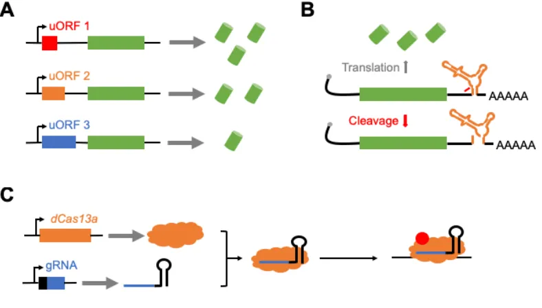
Short uORFS are short sequences containing a start and termination codon (Figure 1.2A) that are occasionally found upstream of coding genes in various eukaryotic systems, including yeast, mice, and humans [58,59]. Studies have shown that synthetic uORFs can be used to regulate gene expression in model systems, and that the strength of reduction of gene expression was based on sequence length and content of the uORF [60,61]. These uORFs have also been discovered in Drosophila [62,63], and has been shown to regulate gene expression [64,65]. While uORFs seem promising as gene regulatory tools, preliminary work from a former graduate student showed results indicating that these tools may have weak regulatory ability in Drosophila.
1.2.2 - Self-cleaving ribozymes
Self-cleaving ribozymes are RNA sequences that contain self-enzymatic activity (reviewed in [66]). If placed in the untranslated regions or an intron of an RNA transcript, previous work has shown that this tool can prevent gene expression through self-cleavage and subsequent degradation (Figure 1.2B) [67–69]. While self-cleaving ribozymes have been used to regulate gene expression in bacteria, yeast, and mammalian cells [67–69], it has yet to be used in Drosophila as a tool to regulate gene expression. Work in Chapter 2 of this thesis provide evidence of self-cleaving ribozymes as viable tool to regulate gene expression in mammalian cells and Drosophila, and potentially in other model systems.
1.2.3 - CRISPR-Cas13a
Using dCas13a in Drosophila and other multicellular systems could potentially lead to a genetic tool that regulates gene expression. While Cas13a has shown to cleave non-specific RNA in bacteria [70,71], mutating non-specific domains has inhibited this effect. However, when this nuclease was implemented in mammalian cells, non-specific RNA cleavage was not observed and showed enhanced RNA targeting specificity compared to RNAi [72]. Thus, this nuclease has the potential to be used as a tool for RNA cleavage a synthetic RNA-binding protein.
1.4 - CONCLUSIONS
1.5 - REFERENCES
[1] L. Guzman, D. Belin, M.J. Carson, J. Beckwith, Tight regulation, modulation, and high-level expression by vectors containing the arabinose PBAD promoter, J Biotechnol. 177 (1995) 4121–4130.
[2] L. Yen, J. Svendsen, J.-S. Lee, J.T. Gray, M. Magnier, T. Baba, R.J. D’Amato, R.C. Mulligan, Exogenous control of mammalian gene expression through modulation of RNA self-cleavage, Nature. 431 (2004) 471–476. doi:10.1038/nature02844.
[3] H.M. Salis, E.A. Mirsky, C.A. Voigt, Automated design of synthetic ribosome binding sites to control protein expression, Nat. Biotechnol. 27 (2009) 946–950.
[4] K.E. McGinness, T.A. Baker, R.T. Sauer, Engineering controllable protein degradation, Mol. Cell. 22 (2006) 701–707.
[5] T.S. Gardner, C.R. Cantor, J.J. Collins, Construction of a genetic toggle switch in Escherichia coli, Nature. 403 (2000) 339–342.
[6] M.B. Elowitz, S. Leibler, A synthetic oscillatory network of transcriptional regulators, Nature. 403 (2000) 335–338.
[7] C.C. Guet, M.B. Elowitz, W. Hsing, S. Leibler, Combinatorial synthesis of genetic networks, Science. 296 (2002) 1466–1470.
[8] J.Y. Qin, L. Zhang, K.L. Clift, I. Hulur, A.P. Xiang, B.Z. Ren, B.T. Lahn, Systematic comparison of constitutive promoters and the doxycycline-inducible promoter, PLoS One. 5 (2010) 3–6.
[9] D.A. Elliott, A.H. Brand, The gal4 system : a versatile system for the expression of genes, Methods Mol Biol. 420 (2008) 79–95.
[10] A.H. Brand, N. Perrimon, Targeted gene expression as a means of altering cell fates and generating dominant phenotypes, Development. 118 (1993) 401–415. [11] S.E. McGuire, G. Roman, R.L. Davis, Gene expression systems in drosophila: a
synthesis of time and space, Trends Genet. 20 (2004) 384–391.
[12] J. Ma, M. Ptashne, The carboxy-terminal 30 amino acids of GAL4 are recognized by GAL80, Cell. 50 (1987) 137–142.
Bicoid mediate its transcriptional downregulation by the Torso pathway, Development. 128 (2001) 2281–2290.
[15] O. Egriboz, S. Goswami, X. Tao, K. Dotts, C. Schaeffer, V. Pilauri, J.E. Hopper, Self-association of the Gal4 inhibitor protein Gal80 is impaired by Gal3: Evidence for a new mechanism in the GAL gene switch, Mol. Cell. Biol. 33 (2013) 3667–3674. [16] S.L. Lai, T. Lee, Genetic mosaic with dual binary transcriptional systems in
Drosophila, Nat. Neurosci. 9 (2006) 703–709.
[17] S.J. Triezenberg, R.C. Kingsbury, S.L. Mcknight, Functional dissection of VP16, the trans-activator of herpes simplex virus immediate early gene expression, Genes Dev. 2 (1988) 718–729.
[18] C.J. Potter, B. Tasic, E. V. Russler, L. Liang, L. Luo, The Q system: A repressible binary system for transgene expression, lineage tracing, and mosaic analysis, Cell. 141 (2010) 536–548.
[19] A. Del Valle Rodríguez, D. Didiano, C. Desplan, Power tools for gene expression and clonal analysis in Drosophila, Nat. Methods. 9 (2012) 47–55.
[20] M. Gossen, H. Bujard, Tight control of gene expression in mammalian cells by tetracycline-responsive promoters, Proc Natl Acad Sci. 89 (1992) 5547–5551. [21] P. Weinmann, M. Gossen, W. Hillen, H. Bujard, C. Gatz, A chimeric transactivator
allows tetracycline-responsive gene expression in whole plants, Plant J. 5 (1994) 559–569.
[22] L. Hennighausen, R.J. Wall, U. Tillmann, M. Li, P.A. Furth, Conditional gene expression in secretory tissues and skin of transgenic mice using the MMTV-LTR and the tetracycline responsive system, J. Cell. Biochem. 59 (1995) 463–472. [23] A. Kistnert, M. Gossentt, F. Zimmermannt, J. Jerecict, C. Ullmer, H. Lubbert, H.
Bujardt1, Doxycycline-mediated quantitative and tissue-specific control of gene expression in transgenic mice, Proc Natl Acad Sci U S A. 93 (1996) 10933–10938. [24] B. Bello, D. Resendez-Perez, W.J. Gehring, Spatial and temporal targeting of gene expression in drosophila by means of a tetracycline-dependent transactivator system, Development. 125 (1998) 2193–2202.
10780.
[26] W. Hillen, A. Wissmann, Tet repressor-tet operator interaction, in: W. Saenger, U. Heinemann (Eds.), Protein-Nucleic Acid Interact., Macmillan Education UK, London, 1989: pp. 143–162.
[27] M. Gossen, S. Freundlieb, G. Bender, G. Müller, W. Hillen, H. Bujard, Transcriptional activation by tetracyclines in mammalian cells, Science. 268 (1995) 1766–1769.
[28] C. Grabher, J. Wittbrodt, Efficient activation of gene expression using a heat-shock inducible Gal4/Vp16-UAS system in medaka, BMC Biotechnol. 4 (2004) 1–6. [29] R.C. Wilson, J.A. Doudna, Molecular mechanisms of RNA interference, Annu. Rev.
Biophys. 42 (2013) 217–239.
[30] L.H. Hartwell, R.K. Mortimer, J. Culotti, M. Culotti, Genetic control of the cell division cycle in yeast: V. Genetic analysis of cdc mutants, Genetics. 74 (1973) 267–286. [31] H.R. Horvitz, Genetic control of programmed cell death in the nematode
Caenorhabditis elegans, Cancer Res. 59 (1999) 1701s–1706s.
[32] C. Nüsslein-Volhard, E. Wieschaus, Mutations affecting segment number and polarity in Drosophila, Nature. 287 (1980) 795–801.
[33] D. St Johnston, C. Nüsslein-Volhard, The origin of pattern and polarity in the Drosophila embryo, Cell. 68 (1992) 201–219.
[34] S. Grimm, The art and design of genetic screens: Mammalian culture cells, Nat. Rev. Genet. 5 (2004) 179–189.
[35] R. Bernards, T.R. Brummelkamp, R.L. Beijersbergen, shRNA libraries and their use in cancer genetics, Nat. Methods. 3 (2006) 701–706.
[36] M. Boutros, J. Ahringer, The art and design of genetic screens: RNA interference, Nat. Rev. Genet. 9 (2008) 554–566.
[37] J.R. Kennerdell, R.W. Carthew, Use of dsRNA-mediated genetic interference to demonstrate that frizzled and frizzled 2 act in the wingless pathway, Cell. 95 (1998) 1017–1026.
[39] R.W. Williams, G.M. Rubin, ARGONAUTE1 is required for efficient RNA interference in Drosophila embryos, Proc Natl Acad Sci. 99 (2002) 6889–6894. [40] G. Dietzl, D. Chen, F. Schnorrer, K.C. Su, Y. Barinova, M. Fellner, B. Gasser, K.
Kinsey, S. Oppel, S. Scheiblauer, A. Couto, V. Marra, K. Keleman, B.J. Dickson, A genome-wide transgenic RNAi library for conditional gene inactivation in Drosophila, Nature. 448 (2007) 151–156.
[41] J.Q. Ni, M. Markstein, R. Binari, B. Pfeiffer, L.P. Liu, C. Villalta, M. Booker, L. Perkins, N. Perrimon, Vector and parameters for targeted transgenic RNA interference in Drosophila melanogaster, Nat. Methods. 5 (2008) 49–51.
[42] J.Q. Ni, R. Zhou, B. Czech, L.P. Liu, L. Holderbaum, D. Yang-Zhou, H.S. Shim, R. Tao, D. Handler, P. Karpowicz, R. Binari, M. Booker, J. Brennecke, L.A. Perkins, G.J. Hannon, N. Perrimon, A genome-scale shRNA resource for transgenic RNAi in Drosophila, Nat. Methods. 8 (2011) 405–407.
[43] R. Barrangou, C. Fremaux, H. Deveau, M. Richards, P. Boyaval, S. Moineau, D. Romero, P. Horvath, CRISPR provides acquired resistance against viruses in prokaryotes, Science. 315 (2007) 1709–1712.
[44] S.J. Brouns, M.M. Jore, M. Lundgren, E.R. Westra, R.J. Slijkhuis, A.P. Snijders, M.J. Dickman, K.S. Makarova, E.V. Koonin, J. van der Oost, Small CRISPR RNAs guide antiviral defense in prokaryotes, Science. 321 (2008) 960–964.
[45] C.R. Hale, P. Zhao, S. Olson, M.O. Duff, B.R. Graveley, L. Wells, R.M. Terns, M.P. Terns, RNA-guided RNA cleavage by a CRISPR RNA-Cas protein complex, Cell. 139 (2009) 945–956.
[46] J.E. Garneau, M.È. Dupuis, M. Villion, D.A. Romero, R. Barrangou, P. Boyaval, C. Fremaux, P. Horvath, A.H. Magadán, S. Moineau, The CRISPR/cas bacterial immune system cleaves bacteriophage and plasmid DNA, Nature. 468 (2010) 67– 71.
[47] A.R. Bassett, C. Tibbit, C.P. Ponting, J.L. Liu, Highly Efficient Targeted Mutagenesis of Drosophila with the CRISPR/Cas9 System, Cell Rep. (2013). [48] S.J. Gratz, A.M. Cummings, J.N. Nguyen, D.C. Hamm, L.K. Donohue, M.M.
[49] E.R. Westra, P.B.G. van Erp, T. Künne, S.P. Wong, R.H.J. Staals, C.L.C. Seegers, S. Bollen, M.M. Jore, E. Semenova, K. Severinov, W.M. de Vos, R.T. Dame, R. de Vries, S.J.J. Brouns, J. van der Oost, CRISPR immunity relies on the consecutive binding and degradation of negatively supercoiled invader DNA by Cascade and Cas3, Mol. Cell. 46 (2012) 595–605.
[50] L.A. Gilbert, M.H. Larson, L. Morsut, Z. Liu, G.A. Brar, S.E. Torres, N. Stern-Ginossar, O. Brandman, E.H. Whitehead, J.A. Doudna, W.A. Lim, J.S. Weissman, L.S. Qi, CRISPR-mediated modular RNA-guided regulation of transcription in eukaryotes, Cell. 154 (2013) 442–451.
[51] M.L. Maeder, S.J. Linder, V.M. Cascio, Y. Fu, Q.H. Ho, J.K. Joung, CRISPR RNA-guided activation of endogenous human genes, Nat. Methods. 10 (2013) 977–979. [52] L.S. Qi, M.H. Larson, L.A. Gilbert, J.A. Doudna, J.S. Weissman, A.P. Arkin, W.A. Lim, Repurposing CRISPR as an RNA-guided platform for sequence-specific control of gene expression, Cell. 152 (2013) 1173–1183.
[53] S. Konermann, M.D. Brigham, A.E. Trevino, J. Joung, O.O. Abudayyeh, C. Barcena, P.D. Hsu, N. Habib, J.S. Gootenberg, H. Nishimasu, O. Nureki, F. Zhang, Genome-scale transcriptional activation by an engineered CRISPR-Cas9 complex, Nature. 517 (2015) 583–588.
[54] B. Zetsche, J.S. Gootenberg, O.O. Abudayyeh, A. Regev, E. V Koonin, F. Zhang, I.M. Slaymaker, K.S. Makarova, P. Essletzbichler, S.E. Volz, J. Joung, J. Van Der Oost, Cpf1 is a single RNA-guided endonuclease of a class 2 CRISPR-Cas system, Cell. 163 (2015) 759–771.
[55] F. Port, S.L. Bullock, Augmenting CRISPR applications in Drosophila with tRNA-flanked sgRNAs, Nat. Methods. 13 (2016) 852–854.
[56] M.A. Moreno-Mateos, J.P. Fernandez, R. Rouet, C.E. Vejnar, M.A. Lane, E. Mis, M.K. Khokha, J.A. Doudna, A.J. Giraldez, CRISPR-Cpf1 mediates efficient homology-directed repair and temperature-controlled genome editing, Nat. Commun. 8 (2017) 1–9.
[58] C. Lawless, R.D. Pearson, J.N. Selley, J.B. Smirnova, C.M. Grant, M.P. Ashe, G.D. Pavitt, S.J. Hubbard, Upstream sequence elements direct post-transcriptional regulation of gene expression under stress conditions in yeast, BMC Genomics. 10 (2009) 1–20.
[59] S.E. Calvo, D.J. Pagliarini, V.K. Mootha, Upstream open reading frames cause widespread reduction of protein expression and are polymorphic among humans, Proc Natl Acad Sci. 106 (2009) 7507–7512.
[60] P.P. Mueller, B.M. Jackson, P.F. Miller, A.G. Hinnebusch, The first and fourth upstream open reading frames in GCN4 mRNA have similar initiation efficiencies but respond differently in translational control to change in length and sequence., Mol. Cell. Biol. 8 (1988) 5439–5447.
[61] J.P. Ferreira, K.W. Overton, C.L. Wang, Tuning gene expression with synthetic upstream open reading frames, Proc. Natl. Acad. Sci. 110 (2013) 11284–11289. [62] C.A. Hayden, G. Bosco, Comparative genomic analysis of novel conserved peptide
upstream open reading frames in Drosophila melanogaster and other dipteran species, BMC Genomics. 9 (2008) 1–14.
[63] E. Ladoukakis, V. Pereira, E.G. Magny, A. Eyre-Walker, J.P. Couso, Hundreds of putatively functional small open reading frames in Drosophila, Genome Biol. 12 (2011) R118.
[64] J. Medenbach, M. Seiler, M.W. Hentze, Translational control via protein-regulated upstream open reading frames, Cell. 145 (2011) 902–913.
[65] S. Schleich, K. Strassburger, P.C. Janiesch, T. Koledachkina, K.K. Miller, K. Haneke, Y.-S. Cheng, K. Küchler, G. Stoecklin, K.E. Duncan, A.A. Teleman, DENR–MCT-1 promotes translation re-initiation downstream of uORFs to control tissue growth, Nature. 512 (2014) 208–212.
[66] A.R. Ferré-D’Amaré, W.G. Scott, Small Self-cleaving Ribozymes, Cold Spring Harb. Perspect. Biol. 2 (2010) a003574.
[67] K.H. Link, L. Guo, T.D. Ames, L. Yen, R.C. Mulligan, R.R. Breaker, Engineering high-speed allosteric hammerhead, Biol. Chem. 388 (2007) 779–786.
14288.
[69] L. Yen, J. Svendsen, J.S. Lee, J.T. Gray, M. Magnier, T. Baba, R.J. D’Amato, R.C. Mulligan, Exogenous control of mammalian gene expression through modulation of RNA self-cleavage, Nature. 431 (2004) 471–476.
[70] O.O. Abudayyeh, J.S. Gootenberg, S. Konermann, J. Joung, I.M. Slaymaker, D.B.T. Cox, S. Shmakov, K.S. Makarova, E. Semenova, L. Minakhin, K. Severinov, A. Regev, E.S. Lander, E. V. Koonin, F. Zhang, C2c2 is a single-component programmable RNA-guided RNA-targeting CRISPR effector, Science. 353 (2016) aaf5573.
[71] A. East-Seletsky, M.R. O’Connell, S.C. Knight, D. Burstein, J.H.D. Cate, R. Tjian, J.A. Doudna, Two distinct RNase activities of CRISPR-C2c2 enable guide-RNA processing and RNA detection, Nature. 538 (2016) 270–273.
CHAPTER 2: Tunable self-cleaving ribozymes for modulating gene expression in eukaryotic systems
Thomas Jacobsen, Gloria Yi, Hadel A. Asafen, Ashley A. Jermusyk, Chase L. Beisel, and Gregory T. Reeves
Original publication:
T. Jacobsen, G. Yi, H.A. Asafen, C.L. Beisel, G.T. Reeves, Tunable self-cleaving ribozymes for modulating gene expression in eukaryotic systems, Manuscript submitted for publication. (2019).
ABSTRACT
Advancements in the field of synthetic biology have been possible due to the development of genetic tools that are able to regulate gene expression. However, the current toolbox of gene regulatory tools for eukaryotic systems have been outpaced by those developed for simple, single-celled systems. Here, we engineered a set of gene regulatory tools by combining self-cleaving ribozymes with various upstream competing sequences that were designed to disrupt ribozyme self-cleavage. As a proof-of-concept, we were able to tune the expression of GFP in mammalian cells, and then showed the feasibility of these tools in Drosophila embryos. For each system, the fold-reduction of gene expression was influenced by the location of the self-cleaving ribozyme/upstream competing sequence (i.e. 5’ untranslated region (UTR) vs. 3’UTR) and the competing sequence used. Together, this work provides a set of genetic tools that can be used to tune gene expression across various eukaryotic systems.
2.1 - INTRODUCTION
Synthetic biology is an interdisciplinary field that relies on biologists, engineers, mathematicians, and others to create novel biological systems by engineering and interchanging genetic parts derived from nature [1,2]. This has led to the advancements of various fields in medicine, molecular biology, and biotech. The ability to construct and analyze these systems has increased due to the availability of gene regulatory tools. Previous work has shown that these tools have the ability to regulate different steps of gene expression, including transcription [3], mRNA processing and stability [4], translation [5], and protein synthesis/stability [6]. This ability has been particularly useful in the construction of synthetic gene circuits, such as counting devices [7], patterning devices [8], toggle switches [9], and gene oscillators [10], as well as the production of novel drugs, therapeutics, and biofuels.
While gene regulatory tools have been developed for various model systems, the development of these tools in eukaryotic systems has been outpaced compared to those developed in single-celled systems like bacteria and yeast. Initially, the development of gene regulatory tools in eukaryotic systems had been focused on transcriptional control [1]. The tools to regulate transcription include the use of naturally-occurring (e.g. LacI, TetR, Gal4) and synthetic (e.g. zinc fingers, transcription activator-like effectors) transcription factors that have the ability to activate or inhibit gene expression [11–16]. Later, other methods of gene regulation have been developed to control translation (upstream open reading frames (uORFs), microRNAs, aptamers) and protein turnover [17–23]. More recently, clustered regularly interspaced short palindromic repeats (CRISPR)nucleases have been repurposed to act as synthetic transcription factors that have the ability to target virtually any gene of interest [24,25]. Even with these tools available, more powerful tools are needed to precisely control gene expression within eukaryotic systems.
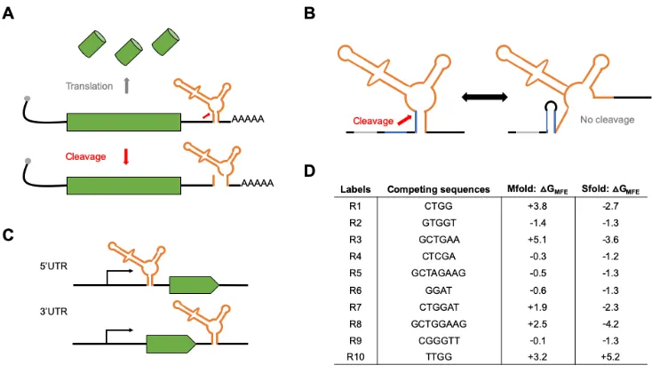
Figure 2.1: Gene regulatory tools based on self-cleaving ribozymes. (A) Inserting self-cleaving ribozymes in the 3’ untranslated region (UTR) of a gene leads to cleavage (red arrow) and subsequent mRNA transcript destabilization/decay and inhibition of protein synthesis. (B) Conceptual design of tunable self-cleaving ribozymes. A competing sequence (blue) is placed directly upstream of the ribozyme (orange). Base-pairing of this sequence with a part of the ribozyme stem prevents ribozyme self-cleavage. The ribozyme is flanked by insulating sequences (gray) to aid in preventing base-pairing interactions between the ribozyme and other sequences in the 3’UTR. (C) Schematic of the constructs used to test the ribozyme constructs in mammalian cells and Drosophila. We placed the ribozyme (orange) either in the 5’UTR or 3’UTR of the reporter genes used (green). (D) List of the competing sequences used in this study, along with their labels used in Figures 2 and 4. Also listed are the free energy differences between the minimal free energy structures of ribozymes in a cleavable and non-cleavable conformation using each competing sequence derived Mfold and Sfold.
Together, these results show that self-cleaving ribozymes combined with upstream competing sequences can modulate gene expression in eukaryotic systems, and that other factors, besides ribozyme self-cleavage and base-pair interactions, influence gene expression.
2.2 - MATERIALS AND METHODS
2.2.1 - Strains, plasmids, oligonucleotides, and fly lines.
All strains, plasmids, oligos, gBlocks, and fly lines used in this work can be found in Supplementary Table 2.1. All PCR amplifications were performed using Q5 Hot Start High-Fidelity 2X Master Mix (NEB, Cat: M0494S) unless specified. All fly lines were generated using site-specific PhiC31-mediated insertion from Genetivision.
We used the pcDNA3.1(+) mammalian expression vector (Thermo Fisher, Cat: V79020) for expression of GFP in HEK293T cells. For this study we used the hammerhead self-cleaving ribozyme from Schistosoma mansoni as it has been associated with a high catalytic activity in vitro and in vivo [29,30]. We first built the active ribozyme constructs by first amplifying GFP and inserting it into the NotI and PstI sites of pCB1180. The inactive ribozyme constructs were built by creating a single point mutation that abolishes catalytic activity of the ribozyme [31]. Then, annealed/phosphorylated oligos containing the inactive and active ribozymes were inserted into the XhoI and NotI sites, located in the 5’ untranslated region (UTR), to make pCB1134/1135. To insert these ribozyme/GFP sequences into pcDNA3.1(+), we PCR amplified the ribozyme-GFP sequence from pCB1134/1135 and inserted it into the HindIII and XbaI restriction sites in pcDNA3.1(+) to create pCB1136/1137. The upstream competing sequences were inserted into pCB1136/1137 by linearizing the plasmids with EcoRI and XhoI and then ligating with phosphorylated/annealed oligos containing the competing sequences of interest (Figure 2.1D). For insertion of the ribozyme/upstream competing sequences in the 3’UTR of GFP, the ribozyme and upstream competing sequences were PCR amplified from the previously built 5’UTR constructs and inserted into the XbaI site of pCB1133.
Center, Cat: 1419) for creating the transgenic fly lines containing the lacZ reporter. To generate the ribozyme constructs, we first removed the UAS-hsp70 sequence using the HindIII and KpnI restriction sites and added the hunchback (hb) proximal enhancer (hbpe), the eve minimal promoter, and the lacZ reporter to create pCB1181. Expressing lacZ from the hbpe creates a well-established domain of hb to easily study the effects from the self-cleaving ribozymes [32–34]. For the insertion of the self-cleaving ribozymes into the 5’UTR and 3’UTR of lacZ, the StuI and KpnI restriction sites of pCB1181 were used, respectively. To insert the upstream competing sequences, both the EcoRI and AvrII sites were added upstream of the ribozyme sequence for ligation with phosphorylated/annealed oligos containing the competing sequences of interest.
2.2.2 - Predicting secondary structures of self-cleaving ribozymes/upstream competing sequences.
The online tools Mfold and Sfold were used to predict the minimal free energy (MFE) structures of the ribozymes lacking or containing an upstream competing sequence using the default settings [35,36]. We extracted the DG of the structures associated with the lowest free energy of a ribozyme in a cleavable and non-cleavable conformation. The DG of each upstream competing sequence was calculated as the difference between the DG of the cleaved and non-cleaved structures. See Supplementary Figure 2.1 for a representative secondary structure of ribozymes in a cleaving or non-cleaving conformation.
2.2.3 - Transient transfections of pcDNA3.1(+)-ribozyme constructs.
2.2.4 - Flow cytometry analysis of transfected HEK293T cells.
We trypsinized the transiently transfected HEK293T cells using trypsin-EDTA (Thermo Fisher, Cat: 25200056) and resuspended them in 500mL 1xPBS (Fisher Scientific, Cat: MT21040CV). The cells were analyzed for fluorescence using the Accuri C6 Flow Cytometer with CFlow plate sampler (Becton Dickinson). The events were gated based on the forward scatter and side scatter, with fluorescence measured in FL2-H, using the 533/30 filter, from at least 10,000 gated events. The fold-reduction of GFP was calculated as the ratio of the fluorescence values for the cells transfected with an inactive ribozyme with a specific competing sequence over that of an active ribozyme with the same competing sequence.
2.2.5 - Fluorescent in situ hybridization of Drosophila embryos.
All embryos were aged to 2-4 hours from laying and then fixed using 37% formaldehyde following standard protocols [37]. Fluorescent in situ hybridization (FISH) was combined with fluorescent immunostaining following standard protocols [37]. Briefly, fixed embryos were washed in 1xPBS buffer supplemented with 0.05% Tween-20, and then hybridized with a fluorescein (ftc)-conjugated anti-sense lacZ probe at 55°C. The embryos were washed and incubated with the rabbit anti-histone (Abcam, Cat: ab1791) (1:10,000 dilution) and goat anti-ftc (Rockland, Cat: 600-101-096) (1:5,000 dilution) primary antibodies overnight at 4°C. Embryos were then washed and incubated for 1.5 hours with fluorescent donkey anti-rabbit-546 (Invitrogen, Cat: A10040) (1:500 dilution) and donkey anti-goat-647 (Invitrogen, Cat: A21447) (1:500 dilution) secondary antibodies at room temperature. Finally, the embryos were washed and stored in 70% glycerol at -20°C prior to being imaged. All prepared embryos were imaged within two weeks of protocol completion.
2.2.6 – Imaging and analysis of embryos.
potential variability of laser strength between imaging sessions. The prepared embryos were mounted laterally using 70% glycerol using two pieces of double-sided tape. A Zeiss LSM 710 microscope was used to acquire 15-25 z-slices 45-60 µm apart at 40x magnification.
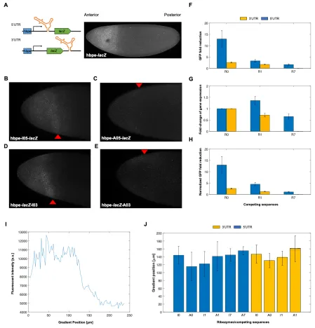
Using Fiji, the z-max intensity projection for each embryo was measured for its fluorescence intensity. The hb expression domain was used as the cutoff for signal (see Figure 2.3A), as the expression profile of lacZ should match the endogenous hb expression pattern due to expression from the hbpe. The fluorescent signal was obtained by measuring the intensity from the anterior pole to the edge of hb domain using the tools available in Fiji. After measuring signal, background noise was measured as the intensity outside of the hb expression pattern. The fold-reduction of lacZ was calculated as the ratio of the fluorescence values for the embryos with an inactive ribozyme with a specific competing sequence over that of an active ribozyme with the same competing sequence. Refer to Supplementary Document 2.1 for an in-depth protocol.
With the same embryos, the width of the lacZ gradient was compared with the active and inactive ribozyme constructs. For this analysis, we used a supervised MATLAB script to first locate/orient the embryo, and then shape the embryos’ periphery boundary. We then measured the fluorescence of the embryo across the anterior-posterior axis (see supplementary material for MATLAB scripts). To measure the distance from the anterior pole to the boundary of the lacZ domain, we selected three points along the y-axis and extracted the width corresponding to 50% loss of the maximum intensity. We selected three different y-values to account for asymmetrical lacZ gradients (Supplementary Figure 2.3). The median of the three values was used to represent the measurement of the lacZ gradient.
2.3 - RESULTS
2.3.1 - Designing self-cleaving ribozymes containing tunable upstream competing sequences.
activity in vitro and in vivo [29,30]. Though these ribozyme constructs can be placed in various locations within a transcript, we chose to test two specific locations: the 5’ and 3’UTR of the reporter genes tested (Figure 2.1C). The competing sequences were placed upstream of the ribozyme to ensure that transcription of the ribozyme before the competing sequence did not result in self-cleavage prior to the transcription of the competing sequence. Insulating sequences were flanked upstream of the ribozyme/competing sequence to limit ribozyme misfolding due to flanking sequences (Figure 2.1B). Finally, we designed the competing sequences using Mfold [35] to obtain a set of sequences that were associated with varying levels of predicted folded and misfolded ribozyme structures (Figure 2.1D). Each competing sequence varied in
sequence
sequence length and composition and were associated with different propensities to base-pair with the stem of the ribozyme. Finally, each competing sequence lacked a start codon to prevent premature translation initiation.
2.3.2 - Self-cleaving ribozymes combined with upstream competing sequences can tune gene expression in mammalian cells.
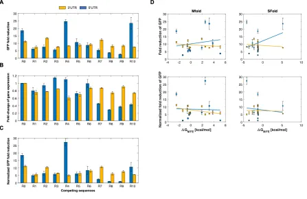
We first sought to test these ribozyme constructs in a mammalian system. To this end, we tested the ribozyme constructs in HEK293T cells. We inserted the self-cleaving ribozymes and 10 different upstream competing sequences in the 5’ UTR or the 3’UTR of GFP to observe how various sequence configurations impacted reporter gene expression. For each ribozyme/competing sequence tested, we used an inactive ribozyme with the same competing sequence to act as a control. As the inactive and active ribozymes differ by a single point mutation [31], the overall structure of the ribozyme was preserved. After transiently transfecting these reporter constructs, the fluorescence of the cells was analyzed by flow cytometry analysis. We found that these ribozyme constructs were able to reduce expression of GFP in HEK293T cells, with fluorescence generally being associated in a bimodal distribution (untransfected cells and cells associated with varying GFP levels) (Supplementary Figure 2.2). When located in the 5’UTR, the ribozymes/upstream competing sequences generally resulted in greater range of fold-reduction levels compared to when located in the 3’UTR (Figure 2.2).
expression ranged from negligible loss (e.g. R2, R6) to ~70% loss (e.g. R8) (Figure 2.2B). Interestingly, the insertion of some upstream competing sequences resulted in increased expression of GFP (e.g. R3, R4). We then accounted for the loss of gene expression due to the insertion of a competing sequence by normalizing the fold-reduction data from Figure 2.2A using the data from Figure 2.2B (Figure 2.2C). While this generally resulted in less fold-reduction of each construct, a wide dynamic range was generally maintained, from almost no fold-reduction to ~25-fold-reduction of gene expression.
After obtaining the experimental data, we then sought to gain insight into the relationship between the fold-reduction of gene expression and the predicted energies of misfolding. To this end, we compared the GFP fold-reduction levels with the predicted free energy differences obtained from Mfold. To obtain these values, the difference between the DG associated with the MFE structure of a ribozyme in a cleavable conformation and the DG associated with the MFE in a non-cleavable conformation was calculated (Supplementary Figure 2.1). While the experimental data from HEK293T cells showed a wide dynamic range of fold-reduction levels, there was a lack of correlation between the experimental data and predicted free energy differences (Figure 2.2D). We then sought to use a different RNA predictive folding algorithm to see if it could better correlate the fold-reduction of gene expression to predicted free energies. Thus, we used Sfold to compare MFE’s to the GFP fold-reduction [36]. Similar to Mfold, there was a lack of correlation between the experimental fold-reductions to the predicted free energies (Figure 2.2D). The lack of a correlation indicates the presence of external factors that influence ribozyme self-cleavage, thus currently making this approach non-predictive. Even so, our data show that ribozyme/upstream competing sequences can be used to tune gene expression in mammalian cells.
2.3.3 - Self-cleaving ribozymes/upstream competing sequences can tune gene expression in Drosophila.
constructs. We first designed Drosophila expression vectors containing the lacZ reporter expressed from the hunchback proximal enhancer (hbpe). This enhancer results in an expression pattern similar to endogenous hb, which has a sharp boundary at roughly 50% anterior-to-posterior (AP) coordinate [32–34]. The hbpe drives expression with a boundary at roughly 33% AP coordinate (Figure 2.3A), which allowed us to quantitatively test these ribozymes in vivo. Similar to the work in HEK293T cells, each ribozyme/upstream competing sequence tested were compared to an inactive ribozyme containing the same competing sequence to act as a negative control. Embryos were first hybridized with an antisense lacZ probe, then imaged by confocal microscopy. We found that the insertion of ribozyme/competing sequences into a transcript expressing lacZ were able to tune lacZ expression levels in Drosophila embryos (Figure 2.3B-E). Unlike with the mammalian cell data, normalizing the fold-reduction data by accounting for the effects of inserting the upstream competing sequences on lacZ expression resulted in only in small changes to the measured dynamic range of fold-reduction values (Figure 2.3). While the fold-reduction values observed in Drosophila were generally less than those observed in HEK293T cells, the correlation of fold-reduction values, compared to the work in mammalian cells, remained the same (i.e. R0 > R1 > R7) and maintained a high dynamic range (~2-14 fold-reduction of lacZ).
2.4 - DISCUSSION
In this work, we engineered a set of genetic tools that were able to modulate gene expression in HEK293T cells and Drosophila. At face value, inserting the ribozymes in the 5’UTR of the reporter genes yielded a greater range of fold-reduction levels compared to the 3’UTR. However, we observed that insertion of upstream competing sequences resulted in the inhibition of gene expression in the absence of ribozyme self-cleavage. This effect was greater when the ribozyme/competing sequence was located in the 5’UTR (Figure 2.2B). After normalizing the fold-reduction levels by accounting for the loss of gene expression, we observed that some ribozyme constructs (most notably the 5’UTR constructs) reduced gene expression more weakly compared to that data prior to normalization (Figure 2.2A/C). In general, the ribozymes/upstream competing sequences were observed to reduce gene expression more strongly in HEK293T cells compared to Drosophila (Figures 2.2 and 2.3), which has also been observed in recent work [41]. This difference could be due to different biological machinery between mammalian and insect models, different experimental assays, or the constructs themselves, as they contain different promoters and reporter genes. Even with the differences in fold-reduction levels between these model systems, these tools maintained a dynamic range of gene expression regulation (~1-25 in HEK293T cells and ~2-14 in Drosophila). While the experimental data did not show a high correlation with the RNA secondary structure predictions (Figure 2.3), we provide a set of gene regulatory tools based on empirical measurements between competing sequences and strength of gene reduction.
interacted with the competing sequence, ribozyme, and/or the insulating sequence. To prevent this phenomenon, the length and/or content of the insulator sequence could be altered. It is also possible that one or more of the competing and/or insulating sequences contain a target sequence for a native biological factor or pathway, such as an endogenous transcription factor, internal ribosome entry site (IRES), or RNAi. While the addition of a specific target sequence is unlikely, novel transcription factors, IRES’, and non-coding RNAs continue to be discovered in eukaryotic systems, including Drosophila [42–48]. Finally, Mfold and/or Sfold may lack the ability to predict the fold-reduction of gene expression associated with the ribozyme constructs. Recent work has shown that hammerhead ribozymes are associated with varying cleavage activities across different model systems (e.g. mammalian vs. yeast) and experimental setups (e.g. in vitro vs. in vivo) [41], which show that cellular context is likely important for the observed activity. Another possibility is that Mfold and Sfold are not accurately capturing RNA folding. While algorithms, such as Mfold and Sfold, have the ability to predict RNA secondary structures, ribozymes can form complex 3D structures (e.g., pseudoknots) that cannot be predicted accurately. Due to these reasons, current predictive RNA folding algorithms may not be sufficient for accurate secondary structure predictions. Improvements on RNA structure prediction models will allow for a more accurate design of competing sequences.
ribozyme/upstream competing sequences. This could prevent interactions between the ribozyme or competing sequence with flanking sequences, resulting in fold-reduction levels only from ribozyme self-cleavage.
2.5 - CONCLUSIONS
We developed a set of tools that were able to tune gene expression in HEK293T cells and Drosophila. While the free energies obtained from the predictive RNA secondary structure tool did not correlate well with fold-reduction of gene expression, the competing sequences used in this work provides a set of genetic tools associated with a wide range of fold-reduction levels. Though tested in mammalian and insect systems, these tools should be applicable in other eukaryotic systems, such as C. elegans, zebrafish, and mice. Previous work has shown that self-cleaving ribozymes are found naturally in these organisms [50–52] and have been used for therapeutic applications [4,53]. These tools will be useful for studies involving synthetic biology, especially for the purposes of building and studying synthetic gene circuits.
2.6 - ACKNOWLEDGMENTS
2.7 - REFERENCES
[1] F. Lienert, J.J. Lohmueller, A. Garg, P.A. Silver, Synthetic biology in mammalian cells: Next generation research tools and therapeutics, Nat. Rev. Mol. Cell Biol. 15 (2014) 95–107.
[2] V. Singh, Recent advancements in synthetic biology: Current status and challenges, Gene. 535 (2014) 1–11.
[3] L. Guzman, D. Belin, M.J. Carson, J. Beckwith, Tight regulation, modulation, and high-level expression by vectors containing the arabinose PBAD promoter, J Biotechnol. 177 (1995) 4121–4130.
[4] L. Yen, J. Svendsen, J.S. Lee, J.T. Gray, M. Magnier, T. Baba, R.J. D’Amato, R.C. Mulligan, Exogenous control of mammalian gene expression through modulation of RNA self-cleavage, Nature. 431 (2004) 471–476.
[5] H.M. Salis, E.A. Mirsky, C.A. Voigt, Automated design of synthetic ribosome binding sites to control protein expression, Nat. Biotechnol. 27 (2009) 946–950.
[6] K.E. McGinness, T.A. Baker, R.T. Sauer, Engineering controllable protein degradation, Mol. Cell. 22 (2006) 701–707.
[7] A.E. Friedland, T.K. Lu, X. Wang, D. Shi, G. Church, J.J. Collins, Synthetic gene networks that count, Science. 324 (2009) 1199–1202.
[8] S. Basu, Y. Gerchman, C.H. Collins, F.H. Arnold, R. Weiss, A synthetic multicellular system for programmed pattern formation, Nature. 434 (2005) 1130–1134.
[9] T.S. Gardner, C.R. Cantor, J.J. Collins, Construction of a genetic toggle switch in Escherichia coli, Nature. 403 (2000) 339–342.
[10] M.B. Elowitz, S. Leibler, A synthetic oscillatory network of transcriptional regulators, Nature. 403 (2000) 335–338.
[11] M. Brown, J. Figge, U. Hansen, C. Wright, K.-T. Jeang, G. Khoury, D.M. Livingston, T.M. Roberts, Lac repressor can regulate expression from a hybrid SV40 early promoter containing a lac operator in animal cells, Cell. 49 (1987) 603–612.
[12] M. Gossen, H. Bujard, Tight control of gene expression in mammalian cells by tetracycline-responsive promoters, Proc Natl Acad Sci. 89 (1992) 5547–5551. [13] M.L. Maeder, S. Thibodeau-Beganny, A. Osiak, D.A. Wright, R.M. Anthony, M.
J.D. Sander, F. Müller-Lerch, F. Fu, J. Pearlberg, C. Göbel, J.P. Dassie, S.M. Pruett-Miller, M.H. Porteus, D.C. Sgroi, A.J. Iafrate, D. Dobbs, P.B. McCray Jr, T. Cathomen, D.F. Voytas, J.K. Joung, Rapid “open-source” engineering of customized zinc-finger nucleases for highly efficient gene modification, Mol. Cell. 31 (2008) 294–301.
[14] R. Morbitzer, P. Römer, J. Boch, T. Lahaye, Regulation of selected genome loci using de novo-engineered transcription activator-like effector (TALE)-type transcription factors, Proc. Natl. Acad. Sci. 107 (2010) 21617–21622.
[15] A. Garg, J.J. Lohmueller, P.A. Silver, T.Z. Armel, Engineering synthetic TAL effectors with orthogonal target sites, Nucleic Acids Res. 40 (2012) 7584–7595. [16] H. Kakidani, M. Ptashne, GAL4 activates gene expression in mammalian cells, Cell.
52 (1988) 161–167.
[17] J. Medenbach, M. Seiler, M.W. Hentze, Translational control via protein-regulated upstream open reading frames, Cell. 145 (2011) 902–913.
[18] J.P. Ferreira, K.W. Overton, C.L. Wang, Tuning gene expression with synthetic upstream open reading frames, Proc. Natl. Acad. Sci. 110 (2013) 11284–11289. [19] Z. Xie, L. Wroblewska, L. Prochazka, R. Weiss, Y. Benenson, Multi-input
RNAi-based logic circuit for identification of specific cancer cells, Science. 333 (2011) 1307–1311.
[20] S. Ausländer, D. Ausländer, M. Müller, M. Wieland, M. Fussenegger, Programmable single-cell mammalian biocomputers, Nature. 487 (2012) 123–127. [21] K. Nishimura, T. Fukagawa, H. Takisawa, T. Kakimoto, M. Kanemaki, An
auxin-based degron system for the rapid depletion of proteins in nonplant cells, Nat. Methods. 6 (2009) 917–922.
[22] K.M. Bonger, L. Chen, C.W. Liu, T.J. Wandless, Small-molecule displacement of a cryptic degron causes conditional protein degradation, Nat. Chem. Biol. 7 (2011) 531–537.
[23] L.A. Banaszynski, L.-C. Chen, L.A. Maynard-Smith, A.G.L. Ooi, T.J. Wandless, A rapid, reversible, and tunable method to regulate protein function in living cells using synthetic small molecules, Cell. 126 (2006) 995–1004.
Lim, Repurposing CRISPR as an RNA-guided platform for sequence-specific control of gene expression, Cell. 152 (2013) 1173–1183.
[25] L.A. Gilbert, M.H. Larson, L. Morsut, Z. Liu, G.A. Brar, S.E. Torres, N. Stern-Ginossar, O. Brandman, E.H. Whitehead, J.A. Doudna, W.A. Lim, J.S. Weissman, L.S. Qi, CRISPR-mediated modular RNA-guided regulation of transcription in eukaryotes, Cell. 154 (2013) 442–451.
[26] A.R. Ferré-D’Amaré, W.G. Scott, Small self-cleaving ribozymes, Cold Spring Harb. Perspect. Biol. 2 (2010) a003574.
[27] K.H. Link, L. Guo, T.D. Ames, L. Yen, R.C. Mulligan, R.R. Breaker, Engineering high-speed allosteric hammerhead, Biol. Chem. 388 (2007) 779–786.
[28] M.N. Win, C.D. Smolke, A modular and extensible RNA-based gene-regulatory platform for engineering cellular function, Proc. Natl. Acad. Sci. 104 (2007) 14283– 14288.
[29] J.M. Carothers, J. a Goler, D. Juminaga, J.D. Keasling, Model-driven engineering of RNA devices to quantitatively program gene expression, Science. 334 (2011) 1716–1719.
[30] G. Ferbeyre, J.M. Smith, R. Cedergren, Schistosome satellite DNA encodes active hammerhead ribozymes, Mol. Cell. Biol. 18 (1998) 3880–3888.
[31] F. Seela, H. Debelak, N. Usman, A. Burgin, L. Beigelman, 1-Deazaadenosine: synthesis and activity of base-modified hammerhead ribozymes, Nucleic Acids Res. 26 (1998) 1010–1018.
[32] W. Driever, C. Nüsslein-Volhard, The bicoid protein is a positive regulator of hunchback transcription in the early Drosophila embryo, Nature. 337 (1989) 138– 143.
[33] R. Lehmann, C. Nüsslein-Volhard, hunchback, a gene required for segmentation of an anterior and posterior region of the Drosophila embryo, Dev. Biol. 119 (1987) 402–417.
[34] M.W. Perry, J.P. Bothma, R.D. Luu, M. Levine, Precision of hunchback expression in the Drosophila embryo, Curr. Biol. 22 (2012) 2247–2252.
[36] Y. Ding, C.Y. Chan, C.E. Lawrence, Sfold web server for statistical folding and rational design of nucleic acids, Nucleic Acids Res. 32 (2004) 135–141.
[37] D. Kosman, C.M. Mizutani, D. Lemons, W.G. Cox, W. McGinnis, E. Bier, Multiplex detection of RNA expression in Drosophila embryos, Science. 305 (2004) 846. [38] L.M. Liberman, G.T. Reeves, A. Stathopoulos, Quantitative imaging of the Dorsal
nuclear gradient reveals limitations to threshold-dependent patterning in Drosophila, Proc. Natl. Acad. Sci. 106 (2009) 22317–22322.
[39] T.A. Carrier, J.D. Keasling, Controlling messenger RNA stability in bacteria: Strategies for engineering gene expression, Biotechnol. Prog. 13 (1997) 699–708. [40] A.A. Jermusyk, N.P. Murphy, G.T. Reeves, Analyzing negative feedback using a synthetic gene network expressed in the Drosophila melanogaster embryo, BMC Syst. Biol. 10 (2016) 85.
[41] L.A. Wurmthaler, B. Klauser, J.S. Hartig, Highly motif- and organism-dependent effects of naturally occurring hammerhead ribozyme sequences on gene expression, RNA Biol. 15 (2018) 231–241.
[42] R.S. Young, A.C. Marques, C. Tibbit, W. Haerty, A.R. Bassett, J.L. Liu, C.P. Ponting, Identification and properties of 1,119 candidate LincRNA loci in the Drosophila melanogaster genome, Genome Biol. Evol. 4 (2012) 427–442.
[43] S. Inagaki, K. Numata, T. Kondo, M. Tomita, K. Yasuda, A. Kanai, Y. Kageyama, Identification and expression analysis of putative mRNA-like non-coding RNA in Drosophila, Genes to Cells. 10 (2005) 1163–1173.
[44] J.L. Tupy, A.M. Bailey, G. Dailey, M. Evans-Holm, C.W. Siebel, S. Misra, S.E. Celniker, G.M. Rubin, Identification of putative noncoding polyadenylated transcripts in Drosophila melanogaster, Proc. Natl. Acad. Sci. 102 (2005) 5495– 5500.
[45] M.T. Marr II, J.A. D’Alessio, O. Puig, R. Tjian, IRES-mediated functional coupling of transcription and translation amplifies insulin receptor feedback, Genes Dev. 21 (2007) 175–183.