This author’s accepted manuscript may be used for non-commercial purposes in accordance with Wiley Terms and Conditions for Self-Archiving.
The full details of the published version of the article are as follows:
TITLE: Does the computed tomographic appearance of the lung differ between young and old dogs?
AUTHORS: Natasha L. Hornby, Christopher R. Lamb
JOURNAL: Vet Radiology & Ultrasound
PUBLISHER: Wiley
PUBLICATION DATE: November 2017
Does the computed tomographic appearance of the lung differ between young and old dogs? 1
Natasha L. Hornby and Christopher R. Lamb 2
Department of Clinical Sciences and Services, The Royal Veterinary College, University of 3
London 4
5
Address correspondence to: C. R. Lamb, Department of Clinical Sciences and Services, The Royal 6
Veterinary College, Hawkshead Lane, North Mymms, Hertfordshire AL9 7TA, UK. 7
Tel: 01707-666234 8
Email: [email protected] 9
Key words: aging, atelectasis, computed tomography, dog, lung 10
Running head: CT of aged lung 11
Funding Sources: The authors received no financial support for the research, authorship or 12
Abstract 14
In computed tomographic (CT) images of humans, decreased lung attenuation, bronchial 15
dilation and/or thickening, air trapping, cysts, and thickened interlobular septa have been 16
associated with increasing age. To determine if there are differences in the CT appearance of 17
the lungs of young and old dogs that could affect interpretation of diagnostic studies, 18
pulmonary CT images of dogs with conditions unrelated to the thorax were reviewed 19
retrospectively in a case-control study. CT studies of 42 young dogs (range 0.3-4.8 years) and 47 20
old dogs (range 9-15.1 years) were jumbled and reviewed by an observer blinded to dog age. CT 21
was performed under sedation in 62 (70%) dogs and under general anesthesia in 27 (30%). 22
Heterotopic bone was more prevalent (62% versus 14%) in old dogs. Lung collapse was 23
significantly associated with old age, greater body weight, and anesthesia. There were no 24
significant differences in median lung attenuation or occurrence of ground glass pattern, cysts, 25
bronchial thickening, bronchial dilation, or degree of tracheal calcification. No examples of 26
reticular pattern, emphysema, pleural thickening or septal thickening were observed in any 27
dog. Despite previous studies describing age-related changes in the radiographic appearance of 28
the lungs of old dogs, it appears that there are minimal observable differences in CT images. 29
Old dogs are more likely to have visible foci of heterotopic bone and may be more prone to 30
lung lobe collapse than young dogs, but neither of these differences should contribute to 31
Introduction 33
Age-related changes in the morphology of the lung have been described in humans1, dogs2,3, 34
and rats.4 A prominent age-related feature observed in dogs was accumulation of macrophages 35
containing dust and pigment in the respiratory bronchioles and alveolar openings.2 It was 36
hypothesized that exposure to dust and reduction in function of the mucociliary escalator over 37
time resulted in accumulation of dust and pigment in the lungs,2 but this was not confirmed. 38
There was also an increase in the relative volume (‘volume density’) of alveolar ducts, 39
progressive calcification of the bronchial cartilage, and increased size of the bronchial glands.2 40
Minimal emphysema was observed and fibrosis was sparse, mainly associated with foci of 41
pneumonitis.2 42
Correlations between morphologic changes in the lungs and the radiographic appearance of the 43
lung were described in 100 aged dogs that had no pathologic evidence of pulmonary or 44
cardiovascular disease.3 Emphysema and alveolar dilatation were reported, but more emphasis 45
was placed on findings of pleural fibrosis and interstitial fibrosis.3 Accumulation of peribronchial 46
collagen over time causing progressive pulmonary fibrosis has also been emphasized in studies 47
of aging rats.4 Together with fibrosis, thickening of the alveolar walls and calcification of 48
bronchial cartilage in dogs were associated with radiographic signs including pleural thickening, 49
increased linear markings, and increased opacity of the tracheal and bronchial walls.3 Foci of 50
heterotopic bone in the lungs of many older dogs produced small nodular opacities in 51
radiographs.3 Based on these findings, increased lung attenuation, pleural and septal thickening 52
images of the lungs of aged dogs; however, there are no published studies of age-related 54
changes in pulmonary CT images of dogs. 55
Changes associated with normal aging in the lungs of humans have been studied extensively, 56
one aim being to avoid over-diagnosis of age-related changes as signs of clinically significant 57
disease. Age-related changes seen in pulmonary CT images of humans include decreased lung 58
attenuation as a result of dilatation of alveoli and/or emphysema, air-trapping, cysts, a sub-59
pleural reticular pattern, bronchial dilation, bronchial thickening, and thickening of interlobular 60
septa without inflammation.5-9 CT signs usually associated with interstitial lung disease may also 61
be seen in healthy elderly humans, and radiologists have been cautioned not to over-interpret 62
such findings in asymptomatic patients.5 63
The aim of the present study was to determine if there are differences in the CT appearance of 64
the lungs of young and old dogs that could affect interpretation of diagnostic studies. 65
66
Method and Materials 67
Ethical approval was granted by the Clinical Research Ethical Review Board at the Royal 68
Veterinary College. For this retrospective case-control study, the medical records at the Queen 69
Mother Hospital for Animals (QMHA) in the period 2012-2016 were searched for cases that 70
satisfied the following inclusion criteria: dogs that had diagnosis of a non-malignant disease or 71
condition unrelated to the thorax; had non-contrast CT images of the lung acquired using a high 72
resolution (sharp or lung) algorithm; and were either less than 5 years old or more than 9 years 73
pleural, bronchial, pulmonary disease, malignant neoplasia or systemic inflammatory disease 75
were not included. 76
For each dog, age at the time of CT, gender, body weight and diagnosis were recorded. On the 77
basis of body weight, dogs were divided in small (<10kg), medium (10-25kg), large (26-40kg), 78
and giant (>40kg) categories. Use of sedation or anesthesia for CT was also recorded. For all 79
sedated dogs, CT images were acquired during free breathing, whereas anaesthetised dogs had 80
brief manual hyperventilation with CT images acquired during the subsequent expiratory pause. 81
As part of the inclusion criteria, all CT images were acquired using the same multi-slice scanner 82
(MX8000 IDT, Phillips Best, The Netherlands). Studies lacking optimal settings for lung 83
examination were excluded. For the purposes of this study, optimal settings were helical 84
acquisition, slice thickness 2mm for small dogs and 3mm for other dogs, matrix 512x512, and 85
high frequency (‘sharp’ or ‘lung enhanced’) reconstruction algorithm. All CT images were 86
reviewed by a board-certified radiologist (CRL) without knowledge of the age, breed or history 87
of the subjects. Images were reviewed in a lung window (level -500HU, width 2000HU) using a 88
proprietary Digital Imaging in Communications and Medicine (DICOM) viewer (OsiriX 64-bit, 89
version 5.2.2, Pixmeo, Switzerland). For each dog, the lungs were also examined using 90
maximum- and minimum-intensity projections with slab thickness 5mm for small dogs and 91
8mm for other dogs. Dogs whose CT images were affected by motion blur were excluded. 92
CT images were evaluated for the following features: mean lung attenuation (Hounsfield units, 93
HU) based on the median of three measurements made using a circular region of interest of 94
minimum area 40mm2 placed in different lung lobes and avoiding bronchi or large vessels; 95
(absent/present); pulmonary cysts (absent/present, diameter of largest cyst); visceral pleural 97
thickening (absent, affecting one lobe, affecting multiple lobes); interlobular septal thickening 98
(absent, affecting one lobe, affecting multiple lobes); bronchial thickening (none, slight, 99
marked); bronchial dilation (median of bronchus: pulmonary artery ratio measurements in the 100
right caudal lobe in three adjacent CT slices); degree of tracheal ring calcification (the median of 101
three HU measurements made by placing a point sample on a tracheal cartilage ring 102
immediately cranial to the origin of the lobar bronchi); foci of heterotopic bone (absent, 103
affecting one lobe, affecting multiple lobes); and pulmonary collapse (number of affected lobes 104
and distribution of collapse was recorded: lobe tip only, peripheral, bronchocentric, entire 105
lobe). The term tip is used here to indicate the pointed extremity of a lung lobe in transverse CT 106
images. Diagnosis of heterotopic bone was based on finding pulmonary hyperdense foci ranging 107
from sub-millimeter rounded foci to larger irregularly-shaped densely ossified foci. Diagnosis of 108
lung collapse was based on finding increased lung attenuation accompanied by reduced volume 109
of the affected lung. Terminology followed recommendations for thoracic imaging by the 110
Fleischner Society.10 111
Statistical analyses were done by a statistician (Yu-Mei Chang) using a commercial statistical 112
software package (SPSS 22, IBM) Significance of differences in median lung attenuation, 113
bronchus: pulmonary artery ratio and tracheal calcification between young and old dogs were 114
tested using the Mann-Whitney test. Differences in prevalence of ground glass opacity, 115
bronchial thickening and cysts were tested using Fisher’s exact test. Differences in occurrence 116
of heterotopic bone and body weight of dogs were tested using Kendall’s tau-c test. 117
body weight, and anesthesia were tested using binary logistic regression and results 119
summarized as odds ratio (OR) and 95% confidence interval (CI). Differences with p<0.05 were 120
considered significant. 121
122
Results 123
CT images of 42 young dogs (age range 0.3-4.8 years) and 47 old dogs (age range 9-15.1 years) 124
were reviewed. All dogs were scanned in sternal recumbency. Characteristics of young and old 125
dog groups are summarized in Table 1. CT features observed in young and old dogs are 126
summarized in Table 2. Heterotopic bone was more prevalent (62% versus 14%; p<0.001) in old 127
dogs. 128
Signs of lung collapse were identified in 40 (46%) dogs. Based on regression analysis, 129
occurrence of lung collapse was significantly associated with age (OR 3.7, CI 1.4-9.5, p=0.007), 130
body weight (OR 1.9, CI 1.1-3.3, p=0.02), and anesthesia (OR 3.1, CI 1.1-8.8, p=0.03). Lung 131
collapse affected the right middle lobe in 22 dogs, left cranial lobe in 21 dogs, right cranial lobe 132
in 18 dogs, left caudal lobe in 7 dogs, accessory lobe in 6 dogs, and right caudal lobe in 4 dogs. 133
Lung collapse affected a single lobe in 21 (24%) dogs and multiple lobes in 19 (22%) dogs. In 134
58/68 (85%) affected lobes collapse was limited to the tip of the lobe, in 7 (1%) lobes it was 135
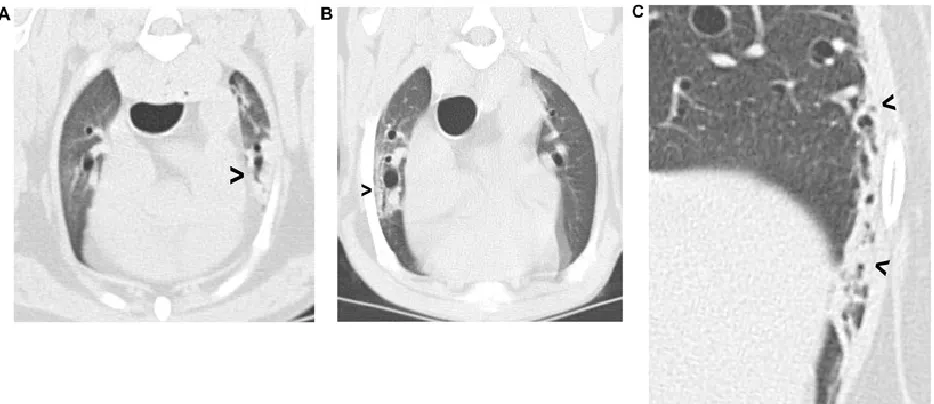
bronchocentric, and in 3 (5%) lobes it was peripheral (figure 1). 136
There were no significant differences in median lung attenuation or occurrence of ground glass 137
pattern, cysts, bronchial thickening, bronchial dilation, or degree of tracheal calcification 138
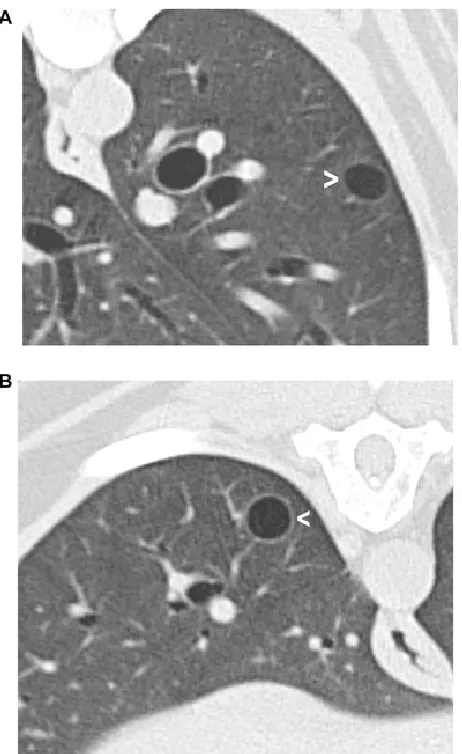
multiple (2-9) in 3 dogs, and the largest ranged in size from 2-11mm diameter (figure 2). No 140
examples of reticular pattern, emphysema, pleural thickening or septal thickening were 141
observed in any dog. 142
143
Discussion 144
In this study there were minimal observable differences between the lungs of young and old 145
dogs. Heterotopic bone was more prevalent in old dogs, which agrees with previous 146
observations.3 Lung collapse was also observed more frequently in old dogs, although the 147
underlying reason for this difference cannot be determined by the present study. For 148
physiological reasons, the right middle lobe is most prone to collapse in the dog.11 In the 149
present study, collapse of the right middle lobe and the right and left cranial lobes occurred 150
with similar frequencies. In the majority of dogs, lung collapse affected only the tip of the lobe. 151
Reduced collateral ventilation because of subclinical disease, such as chronic bronchitis with 152
excessive mucus production, is liable to exacerbate lung collapse.12,13 The increased tendency 153
for lung collapse in older dogs could reflect an increasing prevalence of subclinical lung disease 154
over their lifetime. The observation that lung collapse was more frequent in dogs that had CT 155
under general anesthesia than in dogs that were sedated could reflect the use of high inspired 156
pO2 for anesthesia, which has also been associated with a higher prevalence of lung collapse 157
than when using moderate inspired pO2 (‘medical air’).14 It is uncertain why lung collapse was 158
more frequent in larger dogs. It is known that intermittent positive pressure ventilation to 159
this may reflect the larger gravitational gradient that occurs within the lung of larger patients, 161
as observed in anesthetized horses.16 162
There were no significant differences in median lung attenuation or occurrence of ground glass 163
pattern, cysts, bronchial thickening, bronchial dilation, or degree of tracheal calcification 164
between young and old dogs. These results are in contrast with those of a previous study in 165
which the lungs of old dogs had radiographic signs including pleural thickening, increased linear 166
markings, and increased opacity of the tracheal and bronchial walls.3 Although the results of CT 167
in one group of dogs cannot be compared directly with results of radiography in a different 168
group of dogs, it seems likely that CT would have detected signs of pleural thickening, increased 169
linear markings and increased opacity of the tracheal and bronchial walls had they been 170
present. In fact, based on ability to directly measure lung attenuation using CT and other canine 171
studies that found increased sensitivity of CT for lung lesions17,18, CT might be expected to 172
reveal more age-related changes than radiography. Therefore, the relative lack of CT signs in 173
the present study could reflect a true difference in the condition of the lungs of the dogs used 174
in these respective studies, possibly reflecting different genetic or environmental factors. If so, 175
this could limit the generalizability of our results, which may not be applicable in other 176
locations. 177
Attenuation measurements of tracheal rings were used in the present study as an indirect 178
indicator of bronchial wall calcification. Tracheal rings were measured instead of bronchial walls 179
because they are normally thicker than bronchial walls, hence any attenuation measurements 180
The process of aging occurs over a shorter period of time in dogs than in humans1, and It is 182
generally accepted that dogs of large and giant breeds age more rapidly than dogs of small or 183
medium breeds. The smaller number of giant breed dogs in the aged group in the present study 184
reflects this phenomenon. Non-contiguous age ranges were used in this study to ensure 185
separation of young and aged dogs, which could intermingle if a heterogeneous sample of dogs 186
were classified using a single calendar age threshold alone. 187
In this study, examination of the CT images included quantitative measurements and search for 188
multiple possible lesions based on previous studies of CT signs in aged humans5-9; however, few 189
examples were found of ground glass pattern, bronchial thickening or bronchial dilation, and no 190
signs of reticular pattern, emphysema, pleural thickening or septal thickening were identified in 191
any dog. Hence, finding any of these features in CT images of a dog with respiratory signs 192
suggests the presence of non-age-related lung pathology that is clinically relevant. 193
Cysts were found in 6 (7%) dogs in this study. There is some overlap in use of the terms cyst and 194
bulla, although in humans bulla is used for lesions greater than 1cm, and usually several cm in 195
diameter.10 Lung cysts (and bullae), which can be subclinical, should be distinguished from both 196
congenital lobar emphysema19,20 and superficial bullous emphysema, which is associated with 197
non-traumatic (‘spontaneous’) pneumothorax.21,22 198
The main limitation of the present study was the lack of pathologic examination of lungs, which 199
prevents histologic proof of the nature of lesions identified by CT, and allows the possibility that 200
there were age-related lung changes that were undetected by CT. All CT studies reviewed in 201
this study were obtained using settings suitable for clinical assessment of the lung of dogs, but 202
histologic examination of precisely orientated lung sections. Further studies are indicated to 204
better examine imaging-pathologic correlations in the aging canine lung. The number of dogs 205
available for study was limited by the need to avoid including dogs likely to have had pathologic 206
lung lesions. Use of a single observer to review the CT is also a limitation, but this is not 207
considered to be a major problem because of the relatively homogenous nature of the study 208
sample and use of comprehensive, pre-considered assessment criteria based on previous 209
studies of humans.5-9 210
In conclusion, despite previous studies describing age-related changes in the radiographic 211
appearance of the lungs of old dogs, it appears that there are minimal observable differences in 212
CT images. Old dogs are more likely to have visible foci of heterotopic bone and may be more 213
prone to lung lobe collapse than young dogs, but neither of these differences should contribute 214
to misdiagnosis of pulmonary disease. 215
216
List of Author Contributions 217
Category 1 218
(a) Conception and Design 219
Author name (s)Natasha L. Hornby and Christopher R. Lamb 220
(b) Acquisition of Data 221
(c) Analysis and Interpretation of Data Author name (s) Natasha L. Hornby and Christopher R. 223
Lamb 224
Category 2 225
(a) Drafting the Article 226
Author name (s)Natasha L. Hornby and Christopher R. Lamb 227
(b) Revising Article for Intellectual Content Author name (s) Natasha L. Hornby and Christopher 228
R. Lamb 229
Category 3 230
(a) Final Approval of the Completed Article Author name(s)Natasha L. Hornby and Christopher 231
R. Lamb 232
233
Acknowledgement 234
References 236
1. Pinkerton KE, Green FHY. Normal aging of the lung. In: Harding RD, Pinkerton KE and 237
Plopper CG (Eds). The lung: development, ageing and the environment. London: Elsevier 238
Academic Press, 2004, pp213-233. 239
2. Robinson NE and Gillespie JR. Morphologic features of lungs of aging beagle dogs. Am Rev 240
Respir Dis 1973; 108: 1192-1199. 241
3. Reif JS and Rhodes WH. The lungs of aged dogs. A radiographic-morphologic correlation. J 242
Am Vet Radiol Soc 1966; 7:5-11. 243
4. Calabresi C, Arosio B, Galimberti L, et al. Natural aging, expression of fibrosis-related genes 244
and collagen deposition in rat lung. Experimental Gerontology 2007; 42:1003-1011. 245
5. Copley SJ, Wells AM, Hawtin KH, et al. Lung morphology in the elderly: comparative CT 246
study of subjects over 75 years old versus those under 55 years old. Radiology 2009; 247
251:566-573. 248
6. Copley SJ. Morphology of the ageing lung on computed tomography. J Thorac Imaging 2016; 249
31:140-150. 250
7. Leelakanok N and Piyavisetpat N. CT features of normal lung change in asymptomatic 251
elderly patients. Asian Biomed 2015; 9:613-623. 252
8. Lowery EM, Brubaker AL, Kuhlmann E and Kovacs EJ. The aging lung. J Clin Interv Aging 253
2013; 8: 1489-1496. 254
9. Verbeken EK, Clement JM and Van De Woestijne KP. The senile lung – comparison with
255
10. Hansell DM, Bankier AA, MacMahon H, McLoud TC, Muller NL, Remy J. Fleischner Society: 257
glossary of terms tor thoracic imaging. Radiology 2008; 246: 697-722. 258
11.Robinson NE. Lobar variations in the mechanics of collateral ventilation in intact dog lungs. 259
Am Rev Respir Dis 1981; 124:68-71. 260
12.Robinson NE. Some functional consequences of species-differences in lung anatomy. Adv 261
Vet Sci Comp Med 1982; 26:1-33. 262
13.Lord PF, Gomez JA. Lung lobe collapse. Pathophysiology and radiologic significance. Vet 263
Radiol 1985;26:187-195. 264
14.Staffieri F, Franchini D, Carella GL, et al. Computed tomographic analysis of the effects of 265
two inspired oxygen concentrations on pulmonary aeration in anesthetized and 266
mechanically ventilated dogs. Am J Vet Res 2007; 68:925-931. 267
15.Grimm KA, Lamont LA, Tranquilli WJ, Greene SA, Robertson SA (Eds). Veterinary Anesthesia 268
and Analgesia, The 5th Edition of Lumb and Jones. Oxford: Wiley-Blackwell, 2015, p543.
269
16.Moens Y, Schramel JP, Tusman G, Ambrisko TD, Solà J, Brunner JX, et al. Variety of non-270
invasive continuous monitoring methodologies including electrical impedance tomography 271
provides novel insights into the physiology of lung collapse and recruitment – case report of 272
an anaesthetized horse. Vet Anaesth Analg 2014;41: 196–204. 273
17.Nemanic S, London CA, Wisner ER. Comparison of thoracic radiographs and single breath-274
hold helical CT for detection of pulmonary nodules in dogs with metastatic neoplasia. J Vet 275
18. Eberle N, Fork M, Von Babo V, Nolte I and Simon D. Comparison of examination of thoracic 277
radiographs and thoracic computed tomography in dogs with appendicular osteosarcoma. 278
Vet Comp Oncol 2010; 9: 131-140. 279
19.Mitchell C, Nykamp S. Congenital lobar emphysema in an old English sheepdog puppy. Vet 280
Radiol Ultrasound 2006; 47:465-467. 281
20.Matsumoto H, Kakehata T, Hyodo T, et al. Surgical correction of congenital lobar 282
emphysema in a dog. J Vet Med Sci 2004; 66: 217-219. 283
21.Brissot HN, Dupre GP, Bouvy BM, et al. Thoracoscopic treatment of bullous emphysema in 3 284
dogs. Vet Surg 2003; 32: 524-529. 285
22.Puerto DA, Brockman DJ, Lindquist C, et al. Surgical and nonsurgical management of and 286
selected risk factors for spontaneous pneumothorax in dogs: 64 cases (1986-1999). J Am Vet 287
Med Assoc 2002; 220: 1670-1674. 288
23.Heikkilä HP, Lappalainen AK, Day MJ, Clercx C and Rajamäki MM. Clinical, bronchoscopic, 289
histopathologic, diagnostic imaging, and arterial oxygenation findings in West Highland 290
white terriers with idiopathic pulmonary fibrosis. J Vet Internal Med 2011;25: 433–439. 291
24.Mai C, Verleden SE, McDonough JE, et al. Thin-section CT features of idiopathic pulmonary 292
Table 1. Characteristics of young and old dog groups 294
Young dogs (n=42) Old dogs (n=47)
Median (range) age (y) 3 (0.3-4.8) 11 (9.0-15.1)
Males/ females 28 (67%)/ 14 (33%) 31 (66%)/ 16 (34%)
Body weight range
Small (<10kg)
Medium (10-25kg)
Large (26-40kg)
Giant (>40kg)
10 (24%)
15 (36%)
12 (28%)
5 (12%)
12 (25%)
20 (43%)
13 (28%)
2 (4%)
Diagnostic category
Upper respiratory
Immune-mediated
Alimentary
Neurologic
Orthopedic
Urinary
Benign neoplasm
Oral conditions
Endocrinopathy
19 (45%)
11 (26%)
4 (10%)
4 (10%)
2 (5%)
2 (5%)
0
0
0
12 (25%)
1 (2%)
3 (6%)
3 (6%)
0
2 (4%)
17 (36%)
6 (13%)
3 (6%)
CT under anesthesia 12 (29%) 15 (32%)
Table 2. CT features of the lungs of young and old dogs 296
Young dogs (n=42) Old dogs (n=47)
Median (range) lung attenuation (HU) -788 (-605 to -878) -793 (-679 to -900)
Ground glass opacity 7 (17%) 8 (17%)
Cysts 1 (2%) 5 (10%)
Bronchial thickening 5 (12%) 5 (11%)
Median (range) bronchus: pulmonary artery ratio
0.66 (0.41-1.06) 0.63 (0.40-1.46)
Median (range) tracheal calcification (HU) 224 (65-577) 236 (107-512)
Heterotopic bone
In one lobe
In multiple lobes
3 (7%)
3 (7%)
6 (13%)
23 (49%)
Pulmonary collapse
None
One lung lobe affected
Multiple lobes affected
29 (69%)
9 (21%)
4 (10%)
20 (43%)
12 (26%)
15 (32%)
Legend 298
Figure 1. Examples of pulmonary collapse. A) Collapse of the ventral tip of the left cranial lobe 299
(arrowhead); B) bronchocentric collapse (arrowhead) affecting the right cranial lobe; C) 300
peripheral pulmonary collapse (arrowheads) affecting the lateral aspect of the left caudal lobe. 301
302
Figure 2. Examples of lung cysts. A) 10mm diameter cyst (arrowhead) in the left caudal lobe of a 304
1-year-old golden retriever that had CT as part of the work-up for nasal panniculitis. This was 305
one of 3 pulmonary cysts in this dog; B) 8mm diameter cyst (arrowhead) in the right caudal lobe 306
of a 14-year-old English springer spaniel that had CT as part of the work-up for 307
hyperadrenocorticism. 308