University of Pennsylvania
ScholarlyCommons
Publicly Accessible Penn Dissertations
1-1-2015
Non-Canonical Roles for RAG1 in Lymphocyte
Development
Julie Horowitz
University of Pennsylvania, [email protected]
Follow this and additional works at:
http://repository.upenn.edu/edissertations
Part of the
Allergy and Immunology Commons,
Immunology and Infectious Disease Commons,
and the
Medical Immunology Commons
This paper is posted at ScholarlyCommons.http://repository.upenn.edu/edissertations/1064
For more information, please [email protected].
Recommended Citation
Horowitz, Julie, "Non-Canonical Roles for RAG1 in Lymphocyte Development" (2015).Publicly Accessible Penn Dissertations. 1064.
Non-Canonical Roles for RAG1 in Lymphocyte Development
Abstract
The RAG1/RAG2 (RAG) endonuclease recombines accessible antigen receptor (AgR) genes through DNA double strand break (DSB) intermediates to generate a diverse AgR repertoire. RAG-mediated DSBs signal changes in expression of genes encoding proteins involved in cellular survival, lymphocyte differentiation, and AgR selection. RAG proteins are each comprised of "core" endonuclease domains and dispensable "non-core" regions. Humans with mutations in non-core RAG1 regions exhibit fatal primary immunodeficiencies, and mice expressing truncated core, but not full-length, Rag1 protein (Rag1C/C mice) exhibit impaired early lymphocyte development associated with reduced levels of AgR gene rearrangements. In addition to serving with RAG2 as the V(D)J endonuclease, the RAG1 protein has been proposed to utilize non-core regions to regulate V(D)J recombination by enhancing AgR locus accessibility, promoting efficient RAG endonuclease activity, and/or generating RAG DSB-induced survival and differentiation signals. The experiments described in this thesis use a variety of mouse genetic models to investigate how mutations in RAG1 protein that preserve RAG endonuclease activity impair V(D)J recombination efficiency and lymphocyte development. First, these studies have identified roles for non-core Rag1 regions in regulating normal Alpha/Beta T cell development and TCRB recombination by promoting VB rearrangements and diverse usage of VB gene segments in both primary and secondary VB rearrangements. Second, these studies show that non-core Rag1 regulates normal B cell development by inducing transcriptional activation of the pro-survival kinase Pim2 in response to RAG cleavage and by promoting the survival of developing IgKappa+ and IgLambda+ B cells. These studies have also identified roles for Rag1 in enhancing IgKappa and IgLambda locus accessibility in pre-B cells prior to RAG cleavage. Collectively, the data presented in this thesis demonstrate that RAG1 has critical functions outside of V(D)J recombination that enhance AgR gene segment accessibility, promote V(D)J recombination at multiple AgR loci, and transduce pro-survival signals during AgR recombination to establish a broad AgR repertoire and thereby foster normal lymphocyte development.
Degree Type Dissertation
Degree Name
Doctor of Philosophy (PhD)
Graduate Group Immunology
First Advisor Craig H. Bassing
Keywords
Antigen Receptors, Lymphocyte, RAG, V(D)J Recombination
Subject Categories
NON-CANONICAL ROLES FOR RAG1 IN LYMPHOCYTE DEVELOPMENT
Julie E. Horowitz
A DISSERTATION
in
Immunology
Presented to the Faculties of the University of Pennsylvania
in
Partial Fulfillment of the Requirements for the
Degree of Doctor of Philosophy
2015
Supervisor of Dissertation
_______________________
Craig H. Bassing, Ph.D.
Associate Professor, Pathology and Laboratory Medicine
Graduate Group Chairperson
__________________________
David M. Allman, Ph.D., Associate Professor, Pathology and Laboratory Medicine
Dissertation Committee
Rahul M. Kohli, M.D., Ph.D., Assistant Professor, Medicine (Chair)
Marisa Bartolomei, Ph.D., Professor, Cell and Developmental Biology
Michael P. Cancro, Ph.D., Professor, Pathology and Laboratory Medicine
NON-CANONICAL ROLES FOR RAG1 IN LYMPHOCYTE DEVELOPMENT
COPYRIGHT
2015
Julie E. Horowitz
This work is licensed under the
Creative Commons Attribution-
NonCommercial-ShareAlike 3.0
License
To view a copy of this license, visit
DEDICATION
To my parents, Susan and Peter, who have taken the road less traveled by—
from the South Bronx and West Philly to Riyadh and everywhere in between—and that
ACKNOWLEDGEMENT
I owe a debt of thanks for the time I have spent at Penn, especially:
To Craig Bassing, thank you for your patience, guidance, and support as a mentor. You
taught me to think critically and to fight for my science, pushed me to succeed all the
times I was afraid, and gave me a steady five-year supply of animal crackers, without
which none of the experiments described in this thesis would’ve been analyzed;
To the women of the Bassing Lab, Katherine, Amy, Meg, Sarah, Brenna, and Natalie,
thank you for making the lab a place where I could work hard and laugh harder. You are
phenomenal scientists and even better friends;
To my thesis committee, thank you for listening to hours of V(D)J recombination and for
pushing me to think carefully about my science and my future;
To David G. Schatz for finding RAG1, for being a supporter and advocate of my work,
and for setting a stellar example of how to be a good scientist;
To my extraordinarily good-looking, brilliant, funny, talented siblings, Nicky, Eric, Andy,
and Matt, for being my reality check;
To my good friends, Jaclyn Gidel, Martin Naradikian, and Ellen de Obaldia for showing
me that life should be filled with joy and that happiness comes from being true to
yourself;
And to all the mice, for giving me a place to hide out and think and for keeping me
ABSTRACT
NON-CANONICAL ROLES FOR RAG1 IN LYMPHOCYTE DEVELOPMENT
Julie E. Horowitz
Craig H. Bassing, Ph.D.
The RAG1/RAG2 (RAG) endonuclease recombines accessible antigen receptor
(AgR) genes through DNA double strand break (DSB) intermediates to generate a
diverse AgR repertoire. RAG-mediated DSBs signal changes in expression of genes
encoding proteins involved in cellular survival, lymphocyte differentiation, and AgR
selection. RAG proteins are each comprised of “core” endonuclease domains and
dispensable “non-core” regions. Humans with mutations in non-core RAG1 regions
exhibit fatal primary immunodeficiencies, and mice expressing truncated core, but not
full-length, Rag1 protein (Rag1C/Cmice) exhibit impaired early lymphocyte development
associated with reduced levels of AgR gene rearrangements. In addition to serving with
RAG2 as the V(D)J endonuclease, the RAG1 protein has been proposed to utilize
non-core regions to regulate V(D)J recombination by enhancing AgR locus accessibility,
promoting efficient RAG endonuclease activity, and/or generating RAG DSB-induced
survival and differentiation signals. The experiments described in this thesis use a
variety of mouse genetic models to investigate how mutations in RAG1 protein that
preserve RAG endonuclease activity impair V(D)J recombination efficiency and
lymphocyte development. First, these studies have identified roles for non-core Rag1
regions in regulating normal αβ T cell development and TCRβrecombination by
promoting Vβ rearrangements and diverse usage of Vβ gene segments in both primary
and secondary VβDβJβrearrangements. Second, these studies show that non-core
Rag1 regulates normal B cell development by inducing transcriptional activation of the
developing Igκ+and Igλ+B cells. These studies have also identified roles for Rag1 in
enhancing Igκand Igλlocus accessibility in pre-B cells prior to RAG cleavage.
Collectively, the data presented in this thesis demonstrate that RAG1 has critical
functions outside of V(D)J recombination that enhance AgR gene segmentaccessibility,
promote V(D)J recombination at multiple AgR loci, and transduce pro-survival signals
during AgR recombination to establish a broad AgR repertoire and thereby foster normal
TABLE OF CONTENTS
DEDICATION ... III
ACKNOWLEDGEMENT ... IV
ABSTRACT ... V
TABLE OF CONTENTS ... VII
LIST OF FIGURES ... VIII
CHAPTER 1 ... 1
CHAPTER 2 ... 15
CHAPTER 3 ... 54
CHAPTER 4 ... 92
CHAPTER 5 ... 100
LIST OF FIGURES
Figure 1.1. The structure of antigen receptor loci………..………13
Figure 1.2 The RAG proteins ………...………14
Figure 2.1 Transgenic BCL2 expression partially rescues αβ T cell development in
Rag1C/C mice..………...………40
Figure 2.2 A pre-assembled TCRβ gene completely rescues early αβ T cell
development in Rag1C/C mice..………...………42
Figure 2.3 Rag1C/C mice exhibit altered V
β utilization in primary and secondary
Vβrearrangements……….……….………45
Figure 2.4 TCRβ recombination and TCRβ-mediated Ccnd3-dependent DN thymocyte
proliferative expansion cooperate in αβ T cell development……….………47
Figure 2.5 Rag1C/C mice exhibit altered J
α utilization in Vα-to-Jα rearrangements and
abnormal changes in Vβ repertoire during αβ TCR selection………...………49
Figure 2.6 Replacement of the Vβ14 RSS with the more efficient 3'Dβ1 RSS increases
Vβ14recombination frequency and rescues αβ T cell development in Rag1C/C mice..51
Figure 3.1 Non-core Rag1 promotes Igλ+ B cell development……….80
Figure 3.2 Impaired Igλ+ B cell development in Rag1C/Cmice is not due to decreased
recombinase activity…..………...………82
Figure 3.3 Rag1 protein promotes Igκ+andIgλ+ B cell development..………….……..84
Figure 3.4 Rag1 protein promotes Igλ locus accessibility...………..…………86
Figure 3.5 Rag1 transduces pro-survival signals in pre-B cells undergoing IgL
recombination……….………...………88
Figure 3.6 Ectopic BCL2 expression rescues Igλ+ B cells in Rag1C/C mice………90
CHAPTER 1
Antigen Receptor Assembly and the RAG Recombinase
Antigen Receptor Assembly
In response to infection, antigen-activated lymphocytes clear antigen from the
host and protect against future stimulation with the same antigen. Thus, antigen-specific
lymphocyte activation is the cornerstone of effective adaptive immunity. Antigen
recognition is mediated by antigen receptors (AgRs) expressed on the surface of mature
B and T lymphocytes. The B cell receptor (BCR) is comprised of immunoglobulin heavy
(IgH) and immunoglobulin light (IgL) protein chains (encoded by IgH loci and either Igκ
or Igλloci, respectively), while the T cell receptor (TCR) is comprised of either TCRβ and
TCRα protein chains (encoded by TCRβ and TCRα loci, respectively) or, in a subset of
cells, TCRδ and TCRγ protein chains (encoded by TCRδ and TCRγ loci, respectively).
AgRs are composed of variable regions that mediate antigen recognition as well as
constant regions that mediate effector function in response to antigen stimulation. Most
lymphocytes express a single AgR of unique specificity, yielding a population of
lymphocyte AgRs with ~108 different antigenic specificities (Janeway, 2005).
The lymphocyte-specific RAG1/RAG2 (RAG) endonuclease complex generates
AgR gene diversity by catalyzing the assembly of Ig and TCR genes from germline
variable (V), diversity (D), and joining (J) gene segments and downstream constant (C)
gene segments (Fugmann et al., 2000) (Figure 1.1). The RAG1 protein binds DNA and
exhibits DNA endonuclease activity in the presence of RAG2 protein, a co-factor
essential for RAG cleavage but with no inherent DNA cleavage activity of its own
hetero-tetrameric complex (Kim et al., 2015; Yin et al., 2009), generates DNA
double-strand breaks (DSBs) between individual variable (V), diversity (D), and joining (J) gene
segments and adjacent recombination signal sequences (RSSs). RSSs are composed of
conserved heptamer and nonamer sequences separated by either 12 or 23 base pair
spacer sequences, which are less conserved (Fugmann et al., 2000; Schatz and
Swanson, 2011). The RAG proteins independently localize at a single RSS and then
capture a second RSS, forming a pre-break synaptic complex. RAG synaptic complexes
form between one 12-RSS and one 23-RSS, thus restricting potential V(D)J
rearrangements by the 12/23 rule (Schatz and Ji, 2011). This 12/23 restriction is strongly
enforced at the IgH locus, where VH and JH gene segments have 23-RSSs and DH
segments are flanked by 12-RSSs, ensuring that DH segments are included in IgH gene
assembly and that direct VH-JH recombination does not occur (Schatz and Ji, 2011;
Schatz and Swanson, 2011). Additional mechanisms enforce 12/23 restriction at the
TCRβ locus, where Vβ and Jβ segments are 12/23-compatible (Bassing et al., 2000), to
ensure Dβ segments are included in TCRβ gene assembly. During synapsis, the RAG
protein complex first induces a single-strand DNA nick, which then attacks the second
strand to generate a DNA DSB at each RSS. The resultant hairpin-sealed coding ends
and blunt signal ends remain held together in a post-cleavage complex. RAG DSBs
activate the DNA-dependent protein kinase catalytic subunit, which together with the
Artemis nuclease, forms an endonuclease that processes RAG coding ends for
non-homologous end-joining (Helmink and Sleckman, 2012; Lieber, 2010). RAG DSBs also
activate theataxia telangiectasia mutated (ATM) kinase, which coordinates the cellular
DSB response. ATM substrates stabilize, process, and repair DNA ends, creating V(D)J
coding joins and signal joins (Alt et al., 2013; Boboila et al., 2012; Helmink and
genetic program encompassing cell survival and lymphocyte differentiation genes
(Bednarski and Sleckman, 2012; Bredemeyer et al., 2008; Helmink and Sleckman,
2012). RAG coding ends undergo non-templated nucleotide addition by the terminal
deoxynucleotidyl transferase before joining, while RAG signal ends are simply
end-ligated (Helmink and Sleckman, 2012). The number of possible RAG-mediated V(D)J
joining events coupled with the imprecise manner in which coding ends are processed
together combine to generate AgR diversity.
Antigen Receptor Assembly is Developmentally Regulated
AgRs are assembled in the bone marrow or thymus through a series of ordered,
cell-type and developmental-stage specific V(D)J recombination events (Clark et al.,
2014; Rothenberg, 2014). BCR assembly initiates at the IgH locus, which spans 3 Mb
and is comprised of several regions: the distal 5’ end, consisting of 150 VH gene
segments, a 2 kb region consisting of ~10 VD and 4 JH gene segments, and the 3’
region, consisting of 8 CH genes (Perlot and Alt, 2008) (Figure 1.1). In pre-pro B cells,
RAG accumulates over transcriptionally active D and J gene segments marked by high
levels of tri-methylated histone H3 at lysine 4 (H3K4Me3), acetylated histone H3 at
lysine 9 (H3K9Ac), and RNA polymerase II (RNAPII) binding to initiate DH-to-JH
rearrangements on both IgH alleles (Jung et al., 2006; Schatz and Ji, 2011). These
localized “recombination centers” are proposed to facilitate subsequent VH-to-DJH
recombination eventsvia large-scale looping of the IgH locus that bring distal VH gene
segments in proximity to DJH rearrangements (Chaumeil and Skok, 2012; Degner-Leisso
and Feeney, 2010; Schatz and Ji, 2011) and form independently of RAG (Majumder et
al., 2015). Productive, in-frame VHDJH gene rearrangements together with downstream
CH genes are expressed as IgH proteins that pair with surrogate light chain complexes
pre-BCR leads to cessation of RAG expression and induction of Cyclin D3 protein, which
drives pro-to-pre-B cell differentiation concomitant but independent of proliferation
expansion (Clark et al., 2014; Sicinska et al., 2003). At this time, developmental
stage-specific changes in VH chromatin and VH/DJHlooping likely cooperate to silence
transcription and recombination of VH segments in pre-B cells (Chaumeil and Skok,
2012). The subsequent developmental transition from the large to small Pre-B cell stage
is associated with attenuation of interleukin-7 (IL-7) cytokine signals, cell cycle exit,
re-induction of RAG proteins, transcriptional activation of germline Igκ loci, and consequent
Vκ-Jκ recombination(Clark et al., 2014; Schlissel, 2007). The Igκ locus spans ~3.5 Mb
and is comprised of two regions: the 5’ end, consisting of 140 Vκ gene segments, and
the 3’ end, consisting of 4 functional Jκ gene segments and a single Cκ exon (Schatz
and Ji, 2011) (Figure 1.1). Successful, in-frame Vκ-Jκ rearrangements are expressed as
proteins for pairing with IgH proteins, and the resultant BCRs are tested for
auto-reactivity. Cells that successfully undergo productive Igκ rearrangements and pass
self-tolerance checkpoints exit the bone marrow to complete maturation in the spleen.
However, recognition of self-proteins results in down-regulation of the BCR, re-induction
of RAG transcripts, and secondary Vκ-Jκ rearrangements. Pre-B cells with receptors
that fail to yield productive, non-autoreactive Vκ-Jκ rearrangements exhaust/inactivate
the Igκ locus via RS element deletion and initiate Vλ-Jλ1 rearrangements at Igλ loci
(Clark et al., 2014; Schlissel, 2007). The Igλ locus is ~240 kb and is comprised of two
V-J domains, each containing one or two Vλ gene segments that rearrange to Jλs across
total distances of 30-80 kb (Sanchez et al., 1991; Schlissel, 2007) (Figure 1.1). These
late-stage Igλ rearrangements depend on developmentally regulated Igλ locus
accessibility (Beck et al., 2009) as well as NF-κB-induced Pim2 signals to provide
Bednarski et al., 2012; Derudder et al., 2009; Schlissel, 2007). Cells that successfully
undergo IgL rearrangements (Igκ or Igλ) and pass tolerance checkpoints exit the bone
marrow to complete maturation in the spleen (Luning Prak et al., 2011).
The assembly of αβ T cell receptor genes is similarly coordinated with
programmed differentiation in the thymus (Rothenberg, 2014). The TCRβlocus is ~700
kb and is comprised of two regions: the 5’ end, consisting of 30 Vβ gene segments, and
the 3’ end, consisting of two DβJβ clusters and an additional Vβ14 gene segment at the
extreme 3’ end of the locus (Schatz and Ji, 2011) (Figure 1.1). RAG initiates V(D)J
recombination in CD4/CD8 double negative (DN) thymocytes by generating Dβ-to-Jβ
recombination events on both TCRβ alleles. The DN stage is subdivided into four
developmental stages based on cell surface expression of c-Kit and CD25. Dβ-to-Jβ
recombination is initiated in DN1 (c-Kit+, CD25-) and DN2 (c-Kit+, CD25+) cells. Dβ-to-Jβ
rearrangements on both alleles are completed by the DN3 (c-Kit-, CD25+) stage, where
Vβ-to-DβJβ recombination occurs, one allele at a time (Krangel, 2009). Here,
tissue-specific cis-elements in the TCRβ locus promote large-scale looping that brings distal Vβ
gene segments in proximity to DJβ clusters, facilitating Vβ-to-DJβ rearrangements in DN
cells (Chaumeil and Skok, 2012; Majumder et al., 2015; Shih and Krangel, 2013).
In-frame VβDβJβ joins are expressed as proteins that pair with pre-Tα to form the pre-TCR
signaling complex. Developmental state-specific changes in Vβ chromatin and Vβ/DJβ
looping likely cooperate to silence transcription and recombination of Vβ segments in
pre-T cells.Signals downstream of the pre-TCR inhibit further Vβ rearrangements on
either allele, drive Cyclin D3-mediated proliferation, and promote differentiation through
the DN4 (c-Kit-, CD25-) to the CD4/CD8 double-positive (DP) stage, wherein Vα-to-Jα
Boehmer and Melchers, 2010). The TCRα locus spans ~2 Mb and is comprised of two
regions: the 5’ end, consisting of 100 interspersed Vα/δ gene segments, and the 3’ end,
consisting a Dδ/Jδ array upstream of ~60 Jα gene segments (Figure 1.1). TCRα genes
assemble through Vα-to-Jα rearrangements on both alleles (Chen et al., 2001; Krangel,
2009). Functional TCRα gene rearrangements lead to expression of TCRα chains that
can pair with TCRβ chains. Cell surface-expressed αβ TCRs in CD4/CD8 DP cells are
then selected based on interactions with self-peptide:MHC complexes, which drive
differentiation into either CD4 or CD8 single positive (SP) thymocytes or allow for
additional Vα-to-Jα rearrangements to potentially rescue self-peptide responsive cells
from negative selection (Krangel, 2009). Positively selected SP cells then exit the
thymus to enter the naïve peripheral T cell pool (von Boehmer and Melchers, 2010).
Regulation of V(D)J Recombination by RAG Expression, RAG Binding, AgR Locus
Accessibility
RAG DSBs are essential for AgR assembly yet pose risks to developing
lymphocytes in the form of genomic instability and apoptosis (Helmink and Sleckman,
2012). Accordingly, V(D)J is regulated by several cell-type and stage-specific
mechanisms that both maximize AgR diversity and minimize immune deficiency and
oncogenic lesions in the host. First, V(D)J recombination is restricted to G1-phase of the
cell cycle. The RAG1 protein is expressed throughout the cell cycle. However,
phosphorylation of RAG2 by CyclinA/Ckd2 at threonine 490, results in proteosomal
degradation of RAG2 at the G1/S transition by the Skp2-SCF ubiquitin ligase complex (Li
et al., 1996; Lin and Desiderio, 1993; Lin and Desiderio, 1994). Loss of this regulation
promotes aberrant recombination and results in chromosomal translocations involving
V(D)J recombination,likely preventing cells with unrepaired RAG DSBs from
propagating through the cell cycle.
Second, RAG expression is restricted to two discrete phases of lymphocyte
development. Initial Rag expression in pre-pro B cells and DN2 thymocytes results in
IgH/TCRβgene assembly, respectively (Kuo and Schlissel, 2009). Signaling
downstream of the pre-BCR/TCR results in transcriptional down-regulation of RAG in pro
B/T cells (Amin and Schlissel, 2008; Kuo and Schlissel, 2009) coincident with a period of
cellular proliferation and differentiation. Subsequent RAG re-expression in pre-B/T cells
is permanently silenced in mature, self-tolerant cells by tonic PI3K/Akt signaling
downstream of a functional BCR and by Akt, LAT, and SLP-76 signaling downstream of
a functional TCR. The absence of this regulation, as in constitutive expression of RAG
throughout development, causes severe B cell and T cell lymphopenia, immune
deficiency, and potential increases in genomic stress due to accumulated unrepaired
RAG DSBs (Wayne et al., 1994). Thus, developmental-stage restriction of RAG
expression is critical.
The modulation of RAG expression throughout lymphocyte development is
regulated by a network of transcription factors, enhancers, and other cis-regulatory
elements within the RAG locus that function in a cell-type specific manner (Amin and
Schlissel, 2008; Hsu et al., 2003). The RAG locus contains both RAG1 and RAG2
genes, which are separated by 8 kb and lie in reverse transcriptional orientation. Cell
stage-appropriate expression of RAG is driven by the Rag1 promoter, which is active in
both lymphoid and non-lymphoid cell types, and the lymphocyte-specific Rag2 promoter
(Lauring and Schlissel, 1999). While both promoters share common transcription factor
binding elements, the variable expression of these factors (Bain et al., 1994; Degner et
al., 2009; Kuo and Schlissel, 2009; Reynaud et al., 2008; Verkoczy et al., 2005; Zhang
regulated in cis by the Erag enhancer, which lies ~20 kb upstream of the Rag2 promoter
and blocks early B but not T cell development (Hsu et al., 2003), as well as a silencer
element found between the Rag1 and Rag2 genes that antagonizes an anti-silencer
element ~70 Kb upstream of the Rag2 gene. Deletion of the anti-silencer causes
decreased RAG expression in DP T cells but not B cells (Yannoutsos et al., 2004). Thus
cis-regulatory elements within in the RAG locus contribute the regulation of V(DJ)
recombination via RAG expression.
Finally, the long-standing V(D)J accessibility model links V(D)J recombination to
an open chromatin environment that is both permissive to RAG binding and subsequent
cleavage (Yancopoulos and Alt, 1985). Active AgR loci exhibit increased germline
transcripts, RNAPII binding, histone H3K4me3 and H3K9Ac (Schatz and Ji, 2011), and
DNA hypomethylation (Jung et al., 2006; Krangel, 2007). Recombining AgR loci also
exhibit shifts in nucleosome density and positioning that promote dissociation of RSSs
from chromatin (Cobb et al., 2006; Oltz and Osipovich, 2007; Osipovich et al., 2007;
Schatz and Ji, 2011) and activation of locus-specific cis-elements that enable RAG
binding at D/J gene segments and render D/J RSSs accessible for RAG cleavage (Ji et
al., 2010a; Kondilis-Mangum et al., 2011; Oltz and Osipovich, 2007). Additional
transcription factors like c-Fos and Runx1 in T cells (Cieslak et al., 2014; Wang et al.,
2008) promote RAG binding at Dβ and Dδ gene segments, respectively (Cieslak et al.,
2014; Wang et al., 2008; Zhang et al., 2006b). Recombination is also driven by
large-scale contractions at AgR loci that facilitate recombination of distal AgR gene segments,
mediate ordered recombination, and promote diverse AgR gene usage (Chaumeil and
Skok, 2012; Ebert et al., 2011; Guo et al., 2011; Jhunjhunwala et al., 2009; Shih and
Krangel, 2013; Xiang et al., 2013). Collectively, these observations support a model in
which RAG recruitment over transcriptionally active D and J gene segments initiate
recombination within a focal recombination center (Schatz and Ji, 2011). Thus, the
regulation of V(D)J recombination is intimately linked with RAG accessibility to AgR
genes.
Structure and Biochemical Activities of the RAG Proteins
Until recently, studies investigating the role of the RAG endonuclease complex
used truncated “core” RAG enzymes due to insolubility of the full-length recombinant
proteins (Sadofsky, 2004; Sadofsky et al., 1993). The RAG1 core protein, which lacks
40% of full-length RAG1, is the minimal portion capable of catalyzing V(D)J
recombination with the RAG2 core protein, which lacks 25% of full-length RAG2, in vitro
(Sadofsky, 2004) (Figure 1.2). The carboxy-terminal non-core RAG2 region contains a
PHD domain that binds tri-methylated histone H3 proteins along transcribing genes to
increase V(D)J recombination (Liu et al., 2007; Matthews et al., 2007) and a motif that
restricts RAG2 expression and therefore RAG cleavage to G1 phase cells (Li et al.,
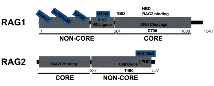
1996) (Figure 1.2). The amino-terminal non-core regions of RAG1include a Really
Interesting New Gene (RING) domain with E3 ubiquitin ligase activity and sequences
that interact with the VprBP kinase, the Roc1/Cul4A/DDB1 ubiquitin ligase complex,
histones H3 and variant H3.3, the MDC1 and Ku70/Ku80 DNA damage response
proteins, and the Gmeb1 transcription factor (Coster et al., 2012; Grazini et al., 2010;
Jones et al., 2011; Kassmeier et al., 2012; Kim et al., 2013; Maitra and Sadofsky, 2009;
Raval et al., 2008) (Figure 1.2). The discoveries of these biochemical activities have led
to hypotheses linking the non-core regions of RAG1 to the modulation of antigen
receptor gene assembly through ubiquitylating proteins to promote Ig/TCR locus
accessibility and through transducing signals following RAG DNA breaks (Dudley et al.,
2003; Grazini et al., 2010; Horowitz and Bassing, 2014; Jones et al., 2011; Sadofsky,
regulating these or other aspects of antigen receptor gene assembly within the context
of developing lymphocytes in vivo remain poorly understood.
Roles for Non-Core RAG1 Regions In Vivo
Although dispensable for V(D)J recombination, non-core RAG regions are
essential for normal lymphocyte development. Human RAG mutations that completely
block V(D)J recombination cause B cell-, T cell-, Natural Killer (NK) cell+ severe
combined immunodeficiency (SCID) associated with a complete lack of circulating B or T
cells (Lee et al., 2014). Hypomorphic RAG mutations that disrupt the biochemical
activities of non-core RAG1 regions but preserve recombination activity cause a variety
of atypical SCID-like conditions, including combined immunodeficiency (CID) with
granuloma and/or autoimmunity, late-onset SCID, and Omenn Syndrome (OS), an
immune deficiency associated with lack of circulating B cells, oligoclonal TCR repertoire,
autoimmunity, T cell infiltration of the skin and gut, high serum IgE levels, and
erythroderma (Aleman et al., 2001; Santagata et al., 2000; Wong and Roth, 2007).
However, how mutations in non-core RAG regions that preserve V(D)J recombination
activity but cause autoimmunity and altered AgR repertoire remain poorly understood.
Untreated OS is typically fatal (Aleman et al., 2001), and current therapies are limited to
hematopoietic stem cell transplantation from healthy donors (Caglayan Sozmen et al.,
2015). Further, the late onset and immune and autoimmune phenotypes observed in
these atypical-SCID and OS patients may complicate treatment (Jspeert et al., 2014).
Thus, defining how RAG mutations that preserve recombination activity but compromise
lymphocyte development may generate novel and/or targeted therapeutics for SCID and
OS patients.
While dispensable in vitro, the non-core regions of Rag1 promote efficient V(D)J
exhibit a partial block in early lymphocyte development and reduced numbers of mature
B and T lymphocytes that are associated with reduced D-J and V-to-DJ at IgH and
TCRβloci (Dudley et al., 2003). Specifically, Rag1C/Cmice exhibit reduced Dβ-Jβ and
Vβ-DβJβ recombination in non-selected DN3 thymocytes, a partial block at the DN-to-DP
transition, and a 50% reduction in mature splenic T cell numbers as well as reduced DH
-JH and VH-DHJH recombination in pre-B cells, a partial block at the pro-B-to-pre-B cell
transition, and a 50% reduction in mature splenic B cell numbers (Dudley et al., 2003).
These phenotypes implicate non-core Rag1 regions in promoting efficient V(D)J
recombination. Yet, the specific roles of non-core Rag1 domains in the context of V(D)J
recombination or lymphocyte differentiation in vivo are unknown. For instance, because
D-to-J recombination is required for V-to-DJ recombination to occur, and as RAG binding
over D and J segments is proposed to promote V-to-DJ recombination, Rag1C/C mouse
phenotypes may be due to defects in D-to-J, V-to-DJ, or both steps. Data shown in
Chapter 2 of this thesis shows that expression of a pre-assembled endogenous VβDβJβ
allele, but not a DβJβ allele, rescues αβ T cell development in Rag1C/Cmice, indicating
non-core Rag1 regions primarily function during V(D)J recombination to promote Vβ
-to-DJβ recombination. Data in Chapter 2 also demonstrates that Rag1C/Cαβ T cells express
a Vβ repertoire that is skewed during both primary and secondary Vβ-to-DβJβ
recombination and that Rag1C/C thymocytes undergo altered
αβTCR selection,
implicating non-core Rag1 regions in promoting a diverse TCRβ repertoire. Further
experiments in Chapter 2 demonstrate that replacement of a single non-consensus Vβ
RSS for an efficient with a consensus RSS nearly rescues αβ T cell development in
Rag1C/C mice, implicating non-core Rag1 in establishing V
β diversity through overcoming
novel roles for non-core Rag1 regions in primary V(D)J rearrangements and αβ TCR
selection to promote the generation of a diverse naïveαβ TCR repertoire in vivo.
In addition to promoting V(D)J recombination, the non-core regions of RAG1
have been proposed to be important for AgR gene segment accessibility, V(D)J
efficiency, RAG DSB repair, and/or signaling following RAG DSBs. Yet, the specific roles
of these domains in the context of V(D)J recombination or lymphocyte differentiation in
vivo are unknown. Data presented in Chapter 3 of this thesis shows that Rag1C/C mice
have decreased accessibility of Jλ1 gene segments and that the Rag1 protein promotes
accessibility of both Jκ and Jλ gene segments, implicating Rag1 in promoting AgR locus
accessibility independent of Rag cleavage activity. Further, RAG DSBs in pre-B cells
induce hundreds of transcriptional changes in genes encoding proteins associated with
cell survival, lymphocyte differentiation, and AgR selection. These include the NF-κ
B-dependent pro-survival factor Pim2, which promotes Igλ+ B cell development. Data
shown in Chapter 4 of this thesis shows that Rag1C/C mice exhibit impaired late B cell
development with profound defects in Igλ+ B cell development that is associated with
reduced Vλ-to-Jλ recombination. Additional experiments show that Rag1C/C pre-B cells
do not upregulate Pim2 during RAG DSBs and that expression of pro-survival BCL2
protein rescues Igλ+ cell development in Rag1C/C mice, indicating that non-core Rag1
regions promote survival signals in response to RAG DSBs in pre-B cells that promote
the development of Igλ+ cells. Collectively, the data presented in this thesis
demonstrates novel, non-cleavage functions for the RAG endonuclease in establishing a
normal immune repertoire and also implicates RAG1 as a chromatin-modifying factor
that promotes accessibility of AgR genes prior to recombination and as a signal
transduction molecule that promotes the survival and differentiation of developing
Figure 1.1
Figure 1.1 The structure of AgR receptor loci. Schematics indicating mouse AgR
receptor loci, with variable (V) gene segments shown in red, diversity (D) gene segments
are shown in grey, joining (J) gene segments are shown in blue, with constant (C) genes
shown in black. Approximate locus sizes are indicated and numbers of gene segments
per locus are indicated in parentheses, where relevant.
IgH
VH (150)
2.8 Mb DH (9) 60 Kb 1.4 KbJH (4) CH
Ig!
V! (140)
3.2 Mb J
! (4)
1.4 Kb C!
TCRβ
Vβ(30)
380 Kb Dβ1 2.5 KbJβ1 (6) Cβ1 Dβ2 2.5 KbJβ2 (6) Cβ2 Vβ14
TCRα/δ
Vα/δ(100)
1.4 Mb Dδ/Jδ Cδ J64 Kbα (61) Cα
Igλ
Vλ(2) Jλ2 Cλ2 Jλ4 Cλ4 Vλ1 Jλ1 Cλ1
Figure 1.2
Figure 1.2 The RAG proteins. Schematics of RAG1 and RAG2 protein domains.
Indicated “core” regions are critical for V(D)J recombination, while “non-core” regions are
dispensable for this reaction in vitro. The core-RAG1 domain possesses DNA cleavage
activity, catalyzed at the indicated D708A residue, a RSS nonamer-binding domain
(NBD), a RSS heptamer-binding domain (HBD), and sequences that bind the V(D)J
co-factor RAG2. The non-core regions of RAG1 include a RING domain with E3 ligase
activity that can ubiquitylate histones H3 and H4 as well as sequences that bind the
VprBP component of the VprBP/Roc1/Cul4/DDB E3 ligase complex, DNA damage
response proteins Ku70/Ku80, and the transcription factor Gmeb1. The core RAG2
protein interacts with RAG1, and the non-core regions of RAG2 include a PHD domain,
which binds H3K4me3, and the indicated T490 residue, which regulates cell-cycle
CHAPTER 2
Noncore RAG1 Regions Promote Vβ Rearrangements and αβ T Cell Development
by Overcoming Inherent Inefficiency of Vβ Recombination Signal Sequences1
Abstract
The RAG proteins are comprised of core endonuclease domains and non-core
regions that modulate endonuclease activity. Mutation or deletion of non-core RAG
regions in humans causes immunodeficiency and altered TCR repertoire, and mice
expressing core but not full-length Rag1 (Rag1C/C) or Rag2 (Rag2C/C) exhibit
lymphopenia, reflecting impaired V(D)J recombination and lymphocyte development.
Rag1C/Cmice display reduced D-to-J and V-to-DJ rearrangements of TCR
β and IgH loci,
while Rag2 C/Cmice show decreased V-to-DJ rearrangements and altered
Vβ/VΗ repertoire. Since Vβs/VΗs only recombine to DJ complexes, the Rag1
C/C phenotype
could reflect roles for non-core RAG1 regions in promoting recombination during only the
D-to-J step or during both steps. Here, we demonstrate that a pre-assembled TCRβ
gene, but not a pre-assembled DβJβcomplex or the pro-survival BCL2 protein,
completely rescues αβ T cell development in Rag1C/C mice. We find that Rag1C/C mice
exhibit altered Vβ utilization in Vβ-to-DJβrearrangements, increased usage of 3'Jαgene
segments in Vα-to-Jαrearrangements, and abnormal changes in Vβ repertoire during
αβ TCR selection. Inefficient Vβ/VΗrecombination signal sequences (RSSs) have been
hypothesized to cause impaired V-to-DJ recombination on the background of a defective
recombinase as in core-Rag mice. We show that replacement of the Vβ14 RSS with a
1
Originally published in The Journal of Immunology. Horowitz, J.E., and Bassing, C.H. 2014. Noncore RAG1 regions promote Vβ rearrangements and αβ T cell development by overcoming inherent
inefficiency of Vβ recombination signal sequences. J. Immunol. Vol: 192, 4, pp1609-1619. Copyright ©
more efficient RSS increases Vβ14 recombination and rescues αβ T cell development in
Rag1C/C mice. Our data indicate that non-core RAG1 regions establish a diverse TCR
repertoire by overcoming VβRSS inefficiency to promote Vβ recombination and αβ T
Introduction
The lymphocyte-specific RAG1/RAG2 (Fugmann et al., 2000) endonuclease
generates Ag receptor diversity by recombining germline variable (V), diversity (D), and
joining (J) gene segments of T cell receptor (TCR) and immunoglobulin (Ig) loci. RAG
cleavage between two participating gene segments and their adjacent RSSs yields DNA
double strand breaks (DSBs) comprised of hairpin-sealed coding ends and blunt signal
ends (Fugmann et al., 2000; Schatz and Ji, 2011). RAG proteins along with DNA
damage response/repair proteins hold these DNA ends in a stable post-cleavage
complex and facilitate their repair by non-homologous end-joining (NHEJ) factors
(Helmink and Sleckman; Lieber). The combination of possible V(D)J joining events and
the imprecise manner by which coding ends are processed cooperate to generate Ag
receptor diversity.
The RAG1 and RAG2 proteins are each comprised of core endonuclease
domains, defined as the minimal sequences required for DNA cleavage in vitro, and
non-core regions that modulate this activity (Sadofsky, 2004; Schatz and Swanson, 2011).
RAG1 or RAG2 mutations that alter or delete non-core RAG1 or RAG2 region amino
acids and reduce overall V(D)J recombinase activity cause Omenn Syndrome (OS) or
other fatal severe combined immunodeficiencies associated with oligoclonal TCR
repertoire and increased T cell mediated autoimmunity (Notarangelo, 2010; Villa et al.,
2008; Villa et al., 2001; Wong and Roth, 2007). However, the contribution of diminished
recombinase activity to aberrant TCR repertoire and autoreactive T cells in OS patients
remains undetermined (Wong and Roth, 2007). Rag1C/CandRag2C/Cmice each display
reduced numbers of mature T and B cells, reflecting impaired lymphocyte development
beyond the progenitor stages associated with reduced TCRβ and IgH recombination
display reduced levels of both D-to-J and V-to-DJ recombination of TCRβ and IgH loci
(Dudley et al., 2003), Rag2C/Cmice show predominantly decreased V-to-DJ
rearrangements associated with altered Vβ/VH usage (Akamatsu et al., 2003; Liang et
al., 2002). Although no reductions in the levels of V-to-J recombination of Igκ or
TCRαloci were discovered in either Rag1C/Cor Rag2C/Cmice, potential changes in the
utilization of individual Igκ or TCRα gene segments have not been assayed (Akamatsu
et al., 2003; Liang et al., 2002). Considering that Vβs and VHs only recombine to DJ
complexes, the Rag1C/Cphenotype could reflect roles for non-core RAG1 regions in
promoting recombination during only the D-to-J step or during both the D-to-J and
V-to-DJ steps (Dudley et al., 2003). Since Vβ and VH rearrangements are selectively impaired
in Rag2C/Cmice, it has been hypothesized that the Rag2C/Cmouse phenotype is due to
the interaction of a defective recombinase with unique features of Vβ/VH RSSs (Liang et
al., 2002). In support of this notion, Vβ/VH RSSs that more closely resemble those of Dβ
and Vλ/Vα/Vγ/Vδsegments are more efficiently cleaved by core-RAG proteins in vitro
and their associated Vβ/VH segments are recombined and expressed at higher
frequencies in Rag2C/Cmice relative to normal mice (Liang et al., 2002). Yet, neither this
decade-old model nor its prediction that Rag1C/Cmice also would exhibit altered V
β and
VH utilization has been tested.
In humans and mice, αβ T cells develop in the thymus through a differentiation
program that involves the ordered assembly, expression, and selection of TCR genes.
TCRβ genes assemble through Dβ-to-Jβ and then Vβ-to-DβJβ rearrangements in CD4
-CD8- “double-negative” (DN) thymocytes (Krangel, 2009). The D
β-to-Jβ recombination
step initiates in c-Kit+CD25- DN1 cells and continues in c-Kit+CD25+ DN2 cells, while Vβ
contain 31 Vβ segments (Trbv1 through Trbv31) and two Dβ-Jβ-Cβ clusters (
Trbd1-Trbj1-Trbc1 and Trbd2-Trbj2-Trbc2)each with one Dβ and six functional Jβsegments,
secondary Vβ rearrangements can occur to Dβ2Jβ2 (Trbd2Trbj2) complexes on alleles
with primary Vβ rearrangements to assembled Dβ1Jβ1 (Trbd1Trbj1) complexes (Brady
et al., 2010). Utilization of individual Vβ segments in primary Vβ-to-Dβ1Jβ1
rearrangements is biased independent of Vβ position (Wilson et al., 2001). However, Vβ
position relative to a pre-assembled VβDβJβ1Cβ1 gene can influence the usage of Vβ
segments in secondary Vβ-to-Dβ2Jβ2 rearrangements (Brady et al., 2010; Steinel et al.,
2010). Assembly and expression of a functional TCRβ gene generates TCRβ chains that
pair with pre-Tα proteins to form pre-TCRs, which promote survival and differentiation,
down-regulate RAG expression, and induce expression of the Cyclin D3 (Ccnd3) protein
(Sicinska et al., 2003; von Boehmer and Melchers, 2010). Ccnd3 drives proliferation as
DN3 cells down-regulate RAG expression and differentiate into c-Kit-CD25- DN4 and
then CD4+CD8+ “double-positive” (DP) thymocytes. TCR
α genes assemble through Vα
-to-Jαrearrangements on both alleles in DP cells, where Vβ-to-DβJβrecombination is
silenced (Sicinska et al., 2003; von Boehmer and Melchers, 2010). The assembly and
expression of a functional VαJαCαgene generates TCRα chains that can pair with
TCRβ chains to generate ab TCRs, which are selected based on interactions with thymic
epithelial cells (Chen et al., 2001; Krangel, 2009). Positive selection increases
expression of αβ TCRs and promotes differentiation of DP thymocytes into CD4+CD8- or
CD4-CD8+ “single-positive” thymocytes that emigrate from the thymus as mature naive
αβ T cells (von Boehmer and Melchers, 2010). Since TCRα loci contain ~100 Vαand
~50 Jα segments, successive Vα-to-Jαrearrangements can occur until positive
2010). Although pre-TCR selection and thymocyte expansion does not significantly
change Vβrepertoire during DN-to-DP thymocyte differentiation (Krangel, 2009), positive
selection can substantially alter Vβ representation in ab TCRs during DP-to-SP
thymocyte development (Wilson et al., 2001).
The mouse αβ T cell differentiation program provides a useful experimental
model to elucidate roles for non-core RAG1 regions in promoting V(D)J recombination
and controlling TCR gene repertoire. Rag1C/Cmice exhibit reduced D
β-Jβ and Vβ
-DβJβrecombination in DN3 thymocytes and impaired DN3-to-DP thymocyte
development from accumulation of cells at the DN3 stage (Blackman et al., 1989;
Carpenter et al., 2009; Kappler et al., 1987; Kappler et al., 1988; MacDonald et al., 1988;
Wade et al., 1988; Wu et al., 2003). These phenotypes may arise from impaired Dβ
-to-Jβ and/or Vβ-to-DβJβ recombination in the absence of non-core RAG1 regions. Yet,
considering that RAG DSBs induce changes in the expression of proteins involved in
cellular survival, lymphocyte differentiation, and Ag gene receptor selection (Dudley et
al., 2003), these phenotypes also may arise from impaired signaling in response to RAG
DSBs induced in Rag1C/CDN3 thymocytes. To determine how non-core RAG1 regions
promote TCRβ gene assembly and αβ T cell development, we have created and
analyzed Rag1C/Cmice containing a pre-assembled DβJβ complex or pre-assembled
functional TCRβgene, expressing the pro-survival EµBCL2 transgene, or with the 3'Dβ1
RSS in place of the Vβ14 RSS. We demonstrate that this TCRβ gene, but not the DβJβ
complex or BCL2, completely rescues DN3-to-DP thymocyte development in Rag1C/C
mice, indicating that the predominant function of non-core RAG1 regions in
differentiating αβ T cells is to promote Vβ recombination. We show that Rag1C/C mice
cells attempting Vβ recombination nor TCRβ-dependent expansion of DN3 thymocytes
contributes to this abnormal Vβ repertoire. We detect increased usage of 3'Jα segments
in Vα-to-Jα rearrangements and abnormal selection of the Vβ repertoire in Rag1C/Cmice,
revealing that non-core RAG1 regions also function during TCRα recombination in DP
thymocytes. Finally, we show that the 3'Dβ1 RSS increases Vβ14recombination and
partially rescues αβ T cell development in Rag1C/C mice. Collectively, our data indicate
that non-core RAG1 regions establish a diverse αβ TCR repertoire by overcoming Vβ
RSS inefficiency to promote Vβ recombination and αβ T cell development and by
Materials and Methods
Mice
Rag1C/C (Bredemeyer et al., 2008), E
µBCL2 (Dudley et al., 2003), Jβ1DJ/DJ (Strasser et
al., 1991), Vβ1NT/NT (Carpenter et al., 2009), and Ccnd3-/- (Brady et al.) mice were
obtained and utilized to generate the mice described in this study. The germline
Vβ143’Db1RSS mice were generated by Cre-loxP-mediated gene-targeting using W4
mouse embryonic stem cells and the construct previously employed to make chimeric
Vβ143’Db1RSS mice (Sicinska et al., 2003). All experimental mice were on a mixed
129SvEv and C57BL/6 background and were littermate or age-matched mice between
4-6 weeks of age. All experiments were conducted in accordance with national
guidelines and approved by the Institutional Animal Care and Use Committee of the
Children's Hospital of Philadelphia.
Flow Cytometry
Single cell suspensions were stained with antibodies in PBS containing 2% BSA. All
antibodies were purchased from BD Pharmingen. CD4 and CD8 analysis was performed
using anti-CD4 (553653), anti-CD8 (553031), and anti-TCRβ (553174) antibodies. DN
stage analysis was performed on lineage-negative cells stained with a mixture of
PE-conjugated anti-CD4 (553653), anti-CD8α (553033), anti-TCRβ (553172), anti-TCRγ
(553178), anti-B220 (553090), anti-CD19 (553786), anti-CD11b (553311), anti-CD11c
(557401), NK1.1 (553165), and Ter119 (553673) antibodies in addition to
anti-CD25 (552880) and anti-CD117 (553356) antibodies. Vβ analysis was performed using
antibodies against TCRβ (553174) as well as Vβ5 (553189), Vβ6 (553192), Vβ8
acquired on a FACSCalibur (BD Biosciences, San Jose, CA) using CellQuest software
(BD Biosciences) and analyzed using FlowJo software (Tree Star).
PCR
Genomic DNA from sorted DP thymocytes (on 100 ng and 1:5 serial dilutions) was
subjected to long-range PCR with the use of primers and PCR conditions as described
Results
Transgenic BCL2 expression partially rescues early αβ T cell development in
Rag1C/C mice.
To determine whether impaired cellular survival in response to RAG DSBs
causes impaired DN3-to-DP thymocyte development in Rag1C/Cmice, we created and
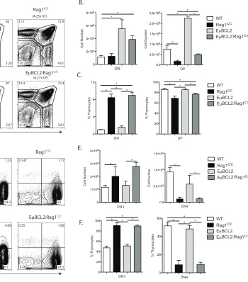
analyzed Rag1C/C mice containing the E
µBCL2 transgene (EµBCL2:Rag1C/C mice) since
expression of the anti-apoptotic BCL2 protein increases survival of DN3 cells attempting
TCRβ rearrangements (Guo et al., 2002; Riegert and Gilfillan, 1999). Consistent with the
pro-survival effect of BCL2, we detected ~3-fold increases in the numbers DN and DP
thymocytes in EµBCL2:Rag1C/C mice compared to Rag1C/C mice, although the difference
in DN thymocyte numbers did not reach statistical significance (Figure 2.1 A and B).
Similarly, we found equivalent numbers of DN3 and DN4 thymocytes in EµBCL2:Rag1C/C
and Rag1C/C mice (Figure 2.1 C and D). We also detected ~4.5-fold decreased numbers
of total and DP thymocytes in EµBCL2:Rag1C/C mice relative to EµBCL2 mice (Figure
2.1 A and B), indicating that BCL2 expression does not completely rescue impaired
DN-to-DP thymocyte development in Rag1C/C mice.These observations indicate that BCL2
expression does notsubstantially enhance the survival of Rag1C/CDN3 thymocytes, yet
has a more pronounced effect on promoting survival of DP thymocytes or cells during
DN-to-DP thymocyte expansion and differentiation in Rag1C/Cmice. Therefore, we
conclude that reduced survival of DN3 thymocytes in response to RAG DSBs is not a
major cause of impaired TCRβ gene assembly and DN-to-DP thymocyte development in
A pre-assembled TCRβ gene completely rescues early αβ T cell development in
Rag1C/C mice.
To determine potential functions of non-core Rag1 regions in promoting Dβ-to-Jβ
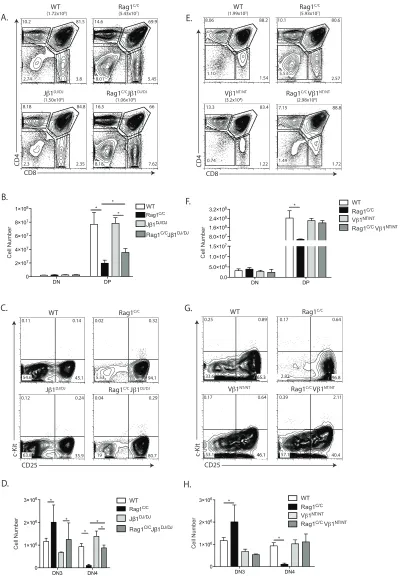
recombination and Vβ-to-DJβ recombination, we generated and analyzed Rag1C/C mice
that contain a pre-assembled DβJβ complex (Jβ1DJ) or pre-assembled functional TCRβ
gene (Vβ1NT) on both TCRβ alleles. The Jβ1DJ allele contains a pre-assembled Dβ1Jβ1.1
(Trbd1Trbj1.1) complex, lacks Dβ2 (Trbd2) and Jβ2 (Trbj2) segments, and must
recombine one of 35 Vβ segments to the Dβ1Jβ1.1 complex to promote αβT lymphocyte
development (Brady et al., 2013) The Vβ1NT allele contains a pre-assembled functional
endogenous Vβ1Dβ1Jβ1.4Cβ1 (Trbv5Trbd1Trbj1.4Trbc1) gene that promotes αβ T cell
development independent of TCRβ recombination We detected ~2-fold more DP
thymocytes in Rag1C/CJ
β1DJ/DJ mice relative to Rag1C/C mice (Figure 2.2 A and B).
However, the DP cell numbers in Rag1C/CJβ1DJ/DJ mice and Rag1C/C mice were 2-fold or
more lower as compared to EµBCL2 and WT mice, respectively (Figure 2.2 B versus
Figure 2.1 B). We also detected ~8-fold more DN4 cells in Rag1C/CJ
β1DJ/DJ mice relative
to Rag1C/C mice (Figure 2.2 C and D), with DN4 thymocyte numbers in Rag1C/CJ
β1DJ/DJ
mice and Rag1C/C mice significantly lower than in E
µBCL2 and WT mice, respectively
(Figure 2.2 D versus Figure 2.1 E). These data demonstrate that a pre-assembled
DβJβ1 complex on both TCRβ alleles partially rescues DN3-to-DN4 and DN-to-DP
thymocyte development in Rag1C/C mice. In Rag1C/CV
β1NT/NT mice relative to Rag1C/C
mice, we observed greater numbers of DP (4-fold more) and DN4 (11-fold more)
thymocytes (Figure 2.2 E - H). Notably, the numbers of DP and DN4 thymocytes in
Rag1C/CV
β1NT/NT mice were similar to those in Vβ1NT/NT and WT mice (Figure 2.2 E - H),
DN3-to-DN4 and DN-to-DP thymocyte development in Rag1C/C mice. Therefore, our data
indicates that non-core RAG1 regions promote both Dβ-to-Jβ and Vβ-to-DJβ
rearrangements and that reduced Vβ-to-DJβ recombination is the major cause of
accumulation of cells at the DN3 stage and impaired DN-to-DP thymocyte development
in Rag1C/Cmice.
Rag1C/C mice exhibit altered V
β utilization in primary and secondary Vβ
rearrangements.
The TCRβ locus architecture permits primary Vβ rearrangements to Dβ1Jβ1
complexes and then secondary Vβ rearrangements to Dβ2Jβ2 complexes, which occur
on the Vβ1NT allele (Carpenter et al., 2009).Our current analysis of thymocyte
development in Rag1C/CJ
β1DJ/DJ and Rag1C/CVβ1NT/NT mice and our previous analysis of
Vβ8 (Trbv13.1, Trbv13.2, and Trbv13.3) and Vβ10 (Trbv4) rearrangements in DN3 cells
of Rag1C/C mice (Brady et al., 2010) demonstrate that primary V
β-to-DJβ rearrangements
are impaired in the absence of non-core Rag1 regions. However, these analyses cannot
address potential function of non-core Rag1 regions in secondary Vβ rearrangements
nor quantify relative usage of individual Vβ segments in Vβ-to-DβJβ rearrangements.
Since DN-to-DP thymocyte differentiation does not significantly alter Vβ repertoire
(Dudley et al., 2003), the use of flow cytometry to monitor Vβ expression on
TCRβintermediate DP thymocytes provides a more sensitive means than PCR to quantify
relative Vβ usage in Vβrearrangements (Wilson et al., 2001). Thus, to determine the
contributions of non-core Rag1 regions in Vβ utilization during primary Vβ-to-DβJβ
rearrangements, we assayed Vβ5 (Trbv12.1 and Trbv12.2), Vβ6 (Trbv19), Vβ8, Vb10,
as only primary Vβ rearrangements occur on the Jb1DJ allele (Brady and Bassing, 2011;
Brady et al., 2010; Wilson et al., 2001; Wu et al., 2003). We observed lower percentages
of Vβ5+ (5-fold less) and Vβ10+ (~3-fold less) DP thymocytes in Rag1C/CJβ1DJ/DJ mice as
compared to Jβ1DJ/DJ mice (Figure A and B). We detected increases in the percentages
of Vβ8+ (2-fold more) and Vβ14+ (3-fold more) DP thymocytes, but no difference in the
percentages of Vβ6+ DP cells, in Rag1C/CJβ1DJ/DJ mice relative to Jβ1DJ/DJ mice (Figure
2.3 A and B). Since only secondary Vβ-to-DβJβ rearrangements involving Vβ10 occur on
the Vβ1NT allele (Carpenter et al., 2009), we next quantified Vβ10 expression on DP
thymocytes of Rag1C/CV
β1NT/NT and Vβ1NT/NT mice to evaluate whether non-core Rag1
regions promote such secondary Vβ rearrangements. We found a 7-fold lower
percentage of Vβ10+ DP thymocytes in Rag1C/CVβ1NT/NT mice as compared to Vβ1NT/NT
mice (Figure 2.3 C and D). Collectively, these data indicate that non-core Rag1 regions
affect Vβ utilization in primary and secondary Vβ rearrangements, at least on Jβ1DJ and
Vβ1NT alleles.
To evaluate whether non-core Rag1 regions affect Vβ utilization in total Vβ
rearrangements on normal TCRβ alleles, we quantified expression of Vβ5, Vβ6, Vβ8,
Vβ10, and Vβ14 on DP thymocytes ofRag1C/C and WT mice. We detected lower
percentages of Vβ5+ (~6-fold less) and Vβ10+ (~2-fold less) DP cells in Rag1C/C mice
relative to WT mice (Figure 2.3 E and F). We also detected ~2-fold increased
percentage of Vβ8+ and ~3-fold increased percentage of Vβ14+ DP thymocytes, but no
difference in the percentage of Vβ6+ DP cells, in Rag1C/C mice as compared to WT mice
(Figure 2.3 E and F). These data demonstrate that non-core Rag1 regions influence Vβ
While we did not observe a major role for impaired survival of Rag1C/C
thymocytes in response to RAG DSBs during TCRβ recombination, altered survival of
DN3 thymocytes during rearrangements of particular Vβ segments could influence
Vβrepertoire in Rag1C/C mice. To investigate this possibility, we quantified the
expression of Vβ5, Vβ6, Vβ8, Vβ10, and Vβ14 on DP thymocytes of EµBCL2:Rag1C/C
and Rag1C/C mice. We detected no significant differences in the frequencies of Vβ6+,
Vβ5+, Vβ10+ or Vβ14+ DP thymocytes between EµBCL2:Rag1C/C and Rag1C/C mice
(Figure 2.3 E and F), however we did observe a slightly higher frequency of Vβ8+ DP
cells in EµBCL2:Rag1C/C mice as compared to Rag1C/C mice (Figure 2.3 E and F). These
data suggest that impaired survival of DN3 cells in response to RAG DSBs does not
cause the altered relative representation of Vβ5, Vβ6, Vβ8, Vβ10, and Vβ14 on DP
thymocytes of Rag1C/C mice.
While Vβ representation is not significantly altered during DN-to-DP thymocyte
differentiation in mice with normal levels of Vβ rearrangements (Brady et al., 2010),
altered expansion of DN cells expressing particular Vβ segments could contribute to the
altered Vb repertoire in DP thymocytes of Rag1C/C mice which have reduced numbers of
thymocytes caused by impaired Vβ-to-DβJβ recombination. To assess this possibility,
we generated and analyzed Rag1C/CCcnd3-/- mice since expression of TCR
β chains drive
DN-to-DP thymocyte expansion through Ccnd3, and Ccnd3-/- mice exhibit normal Vb
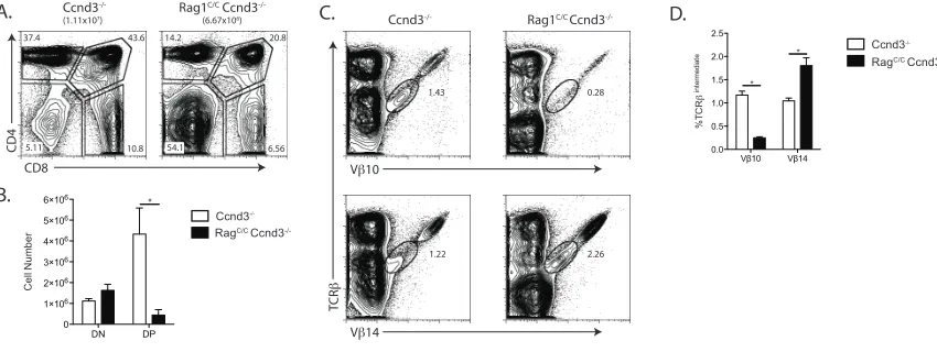
repertoire in DP thymocytes (Wilson et al., 2001). We found equivalent numbers of DN
thymocytes, but ~10-fold lower numbers of DP cells, in Rag1C/CCcnd3-/- mice as
compared to Ccnd3-/- mice (Figure 2.4 A and B), indicating that TCR
β-mediated
DN-to-DP thymocyte development is profoundly impaired in Rag1C/CCcnd3-/- mice relative to
~40-fold as compared to Rag1C/C mice versus ~17 fold for Ccnd3-/- mice relative to WT
mice (Compare Figure 2.4 B and Figure 2.1 B). We also found a ~5-fold lower frequency
of Vβ10+ DP cells but a ~2-fold increase frequency of Vβ14+ DP thymocytes in
Rag1C/CCcnd3-/- mice as compared to Ccnd3-/- mice (Figure 2.4 C and D). Considering
that we observed similar increased and deceased frequencies of Vβ10+ and Vβ14+ DP
thymocytes, respectively, in Rag1C/C mice relative to WT mice (Figure 2.3 E and F), our
data indicate that the altered representation of Vβ10 and Vβ14 on DP thymocytes of
Rag1C/C mice is not caused by differences in TCR
β-mediated proliferation of DN cells
expressing particular Vβ segments. Therefore, based on our quantification of Vβ
expression on DP thymocytes of mice expressing wild-type or core-Rag1 proteins in
combination with other genetic modifications, we conclude that Rag1 non-core regions
control Vβ repertoire at the level of relative Vβ usage in primary and secondaryVβ
-to-DβJβ rearrangements.
Rag1C/C mice exhibit altered Jα utilization in Vα-to-Jα rearrangements and
abnormal changes in Vβ repertoire during ab TCR selection.
In DP thymocytes of wild-type mice, TCRα recombination occurs on both alleles
and normally involves successive rounds of Vα-to-Jα rearrangements on each allele
(Brady and Bassing, 2011; Sicinska et al., 2003). Although both TCRα alleles are
recombined in mature αβ T cells of Rag1C/C mice (Krangel, 2009), whether the absence
of non-core Rag1 regions results in a modest reduction in Vα-to-Jα rearrangements in
DP thymocytes is not known. Since reduced V(D)J recombinase activity in DP cells
leads to increased representation of 5'Jαs and decreased representation of 3'Jαs in Vα
in DP cells of Rag1C/C mice are similarly biased. For this purpose, we used PCR primers
that hybridize to the Vα3 (Trav9-4) family of gene segments or to Jα61 (Traj61), Jα42
(Traj42), Jα17 (Traj17), or Jα4 (Traj4) to amplify Vα3-to-Jα rearrangements involving
these 5' (Jα61, Jα42) or 3' (Jα17, Jα4) Jα gene segments from sort-purified DP cells of
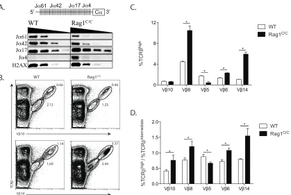
WT or Rag1C/C mice. The levels of PCR products representing V
α3 rearrangements to
Jα61 and Jα42 were reduced in DP thymocytes from Rag1C/C mice relative to WT mice
(Figure 2.5 A). In contrast, the levels of PCR products representing Vα3 rearrangements
to Jα17 and Jα4 were elevated in DP thymocytes from Rag1C/C mice as compared to WT
mice (Figure 2.5 A). These data reveal that loss of non-core Rag1 regions results in
decreased usage of 5'Jαs and increased usage of 3'Jαs in Vα-to-Jα rearrangements.
Although this biased targeting of Vα3 rearrangements toward 3'Jα gene segments is not
consistent with diminished recombinase activity at the TCRαlocus, our results
demonstrate that non-core Rag1 regions control formation of the TCRα gene repertoire
during Vα-to-Jα recombination in DP thymocytes.
Since positive selection of αβ TCRs expressed on DP thymocytes shapes TCRβ
repertoire (Yannoutsos et al., 2004) and Jα repertoire is altered in DP thymocytes of
Rag1C/C mice (Figure 2.5 A), we investigated the impact of non-core Rag1 regions on Vβ
repertoire during DP-to-SP thymocyte development. We detected ~4-fold lower
frequencies of Vβ5+ TCRβhigh SP thymocytes and higher frequencies of Vβ6+ (~2-fold
more) Vβ8+ (~2-fold more), and Vβ14+ (~5-fold more) TCRβhigh SP cells in Rag1C/C mice
relative to WT mice (Figure 2.5 B and C). Considering that we observed ~2-fold lower
frequencies of Vβ10+ DP thymocytes and equivalent frequencies of Vβ6+ DP cells in
Rag1C/C mice relative to WT mice (Figure 2.3 E and F), these data indicate that positive
WT mice. To confirm this notion, we calculated the ratios of the frequencies of cells
expressing Vβ5, Vβ6, Vβ8, or Vβ14 on TCRβhigh versus TCRβintermediate thymocytes in
Rag1C/C and WT mice (Figure 2.5 D). This analysis indicated enhanced selection for
Vβ6+, Vβ8+, Vβ10+, and Vβ14+ cells and increased selection against Vβ5+ cells during
positive selection of DP thymocytes in Rag1C/C mice as compared to WT mice (Figure
2.5 D). Collectively, these data show that loss of non-core Rag1 regions leads to
abnormal changes in Vβ repertoire during αβ TCR selection.
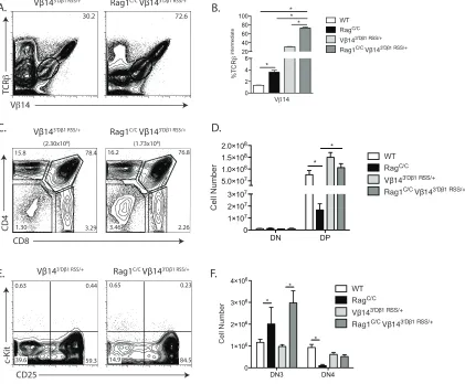
Replacement of the Vβ14 RSS with the more efficient 3'Dβ1 RSS increases Vβ14
recombination frequency and rescues αβ T cell development in Rag1C/C mice.
We have shown that Rag1C/C mice exhibit impaired DN3-to-DP thymocyte
development caused by reduced levels of Vβ-to-DJβ rearrangements, and that this
impaired Vβ recombination is associated with altered recombination frequencies of
individual Vβ segments. A similar phenotype in Rag2C/C mice lead to the hypothesis that
conserved sequence features of Vβ/VH RSSs that make them inefficient relative to 3'Dβ
and Vκ/Vλ/Vδ/Vα RSSs contributes to impaired V-to-DJ recombination on the
background of a diminished recombinase (Blackman et al., 1989; Carpenter et al., 2009;
Kappler et al., 1987; Kappler et al., 1988; MacDonald et al., 1988; Speiser et al., 1989;
Wade et al., 1988). In support of this model, the Vβ8 RSS more closely resembles the
consensus Vκ/Vλ/Vδ/Vα RSS than canonical Vβ RSSs, and Vβ8 exhibits a 1.8-fold
higher rearrangement and expression in Rag1C/C and Rag2C/C mice compared to WT
mice (Liang et al., 2002); Figure 2.3 E and F). In addition, the Vβ5 and Vβ10 RSSs more
closely resemble the consensus Vβ/VH RSS than canonical Vβ RSSs, and Vβ5 and