University of Pennsylvania
ScholarlyCommons
Publicly Accessible Penn Dissertations
1-1-2013
Dynamics of Microeukaryotes and Archaea in the
Mammalian Gut Microbiome
Serena Nicole Dollive
University of Pennsylvania, [email protected]
Follow this and additional works at:
http://repository.upenn.edu/edissertations
Part of the
Bioinformatics Commons
, and the
Microbiology Commons
This paper is posted at ScholarlyCommons.http://repository.upenn.edu/edissertations/749
For more information, please [email protected].
Recommended Citation
Dollive, Serena Nicole, "Dynamics of Microeukaryotes and Archaea in the Mammalian Gut Microbiome" (2013).Publicly Accessible Penn Dissertations. 749.
Dynamics of Microeukaryotes and Archaea in the Mammalian Gut
Microbiome
Abstract
The vertebrate microbiome consists of the bacteria, fungi, archaea, protozoans, and viruses that inhabit the body at diverse locations including the skin, mouth, upper airways, urogenital tract, and digestive tract. These microorganisms are known to synthesize vitamins, interact with and tone the immune system, and
dramatically affect human health. A long list of diseases has been associated with imbalances in commensal microbiome communities. The work presented in this dissertation aims to characterize the microeukaryotic and archaeal components of the gut microbiome through development of wet lab techniques and in silico methods, and apply them to the study of response to antibiotics. These methods provided a picture of the healthy fungal and archaeal communities in the gut, with high prevalence of the yeast Saccharomyces and the archaeon Methanobrevibactor, along with several other species. These new tools were then used to investigate the longitudinal changes that the microbiome undergoes when treated with heavy antibiotics. Using an antibiotic cocktail containing ampicillin, neomycin, vancomycin, and metronidazole in a mouse model, we found that bacterial communities were effectively suppressed and fungi grew out by one to two orders of magnitude. After we discontinued antibiotics, bacterial and fungal cell counts returned to baseline levels within one week, but community composition was still significantly altered. Eight weeks after cessation of antibiotics, fungal community composition was not significantly different from non-treated controls, but several mice continued to have elevated levels of yeasts that had grown out during antibiotic treatment. The bacterial community composition was still significantly different from non-treated controls. Ultimately, this work demonstrated potentially deleterious long term effects of antibiotic use, and emphasizes how strong cage effects can be in mouse studies. The research performed in this dissertation will aid researchers looking to study all three domains of life and take into account the effects of commonly used antibiotics in future microbiome studies.
Degree Type
Dissertation
Degree Name
Doctor of Philosophy (PhD)
Graduate Group
Genomics & Computational Biology
First Advisor
Frederic D. Bushman
Keywords
Archaea, Fungi, Gut, ITS, Microbiome, Microeukaryotes
Subject Categories
Bioinformatics | Microbiology
DYNAMICS OF MICROEUKARYOTES AND ARCHAEA IN THE
MAMMALIAN GUT MICROBIOME
Serena Dollive
A DISSERTATION
in
Genomics and Computational Biology
Presented to the Faculties of the University of Pennsylvania
in
Partial Fulfillment of the Requirements for the
Degree of Doctor of Philosophy
2013
Supervisor of Dissertation
________________________
Frederic D. Bushman, Ph.D., Professor, Microbiology
Graduate Group Chairperson
________________________
Maja Bucan, Ph.D., Professor, Genetics
Dissertation Committee
Christian J. Stoekert, Ph.D., Research Professor of Genetics
Ronald G. Collman, M.D., Professor of Medicine
Hongzhe Li, Ph.D., Professor of Biostatistics
DYNAMICS OF MICROEUKARYOTES AND ARCHAEA IN THE MAMMALIAN GUT MICROBIOME
COPYRIGHT
2013
iii
Dedication
iv
ACKNOWLEDGMENTS
I would first like to thank my advisor, Rick Bushman for his scientific tutelage, support,
and persistent championing for forward motion. Under Rick’s mentorship, I have not just
developed as a scientist but gained a wide set of life skills that will be invaluable going forward. I
would also like to thank our collaborators in the Human Microbiome Project at Penn. I am
especially obliged to Gary Wu and Jim Lewis for their help, support, advice, and insights. I am
also grateful to my thesis committee members: Chris Stoeckert, Ron Collman, Hongzhe Li, and
Paul D. Sniegowski, for their time, knowledge, recommendations, and investment in my graduate
career.
I am thankful for the lessons and skills I have learned from fellow lab members. Chris
Hoffmann taught me nearly all of the wet lab techniques I know today and had an immense
amount of patience with me as a very green rotation student. Through my years in the Bushman
Lab, Chris has been unbelievably helpful and a great friend. I am indebted to Kyle Bittinger for
his advice, cheerleading, and persistent willingness to put aside his work and teach me. I am
thankful to my former baymate Shoshanna Roth who took the time to help me navigate through
the academic world as a beginning graduate student. I am also grateful to Ying-Yu Chen and
Stephanie Grunberg for amount of work and effort they put into the collaborative projects in this
dissertation. I am obliged to the microbiology graduate students in the Bushman lab whose
graduate careers coincided with mine: Emily Charlson, Greg Peterfreund, and Sam Minot.
Having companions with whom I could commiserate and celebrate from classes to dissertation
was incredibly heartening. I am glad for the support and friendship from past and present lab
members: Rohini Sinha, Nirav Malani, Aubrey Bailey, Young Hwang, Jimmy Hu, Sana Attar,
v
Rebecca Custers-Allen, Jennifer Hwang, Christel Chehoud, Erik Clarke, Karen Ocwieja, Mali
Skotheim, Caitlin Greig, and Laurie Zimmerman.
I am also extremely grateful for the support of the GCB faculty and students. Their
enthusiasm and eagerness to help made my graduate student carrier so much more enlightening
and exciting. I am particularly indebted to Maja Bucan, who encouraged and supported me. I am
very appreciative of my fellow GCB graduate students whose friendship and solidarity bettered
my graduate school experience. I am thankful to have had collaborative and congenial
cohort-mates in Ellen Tsai, Aleah Caulin, Nick Stong, and Sira Sriswasdi, whose comradery made
prelims bearable. I am glad to have been able to get to know and spend time with many of my
GCB classmates, including Scott Sherrill-Mix, Rithun Mukherjee, Hannah Dueck, Yih-Chii
Hwang, Sarah Middleton, Varun Aggarwala, Fan Li, Kathleen Sprouffske, Rumen Kostadinov,
Paul Ryvkin, Jun Chen, Anthony Olarerin, Miler Lee, Erik Clarke, Hannah Hutton, Ryan
Coleman, Brett Hannigan, Najaf Shah, and others. I am especially grateful to Hannah Chervitz
and Tiffany Barlow for their continual support and help over the past five years.
I would like to acknowledge the friends I have made at Penn who have made
Philadelephia my home. Friends like Lauren Friedman, Kristy Skowronski, Shanshan Ding,
Yiwen Song, Grace Chao, Katherine O’Brien, Siqing He, Viktoriya Syrovatkina, Judite Costa,
and Anton Kutovoy made the years I have spent at Penn wonderful. I would like to thank my
family: my mother, Janice, my siblings, Blake and Monica, my grandmother, Virginia, my
Aunt Kathy, Uncle John, Uncle Cliff, cousins Brian and Tim, and partner and best friend Tom
vi
ABSTRACT
DYNAMICS OF MICROEUKARYOTES AND ARCHAEA IN THE MAMMALIAN GUT
MICROBIOME
Serena Dollive
Fredrick D. Bushman
The vertebrate microbiome consists of the bacteria, fungi, archaea, protozoans, and
viruses that inhabit the body at diverse locations including the skin, mouth, upper airways,
urogenital tract, and digestive tract. These microorganisms are known to synthesize vitamins,
interact with and tone the immune system, and dramatically affect human health. A long list of
diseases has been associated with imbalances in commensal microbiome communities. The work
presented in this dissertation aims to characterize the microeukaryotic and archaeal components
of the gut microbiome through development of wet lab techniques and in silico methods, and
apply them to the study of response to antibiotics. These methods provided a picture of the
healthy fungal and archaeal communities in the gut, with high prevalence of the yeast
Saccharomyces and the archaeon Methanobrevibactor, along with several other species. These
new tools were then used to investigate the longitudinal changes that the microbiome undergoes
when treated with heavy antibiotics. Using an antibiotic cocktail containing ampicillin, neomycin,
vancomycin, and metronidazole in a mouse model, we found that bacterial communities were
effectively suppressed and fungi grew out by one to two orders of magnitude. After we
discontinued antibiotics, bacterial and fungal cell counts returned to baseline levels within one
week, but community composition was still significantly altered. Eight weeks after cessation of
antibiotics, fungal community composition was not significantly different from non-treated
vii
antibiotic treatment. The bacterial community composition was still significantly different from
non-treated controls. Ultimately, this work demonstrated potentially deleterious long term effects
of antibiotic use, and emphasizes how strong cage effects can be in mouse studies. The research
performed in this dissertation will aid researchers looking to study all three domains of life and
viii
TABLE OF CONTENTS
ACKNOWLEDGMENTS ... IV
ABSTRACT ... VI
TABLE OF CONTENTS ... VIII
LIST OF TABLES ... XI
LIST OF FIGURES ... XII
CHAPTER 1: INTRODUCTION ... 1
History of Microbiome Studies ... 1
Characterization of Microbiome Communities ... 2
The Gut Microbiome ... 3
Fungi ... 5
Protozoa ... 6
Archaea ... 7
Classification of Amplicon Based Sequencing Data ... 10
Antibiotics ... 12
Motivation and Thesis Outline ... 14
References ... 14
CHAPTER 2: A SAMPLE-TO-ANALYSIS PIPELINE FOR ANALYSIS OF
MICROEUKARYOTES IN HUMAN STOOL... 29
2.1 Abstract ... 29
2.2 Introduction ... 29
ix
2.4 Results ... 34
2.5 Discussion ... 50
2.6 Acknowledgements ... 52
2.7 References ... 52
Supplementary Information ... 56
CHAPTER 3: PROTOCOL TO CHARACTERIZE ARCHAEA IN THE
MICROBIOME THROUGH SEQUENCING ... 64
3.1 Abstract ... 64
3.2 Introduction ... 64
3.3 Materials and Methods ... 65
3.4 Results ... 66
3.5 Discussion ... 71
3.6 Acknowledgements ... 72
3.7 References ... 72
CHAPTER 4: PERTURBATION OF FUNGI AND BACTERIA THROUGH
ANTIBIOTICS IN THE MURINE GUT ... 76
4.1 Abstract ... 76
4.2 Introduction ... 77
4.3 Material and Methods ... 78
4.4 Results ... 81
4.5 Discussion ... 96
4.6 Acknowledgements ... 98
4.7 References ... 99
Supplementary Information ... 105
CHAPTER 5: CONCLUSIONS AND FUTURE DIRECTIONS ... 123
x
APPENDIX ... 130
xi
LIST OF TABLES
Sup. Table 2-1.
Samples studied from known eukaryotic organisms.
33
Sup. Table 2-2.
Samples studied from human stool.
34
Sup. Table 2-3.
Oligonucleotides used in Chapter 2.
35
Sup. Table 2-4.
Comparison of BROCC, MARTA, and MEGAN
36
programs.
Table 3-1
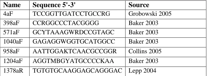
Primer sequences used in Chapter 3.
40
Sup. Table 4-1.
Oligonucleotides used in this chapter.
93
Sup. Table 4-2.
DNA yields and numbers of genomes inferred from
94
ribosomal gene copies data.
xii
LIST OF FIGURES
Figure 2-1.
The eukaryotic ribosomal DNA locus and the targets
9
of amplicons studied.
Figure 2-2.
Classification of
in silico
generated mixed communities
13
of eukaryotic rRNA gene sequences using BROCC.
Figure 2-3.
Analysis of DNA samples from known eukaryotes.
15
Figure 2-4.
Rank-abundance plots of OTUs from stool samples.
19
Figure 2-5.
Comparison of major eukaryotic microbes detected in
21
human stool.
Sup. Figure 2-1.
Comparison of PCR amplification reactions
29
Sup. Figure 2-2.
Analysis of DNA samples from known eukaryotes using 30
BROCC, MARTA, and MEGAN
Sup. Figure 2-3.
Pseudocode of the BROCC program.
31
Sup. Figure 2-4.
Flow chart of BROCC implementation.
32
Figure 3- 1.
Amplification of different primers across macaque and
42
murine stool samples.
Figure 3-2.
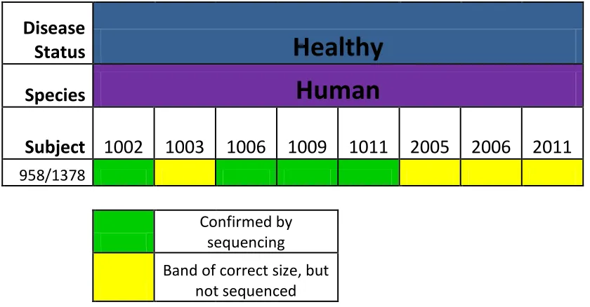
Amplification of human stool samples over the
44
958aF/1378aR primer pair.
Figure 4-1.
Chapter 4 Experimental Diagram.
55
Figure 4-2.
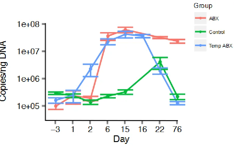
Longitudinal analysis of 16S rRNA gene copies per ng of 56
stool DNA.
Figure 4-3.
Longitudinal analysis of 18S rRNA gene copies per ng of 57
stool DNA.
Figure 4-4.
Diagrams of estimated A) Bacterial and B) Fungal
59
genomes per mouse pellet.
Figure 4-5.
Longitudinal analysis of bacterial lineages inferred
62
from 16S rRNA gene sequencing.
Figure 4-6.
Abundance analysis of observed bacterial lineages.
63
xiii
Figure 4-8.
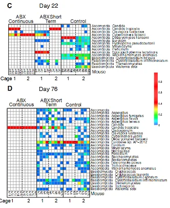
Abundance analysis of observed fungal lineages.
68
Sup. Figure 4-1.
Comparison of microeukaryote lineages specified by the 78
ITS and 18S amplicons.
Sup. Figure 4-2.
Heat maps showing the composition of bacterial
79
communities inferred from 16S sequence data.
Sup. Figure 4-3.
16S and ITS sequences recovered from five samples of
84
mouse chow.
Sup. Figure 4-4.
Heat maps showing the composition of fungal communities 85
inferred from ITS sequence data.
Sup. Figure 4-5.
PCoA analysis distances measures for bacterial and fungal 90
communities.
1
CHAPTER 1: Introduction
History of Microbiome Studies
For over a century, microbial life has been known to live in the healthy human gut [1].
Early methods to study the microbiome were restricted to culture based methods [2] and light
microscopy [1]. These studies were extremely limited, because only a small fraction of bacteria
are culturable [3]. Standard culturing methods may not detect species that are restrictive
anaerobes or auxotrophic. Also, culture based studies are highly biased towards species with the
ability to grow quickly and thrive in the selection media used in a specific study [4]. Basic
microscopic techniques often have difficulty differentiating between species with similar size and
morphology. Advanced phylogenetic stains using Fluorescent In Situ Hybridization (FISH) are
available, but only provide visual information about a microbial community [5].
Microbial community characterization was greatly advanced by the invention of Sanger
sequencing technology in 1977 [6], which did not have the extreme biases seen in culture based
methods and enabled researchers to study a much larger proportion of a microbial population.
Early studies used clone based Sanger sequencing, which provided a larger view of microbiome
communities but was still hampered by low throughput limitations [7]. The advent of high
throughput DNA sequencing in the mid 2000s with early next generation sequencing technologies
such as 454 Pyrosequencing[8], Solexa sequencing [9], and SOLiD sequencing [10], enabled
researchers to produce a large number of reads at a much lower cost than the older Sanger method
and generated read coverages large enough to reflect the true microbial populations in diverse
2
Characterization of Microbiome Communities
There are several strategies used to characterize complex microbial communities. The
most popular method for simple analysis is amplicon based sequencing. Amplicon based
sequencing consists of targeted amplification of a conserved, yet polymorphic region that appears
in all members of the target clade. Ideally, the primer landing sites should be sufficiently
conserved to match the primers and amplify without bias for different species, and the genomic
DNA that lies between the primers must be sufficiently variable between related species to allow
differentiation [11]. Many different genes are used for amplicon based sequencing in different
clades. The 16S ribosomal RNA gene is used almost exclusively in bacteria [12], although other
amplicons have been proposed [13]. The 16S rRNA and rpoB genes [14] are used in archaeal
sequencing. The 18S rRNA gene, 28S rRNA gene, and Internal Transcribed Spacers (ITS 1 and
2) are often used to characterize fungi [15] and protozoa [16,17,18]. Amplicon based sequencing
allows the investigator to identify community members rigorously, but does not provide any
details about community gene content more broadly.
Shotgun metagenomic sequencing has become increasingly popular for characterizing
microbiome communities. Metagenomic sequencing of communities is performed similarly to
genome sequencing: DNA is extracted, shredded, and sequenced [19]. Once sequencing is
complete, data is assembled into contigs through traditional genome assembly methods [20] or
specialized methods, such as de Bruijn graph assembly [21] or binning with self organizing maps
[22]. Data can be analyzed to determine which genes and metabolic capabilities are present in a
community. Nevertheless, taxonomic attribution is difficult with shotgun sequencing, because
many of the genes found may not be present in available databases. Some researchers use 16S
genes mined from sequencing data to estimate the relative abundance of species [19]. Others have
3
species found [23]. Even with improved classification methods, a bias against the detection of
rare community members still exists. In recent years, shotgun sequencing has become
increasingly popular with increases in read length and throughput in Solexa sequencing [19],
which enables better sample coverage and more thorough community characterization.
Recently sequencing of bulk RNA (RNA-Seq) from microbiome samples has become
popular [24]. Such studies allow researchers to determine which genes are being actively
transcribed within a microbial community, which is not possible with DNA sequencing. These
studies can elucidate community dynamics and help determine which community members are
responsible for metabolic activities [25]. Despite these useful insights, RNA-Seq does offer new
bioinformatic challenges. RNAs cannot be assembled into large contigs like genomic DNA: reads
are often mapped back to reference genomes, and analysis quality is more dependent on the
number and quality of reference genomes [26].
The Gut Microbiome
The microbiome consists of the bacteria, archaea, fungi, protozoa, and viruses that live on
and in the human body. It is estimated that microbial cells outnumber human host cells by 10 to 1
[27], and the cumulative gene content is approximately 3.3 million protein coding genes [19].
Commonly studied body sites include the mouth [28], skin [29], upper airways [30], urogenital
tract [31], and digestive tract [32]. Different body sites have been documented to contain unique
communities [33], mostly attributable to differences between sites such as pH, oxygen content,
salinity, temperature, nutrients available at the site, and other factors [33,34,35,36].
The digestive tract is of particular interest, because it contains the majority of commensal
microbiome cells [19]. A majority of the host immune system cells line the digestive tract and
4
[38], ulcerative colitis [39], inflammatory bowel disease [19,40], colorectal cancer [41], and
Clostridium difficile infection [42] have been associated with disruptions in the gut microbiome.
Interestingly, a wide spectrum of non-digestive diseases including diabetes [43], obesity [32],
liver diseases [41], rheumatoid arthritis [44], and autism [45] have also been associated with
changes in the microbiome as well.
Each unique section of the digestive tract has a distinct community [46,47]. pH [47], bile
salt type [27], and oxygen content [48] all vary in different digestive organs, shaping the contents
of the microbial communities. Commonly, stool samples are used to study the gut microbiome,
because they are less invasive than collection methods using an endoscope or surgery, and have
been documented as an adequate surrogate for the communities of the lower gastrointestinal tract
[49].
Most gut microbiome studies have been restricted to analyzing the bacterial communities
exclusively. This is due to the fact that the majority of microbiome cells are bacterial [50] and
robust characterization methods for bacteria have been developed [49]. Several sets of primers
targeting the bacterial 16S are widely used [51]. There is no single core bacterial community in
the gut, but more recent analysis has revealed the presence of enterotypes: sets of specific bacteria
that co-occur in individuals [52]. The initial enterotype study proposed three unique enterotypes
[52], but others have postulated the existence of only two [53]. The two accepted enterotypes are
dominated by Prevotella and Bacteroides, respectively. The more controversial third enterotype is
postulated to be dominated by Ruminococcus [52]. All three enterotypes perform core
microbiome functions including vitamin synthesis, energy regulation, and conditioning the host
immune system [54]. Regardless of the enterotype, the gut bacterial community consists primarily
5
[33]. Several other phyla including Actinobacteria and Fusobacteria appear at very low levels as
well [41].
Fungi
The fungi are a distinct group of micro- and macro-eukaryotes. Both fungi and animals
are members of the opisthokonts, which are characterized by a single flagellum [56] and
conserved insertions in elongation factor-1α and enolase, and supported by several single gene
phylogenies [57]. Fungi have organelles and cell metabolism similar to those in animals, and the
yeast Saccharomyces cerevisiae is often used as a model organism to study genetics and
molecular biology for insights into human biology [57]. Nevertheless, fungi contain a rigid cell
wall structure, which is not seen in animals [58]. Fungal cell walls consist of layers of mannan,
β-glucan, and chitin in different configurations across fungal species and are difficult to lyse with
traditional DNA extraction methods [59]. Many fungal species have variable ploidy. Many
species have haploid, diploid, and polyploid states [60] and genders or mating types within the
haploid state [61]. Also, some fungal species can be multinucleated [62,63]. Fungi have been
observed in nearly all environments with which human come into contact. They can live in soil
[64], water [65], and inside living organisms [66], and can become airborne [67].
Fungi and the Microbiome
Pathogenic fungi are a public health problem and have become increasingly so in the past
several decades with increases in susceptible populations. Specifically, patients with diseases
such as HIV/AIDS [68] and hematological cancers that weaken the immune system [69] are at
elevated risk. Similarly, patients that have had solid organ [70,71] or bone marrow transplants
[72], antibiotic exposure [73], antifungal exposure [73], received systemic corticosteroids [74], or
used a catheter [73] are also at risk. Many fungi can cause invasive fungal infections such as
6
[75,76,77]. Infections can be local and relatively minor, as seen in some cutaneous infections
[78], but many are severe and widespread as in fungal pneumonia [79] or invasive systemic
infection [80]. Fungal infections are particularly serious because they have a higher mortality
rate, over a third of cases in some studies [81,82]. Recently, epidemiologists have seen the rise of
strains of Candida [83], Aspergillus [84], and Cryptococcus spp. [85] that are resistant to
commonly used azole drugs. These isolates are a growing health concern, especially because the
populations of susceptible patients have been increasing [86].
Fungi also inhabit the human body as commensal microbes without causing disease.
Candida spp. have been found in the gut of a majority of humans with culture based methods,
and early sequencing methods indicated that more uncultured fungi are present [87]. The
majority of fungal cells on the skin are Malassezia spp., but culture based evidence suggests that
several other species inhabit the skin including Debaryomyces and Cryptococcus spp. [29].
Culture independent methods indicate that a large diversity of species occupy the oral cavity,
including Candida and Cladosporium spp. [88]. Commensal fungi usually do not cause acute
infection, but in some instances have been linked to disease severity. In patients with ulcerative
colitis, the presence of Candida has been associated with increased disease severity in specific
host genotypes [39]. Outgrowth and infection of commensal Candida commonly occurs when
non-pathogenic yeast forms grow hyphae and invade local tissue. Differences between the host
response to yeast and hyphal states are not well understood, but the host immune system
recognizes the fungal cell wall as a pathogen-associated molecular pattern (PAMP) and has
specific receptors, including Dectin-1 and Dectin-2, that detect and respond to fungi [89].
Protozoa
The Protozoa consist of a diverse paraphyletic group of single celled and multicellular
7
rhizaria, amoebozoa, alveolata, stramenopiles, and excavates [57]. Protozoan organisms are
highly diverse. While all protozoa contain the same basic eukaryotic cell structure, many species
have evolved specialized organelles [90,91,92]. Determination of the protozoal and the
eukaryotic phylogeny has proven difficult, and numerous arrangements of high taxonomic levels
and rootings have been proposed in recent years [57,93,94].
Protozoa and the Microbiome
Many protozoan species are human pathogens and the cause of public health problems in
developing and tropical nations [95]. Many Protozoan diseases such as leishmania [96], malaria
[97], Chagas disease [98], and African sleeping sickness [99] are blood borne pathogens that are
transferred through an insect vector and cause millions of deaths and hundreds of millions
illnesses annually [100]. Other protozoan pathogens have an oral route of infection: Giardia
[101], Entamoeba [102], Blastocystis [103], and Balantidium [104] and cause gastrointestinal
disease.
Protozoans have also been documented to live in the gut microbiome without causing
disease. Several species of amoeba [105] along with recognized pathogens like Giardia [106] and
Blastocystis [107] have been found to live commensally in the gut. Nevertheless, it is generally
believed that commensal protozoans are a minority community component. Commensal
Blastocystis and fungi far outnumber other microeukaryotes [108]. Nevertheless, further studies
are needed to understand the occurrence and role of non-fungal microeukaryotes in the gut
microbiome in health and disease.
Archaea
The archaea are a unique domain of prokaryotic life more closely related to eukaryotes
8
1930s [110], archaeal species were initially classified within bacteria [111] and were not
recognized as belonging in a clade apart from bacteria until 1977, when Carl Woese proposed that
archaea were a distinct lineage through phylogenetic analysis of 16S ribosomal (rDNA)
sequences [112]. Currently, there are two main archaeal kingdoms consisting of the majority of
known species: Euryarchaeota and Crenarchaeota [113]. Additional kingdoms have been
proposed to accommodate fringe species such as Nanoarchaeota (consisting of the species
Nanoarchaeum equitans) [114] and Korarchaeota [115,116] (consisting of Korarchaeum
cryptofilum)[117]. Recent phylogenetic analysis of sequenced archaeal genomes has led to the
proposal of a third main kingdom: Thaumarchaeota, consisting of mesophilic species formerly
classified within Crenarchaeota [118]. More deep branching archaeal kingdoms have been
proposed as recently as 2011 [119], and the taxonomic and phylogenetic structure of the archaea
and its exact relation to the Eukarya are still debated [120].
Cellular analyses of archaea have revealed that the domain has a unique cell structure and
distinct metabolic capabilities. For example, the cell wall architecture is diverse across archaeal
clades but significantly different from the bacterial cell wall [121], often difficult to lyse [122],
and believed to be widely resistant to the enzyme lysozyme [123]. Additionally, archaea have
even evolved exclusive cell wall structures such as the cannulae and hami [121], and archaeal
membranes consist of ether linked lipids instead of ester linked lipids [124]. Methanogenesis is
exclusive to the archaea and requires a complicated cascade of enzymes [125]. Similarly, several
archaeal species have been documented to use highly modified enzymatic pathways in central
glycolysis reactions [126]. The archaea are truly a separate clade from bacteria, confirmed by
9 Archaeal Detection in Environmental Samples
Archaea have been found in a wide range of environments and commonly contain
specific adaptations for their environment. Several species have been found in extremely hot and
acidic environments [127,128]. These organisms have evolved heat and acid resistant enzymes
[129], long, branched, and highly saturated membrane lipids [124], and proton pumps to keep the
pH within the cell up to 5 orders of magnitude lower than the exterior environment [130].
Conversely, archaea have been found in psychrophilic environments below 0°C and are believed
to be able to survive at lower temperatures [131]. These archaea adjust to cold temperatures by
having a cell membrane consisting of less saturated lipids [124] and reducing the use of charged
amino acids [132]. Archaea have also been found in hypersaline environments such as the Dead
Sea [133] and the Great Salt Plains [134]; these archaeal species contain enzymes adapted for
activity in saline environments [135]. In addition to extreme environments, archaea are
commonly found in more mesophilic environments such as freshwater lakes [136], sea water
[137], wetlands [138], and soil [139].
Archaea in the Gut Microbiome
In addition to residing in most environmental locales, archaea have been documented to
live commensally in the microbiome. Methanobrevibacter smithii has been long considered to be
the dominant archaeon in the human gut [50,140], living in a majority of humans [122,141]. M.
smithii is believed to complete energy harvesting by absorbing CO2 produced by other microbes
and converting it to methane [142]. Furthermore, the presence of M. smithii is enriched in the
Ruminococcus-containing enterotypes [52] . Other species within the Methanobrevibacter genus
have been detected in non-human primates [143], ruminants [144], and termites [145].
Additionally, methanogens related to Methanobrevibacter have been found in primates [143],
10
Archaea in the gut are much less diverse than the bacterial inhabitants. Often M. smithii is
the only archaeal species detected [140], and the M. smithii population usually exhibits limited
genetic diversity. In some cases it appears almost clonal in metagenomic assemblies [50]. Several
other archaeal species such as Methanosphaeara stadtmanae [122] and Methanomassiliicoccus
luminyensis [148] have been detected in lower percentages in humans as well.
Classification of Amplicon Based Sequencing Data
The use of amplicon based sequencing creates datasets often containing similar
sequences that need to be differentiated and binned by similarity into Operational Taxonomic
Units (OTUs). Each OTU represents a species within the sequenced microbial community [149].
From each OTU a representative sequence is chosen. A representative sequence can be chosen by
length, identity to other sequences, frequency within the OTU, or at random [150]. After a set of
representative sequences is generated over sequencing data, the representative sequences are put
through a classifier developed for the sequenced amplicon.
16S Classification
The most popular method to classify 16S bacterial and archaeal sequences is through
naïve Bayesian k-mer classifiers. k-mer classifiers work by generating every possible k-length
word in a query sequence and then comparing the frequency of each k-mer in the query to k-mer
distributions in each of the genera represented in a reference database. Each possible k-mer has its
own word specific prior based on its frequency of occurrence in the database, which is in turn
used to calculate the probability of observing a k-mer in a given genus. The probability of a query
belonging to a genus is calculated by multiplying together all the probabilities of observing each
of its k-mers in the genus. This value is computed across all genera, with a naive prior. That is, no
11
genus with the highest probability is assigned to the query sequence [151]. Classifications to
lower taxonomic levels are possible, but most current implementations classify to the genus level.
Different k-mer lengths have been used in different implementations. The RDP
classifier uses 8-mers, because 8 and 9-mers had superior accuracy in initial tests compared to 6
and 7-mers, and 8-mers are more memory efficient [151]. Greengenes uses a 7-mer classifier, but
allows for classifications down to the species level [152,153]. This method has been thoroughly
vetted through “leave one out” testing of defined curated references and found to be robust [151].
Other classification methods relying on BLAST [150] and sequence placement within a reference
phylogeny [154] have also been used for 16S taxonomic attribution, but k-mer classifiers are
preferred because of their accuracy, speed, and lower susceptibility to misclassifications caused
by errors within the reference database. Several large software packages have been published
incorporating multiple classifiers and other statistical and phylogenetic tools for analysis of 16S
sequences [150,154,155].
18S and ITS Classification
Classification of commonly used eukaryotic amplicons is much more difficult than
classification of prokaryotic 16S sequences. The eukaryotic taxonomy is currently in a state of
flux. In the past decade many new genera have been proposed and rearranged, many of which
contain species found in the gut microbiome [156,157,158]. Furthermore, taxonomies have
undergone major rearrangements up to the phylum level in fungi [159,160] and up to the kingdom
level in protozoa [57,161]. Worse yet, until recently it was commonly accepted practice for a
single fungal species to have different names based on its morphology and mating state [15].
Upon recent examination, up to 16 taxonomic names have been found to belong to a single
species [162]. This redundant naming process was discontinued in 2011 [163], but many
12
As in prokaryotes, ribosomal genes are utilized for amplicon based sequencing, but
unlike in prokaryotes, several different genes are commonly used. The 18S ribosomal subunit, the
eukaryotic homolog to the prokaryotic 16S rRNA gene, is popular and there are large curated 18S
databases available [164], but the 18S gene is too conserved for consistent differentiation below
the family level. Like the 18S, well curated resources exist for the 28S rRNA gene. The 28S
rRNA is more variable than the 18S gene, but the Internal Transcribed Spacers (ITS) 1 and 2
offer the best species level resolution and identification, particularly for fungi [15]. Several
groups have used k-mer classifiers for fungal 18S and 28S rRNA genes [150,165]; however
databases are still small compared to bacterial references, and the ITS1 and ITS2 genes are too
variable for k-mer classifiers. Also, phylogenetic tree insertion methods using have been used on
the 18S and 28S genes [154], but they are vulnerable to misclassified sequences in the reference
set and similarly are not feasible for the ITS1 and ITS2 genes. Subsequently, many researchers
have turned to using manually curated BLAST results to classify ITS sequences [39,88].
Antibiotics
Human use of antibiotics has a long history. Evidence of antibiotic use dates back to
antiquity when ancient Nubians, Greeks, and Chinese used antibiotic laced compounds
therapeutically [166,167,168]. More recently, antibiotics were studied in several laboratories in
the late 19th century [169] and were characterized and adapted for clinical use by Alexander
Fleming in 1929. Antibiotics were immediately hailed as a wonder drug for previously incurable
or untreatable bacterial infections [170]. Antibiotics are in use today across the globe as
treatments for a wide range of infections [171].
Antibiotic drugs work by exploiting differences in cellular structure or metabolism between
humans and an infecting pathogen. Many bactericidal antibiotics such as β-lactams [172] target
13
tetracyclines [174], disrupt bacterial protein synthesis. Sulfonamides specifically target folate
synthesis [175]. Like antibacterial agents, antifungal agents exploit differences in fungal and
human cellular biology. However, fungal cells are much more similar to human cells than
bacterial cells, and there are fewer workable drug targets. Azoles target lanosterol 14
α-demethylase, which is a necessary enzyme in cholesterol synthesis. Polyene antifungals create
pores in fungal cell membranes. Development of new classes of antibacterial and antifungal
agents is ongoing [176].
Side effects of antibiotics can range from relatively mild gastrointestinal dysfunction to serious
hematological, cardiac, pulmonary, hepatic, nephritic, and neurological problems[177].
Furthermore, repeated exposures to antimicrobial agents have led strains of many important
pathogens, such as Escherichia coli [178], Clostridium difficile [179], and Mycobacterium
tuberculosis [180], to become drug resistant and difficult to treat.
Ingested and intravenous antibiotics have been demonstrated to dramatically affect the
composition of the gut microbiome community [34]. Treatment for a bacterial infection or
prophylaxis does not just quash the offending pathogen. Dramatic microbiome and phenotypic
changes have been observed in mice [181] and swine [182] treated with even subclinical doses.
Further study is required in order to determine the long term effects of antibiotic use on human
gut health. Antibiotics have been proposed as a possible mechanism to treat disease related to the
gut microbiome, because they are clinically tested and proven to alter the gut community [34].
Several narrow spectrum antibiotics have been proposed as treatments for bacteria like C. difficile
in order to avoid altering surrounding flora as a means of preserving commensal bacteria and
14
Motivation and Thesis Outline
The gut microbiome plays a central role in human health. However, at the start of this thesis
project there existed methods mainly focused on characterizing the bacterial gut flora. It is known
that the bacteria in the gut interact biochemically with archaea and microeukaryotes; therefore,
studying bacteria in isolation provides an incomplete view of the gut community. One of the
primary goals of this work was to pioneer methods to characterize these important communities.
In Chapter 2, I present a sample-to-analysis pipeline to sequence fungal and microeukaryotic
communities from human stool. This work analyzes the effectiveness of established primers and
novel primers, developed by Greg Peterfreund in the Bushman Laboratory, and describes a
classifier that I developed for classifying reads generated from those primers. In Chapter 3, I
introduce a method for successfully amplifying the archaeal 16S gene from stool samples, while
avoiding non-target DNA. Finally, in Chapter 4, I study the longitudinal effects of antibiotic
treatment in the gut microbiome using a mouse model. This study analyzes both the bacterial and
microeukaryotic communities during and after antibiotic treatment. Finally, Chapter 5 describes
the impact of this work and the new possibilities that it enables.
References
1. MacNeal WJ, Latzer LL, Kerr JE (1909) The Fecal Bacteria of Healthy Men. Part I. Introduction and Direct Quantitative Observations. The Journal of Infectious Diseases 6: 123-169.
2. Simonds JP (1915) Classification of the Bacillus Welchii Group of Bacteria. The Journal of Infectious Diseases 16: 31-34.
3. Kell DB, Kaprelyants AS, Weichart DH, Harwood CR, Barer MR (1998) Viability and activity in readily culturable bacteria: a review and discussion of the practical issues. Antonie van
Leeuwenhoek 73: 169-187.
15
5. Hill G, Mitkowski N, Aldrich-Wolfe L, Emele L, Jurkonie D, et al. (2000) Methods for assessing the composition and diversity of soil microbial communities. Applied Soil Ecology 15: 25-36.
6. Sanger F, Nicklen S, Coulson AR (1977) DNA sequencing with chain-terminating inhibitors. Proceedings of the National Academy of Sciences of the United States of America 74: 5463-5467.
7. Hamady M, Knight R (2009) Microbial community profiling for human microbiome projects: Tools, techniques, and challenges. Genome research 19: 1141-1152.
8. Margulies M, Egholm M, Altman WE, Attiya S, Bader JS, et al. (2005) Genome sequencing in microfabricated high-density picolitre reactors. Nature 437: 376-380.
9. Bennett S (2004) Solexa Ltd. Pharmacogenomics 5: 433-438.
10. McKernan KJ, Peckham HE, Costa GL, McLaughlin SF, Fu Y, et al. (2009) Sequence and structural variation in a human genome uncovered by short-read, massively parallel ligation sequencing using two-base encoding. Genome research 19: 1527-1541.
11. Petrosino JF, Highlander S, Luna RA, Gibbs RA, Versalovic J (2009) Metagenomic pyrosequencing and microbial identification. Clin Chem 55: 856-866.
12. Janda JM, Abbott SL (2007) 16S rRNA gene sequencing for bacterial identification in the diagnostic laboratory: pluses, perils, and pitfalls. Journal of clinical microbiology 45: 2761-2764.
13. Garcia-Martinez J, Acinas SG, Anton AI, Rodriguez-Valera F (1999) Use of the 16S--23S ribosomal genes spacer region in studies of prokaryotic diversity. Journal of microbiological methods 36: 55-64.
14. Case RJ, Boucher Y, Dahllöf I, Holmström C, Doolittle WF, et al. (2007) Use of 16S rRNA and rpoB genes as molecular markers for microbial ecology studies. Applied and environmental microbiology 73: 278-288.
15. Schoch CL, Seifert KA, Huhndorf S, Robert V, Spouge JL, et al. (2012) Nuclear ribosomal internal transcribed spacer (ITS) region as a universal DNA barcode marker for Fungi.
Proceedings of the National Academy of Sciences 109: 6241-6246.
16. Lipscomb DL, Farris JS, Källersjö M, Tehler A (1998) Support, ribosomal sequences and the phylogeny of the eukaryotes. Cladistics 14: 303-338.
17. Krabberød AK, Bråte J, Dolven JK, Ose RF, Klaveness D, et al. (2011) Radiolaria divided into Polycystina and Spasmaria in combined 18S and 28S rDNA phylogeny. PloS one 6: e23526.
16
19. Qin J, Li R, Raes J, Arumugam M, Burgdorf KS, et al. (2010) A human gut microbial gene catalogue established by metagenomic sequencing. Nature 464: 59-65.
20. Schloss P, Handelsman J (2005) Metagenomics for studying unculturable microorganisms: cutting the Gordian knot. Genome biology 6: 229.
21. Simpson JT, Durbin R (2012) Efficient de novo assembly of large genomes using compressed data structures. Genome research 22: 549-556.
22. Dick GJ, Andersson AF, Baker BJ, Simmons SL, Thomas BC, et al. (2009) Community-wide analysis of microbial genome sequence signatures. Genome biology 10: R85.
23. Segata N, Waldron L, Ballarini A, Narasimhan V, Jousson O, et al. (2012) Metagenomic microbial community profiling using unique clade-specific marker genes. Nature methods 9: 811-814.
24. Giannoukos G, Ciulla DM, Huang K, Haas BJ, Izard J, et al. (2012) Efficient and robust RNA-seq process for cultured bacteria and complex community transcriptomes. Genome biology 13: R23.
25. McNulty NP, Yatsunenko T, Hsiao A, Faith JJ, Muegge BD, et al. (2011) The impact of a consortium of fermented milk strains on the gut microbiome of gnotobiotic mice and
monozygotic twins. Science translational medicine 3: 106ra106.
26. Turnbaugh PJ, Quince C, Faith JJ, McHardy AC, Yatsunenko T, et al. (2010) Organismal, genetic, and transcriptional variation in the deeply sequenced gut microbiomes of identical twins. Proc Natl Acad Sci U S A 107: 7503-7508.
27. Savage DC (1977) Microbial ecology of the gastrointestinal tract. Annu Rev Microbiol 31: 107-133.
28. Dewhirst FE, Chen T, Izard J, Paster BJ, Tanner AC, et al. (2010) The human oral microbiome. Journal of bacteriology 192: 5002-5017.
29. Grice EA, Segre JA (2011) The skin microbiome. Nature Reviews Microbiology 9: 244-253.
30. Charlson ES, Chen J, Custers-Allen R, Bittinger K, Li H, et al. (2010) Disordered microbial communities in the upper respiratory tract of cigarette smokers. PLoS One 5: e15216.
31. Burton JP, Cadieux PA, Reid G (2003) Improved understanding of the bacterial vaginal microbiota of women before and after probiotic instillation. Applied and environmental microbiology 69: 97-101.
17
33. Costello EK, Lauber CL, Hamady M, Fierer N, Gordon JI, et al. (2009) Bacterial Community Variation in Human Body Habitats Across Space and Time. Science (New York, NY).
34. Pflughoeft KJ, Versalovic J (2012) Human microbiome in health and disease. Annual Review of Pathology: Mechanisms of Disease 7: 99-122.
35. Lauber CL, Hamady M, Knight R, Fierer N (2009) Soil pH as a predictor of soil bacterial community structure at the continental scale: a pyrosequencing-based assessment. Applied and environmental microbiology.
36. Costello EK, Stagaman K, Dethlefsen L, Bohannan BJ, Relman DA (2012) The application of ecological theory toward an understanding of the human microbiome. Science 336: 1255-1262.
37. Furness JB, Kunze WA, Clerc N (1999) II. The intestine as a sensory organ: neural, endocrine, and immune responses. American Journal of Physiology-Gastrointestinal and Liver Physiology 277: G922-G928.
38. Quince C, Lundin EE, Andreasson AN, Greco D, Rafter J, et al. (2013) The impact of Crohn's disease genes on healthy human gut microbiota: a pilot study. Gut.
39. Iliev ID, Funari VA, Taylor KD, Nguyen Q, Reyes CN, et al. (2012) Interactions between commensal fungi and the C-type lectin receptor Dectin-1 influence colitis. Science 336: 1314-1317.
40. Bushman FD, Lewis JD, Wu GD (2013) Diet, the Human Gut Microbiota, and IBD. Anaerobe.
41. Cho I, Blaser MJ (2012) The human microbiome: at the interface of health and disease. Nat Rev Genet 13: 260-270.
42. Khoruts A, Dicksved J, Jansson JK, Sadowsky MJ (2010) Changes in the composition of the human fecal microbiome after bacteriotherapy for recurrent Clostridium difficile-associated diarrhea. Journal of clinical gastroenterology 44: 354.
43. Boerner BP, Sarvetnick NE (2011) Type 1 diabetes: role of intestinal microbiome in humans and mice. Annals of the New York Academy of Sciences 1243: 103-118.
44. Scher JU, Abramson SB (2011) The microbiome and rheumatoid arthritis. Nature Reviews Rheumatology 7: 569-578.
45. Sajdel-Sulkowska EM, Zabielski R (2013) Gut Microbiome and Brain-Gut Axis in Autism— Aberrant Development of Gut-Brain Communication and Reward Circuitry.
18
47. Walter J, Ley R (2011) The human gut microbiome: ecology and recent evolutionary changes. Annual review of microbiology 65: 411-429.
48. Albenberg L, Judge C, Esipova T, Grunberg S, Chen J, et al. (2012) An Oxygen Equilibrium at the Host‐Microbial Interface Determined by Phosphorescent Nanoprobe Technology: P‐257 YI. Inflammatory Bowel Diseases 18: S112-S113.
49. Wu GD, Lewis JD, Hoffmann C, Chen YY, Knight R, et al. (2010) Sampling and
pyrosequencing methods for characterizing bacterial communities in the human gut using 16S sequence tags. BMC Microbiol 10: 206.
50. Gill SR, Pop M, Deboy RT, Eckburg PB, Turnbaugh PJ, et al. (2006) Metagenomic analysis of the human distal gut microbiome. Science (New York, NY) 312: 1355-1359.
51. Baker GC, Smith JJ, Cowan DA (2003) Review and re-analysis of domain-specific 16S primers. Journal of microbiological methods 55: 541-555.
52. Arumugam M, Raes J, Pelletier E, Le Paslier D, Yamada T, et al. (2011) Enterotypes of the human gut microbiome. Nature 473: 174-180.
53. Wu GD, Chen J, Hoffmann C, Bittinger K, Chen YY, et al. (2011) Linking long-term dietary patterns with gut microbial enterotypes. Science 334: 105-108.
54. Backhed F, Ley RE, Sonnenburg JL, Peterson DA, Gordon JI (2005) Host-Bacterial Mutualism in the Human Intestine. Science 307: 1915-1920.
55. Mahowald MA, Rey FE, Seedorf H, Turnbaugh PJ, Fulton RS, et al. (2009) Characterizing a model human gut microbiota composed of members of its two dominant bacterial phyla. Proc Natl Acad Sci U S A 106: 5859-5864.
56. LabyrinthulidsOpalinids B, Raphidophyceae E (2011) A. Opisthokonts. Evolution of Primary Producers in the Sea 2: 77.
57. Keeling PJ, Burger G, Durnford DG, Lang BF, Lee RW, et al. (2005) The tree of eukaryotes. Trends Ecol Evol 20: 670-676.
58. Bartnicki-Garcia S (1968) Cell wall chemistry, morphogenesis, and taxonomy of fungi. Annual Reviews in Microbiology 22: 87-108.
59. Latgé JP (2007) The cell wall: a carbohydrate armour for the fungal cell. Molecular microbiology 66: 279-290.
60. Morrow CA, Fraser JA (2013) Ploidy variation as an adaptive mechanism in human pathogenic fungi. Semin Cell Dev Biol.
19
62. Gerstenberger JP, Occhipinti P, Gladfelter AS (2012) Heterogeneity in mitochondrial
morphology and membrane potential is independent of the nuclear division cycle in multinucleate fungal cells. Eukaryotic cell 11: 353-367.
63. Markina-Iñarrairaegui A, Etxebeste O, Herrero-García E, Araújo-Bazán L, Fernández-Martínez J, et al. (2011) Nuclear transporters in a multinucleated organism: functional and localization analyses in Aspergillus nidulans. Molecular biology of the cell 22: 3874-3886.
64. Miransari M (2011) Interactions between arbuscular mycorrhizal fungi and soil bacteria. Applied microbiology and biotechnology 89: 917-930.
65. Hageskal G, Lima N, Skaar I (2009) The study of fungi in drinking water. Mycological Research 113: 165-172.
66. Savage AE, Zamudio KR (2011) MHC genotypes associate with resistance to a frog-killing fungus. Proceedings of the National Academy of Sciences 108: 16705-16710.
67. Blum G, Eschertzhuber S, Auberger J, Ulmer H, Geltner C, et al. (2012) Airborne fungus exposure prior to hospitalisation as risk factor for mould infections in immunocompromised patients. Mycoses 55: 237-243.
68. Ampel NM (1996) Emerging disease issues and fungal pathogens associated with HIV infection. Emerging infectious diseases 2: 109-116.
69. Chamilos G, Luna M, Lewis RE, Bodey GP, Chemaly R, et al. (2006) Invasive fungal infections in patients with hematologic malignancies in a tertiary care cancer center: an autopsy study over a 15-year period (1989-2003). Haematologica 91: 986-989.
70. Gallis HA, Berman RA, Cate TR, Hamilton JD, Gunnells JC, et al. (1975) Fungal infection following renal transplantation. Archives of internal medicine 135: 1163-1172.
71. Koo S, Kubiak DW, Issa NC, Dietzek A, Boukedes S, et al. (2012) A targeted peritransplant antifungal strategy for the prevention of invasive fungal disease after lung transplantation: a sequential cohort analysis. Transplantation 94: 281-286.
72. Trenschel R, Peceny R, Runde V, Elmaagacli A, Dermoumi H, et al. (2000) Fungal colonization and invasive fungal infections following allogeneic BMT using metronidazole, ciprofloxacin and fluconazole or ciprofloxacin and fluconazole as intestinal decontamination. Bone marrow transplantation 26: 993-997.
73. Ben-Ami R, Olshtain-Pops K, Krieger M, Oren I, Bishara J, et al. (2012) Antibiotic exposure as a risk factor for fluconazole-resistant Candida bloodstream infection. Antimicrobial agents and chemotherapy 56: 2518-2523.
20
75. Wajszczuk CP, Dummer JS, Ho M, Van Thiel DH, Starzl TE, et al. (1985) Fungal infections in liver transplant recipients. Transplantation 40: 347.
76. Mandell W, Goldberg DM, Neu HC (1986) Histoplasmosis in patients with the acquired immune deficiency syndrome. The American journal of medicine 81: 974-978.
77. Roberts CJ (1984) Coccidioidomycosis in acquired immune deficiency syndrome: depressed humoral as well as cellular immunity. The American journal of medicine 76: 734-736.
78. Leyden JJ, Marples RR (1973) Ecologic principles and antibiotic therapy in chronic dermatoses. Archives of dermatology 107: 208-211.
79. Libshitz H, Pagani J (1981) Aspergillosis and mucormycosis: two types of opportunistic fungal pneumonia. Radiology 140: 301-306.
80. Gullo A (2009) Invasive fungal infections. Drugs 69: 65-73.
81. Viscoli C, Girmenia C, Marinus A, Collette L, Martino P, et al. (1999) Candidemia in cancer patients: a prospective, multicenter surveillance study by the Invasive Fungal Infection Group (IFIG) of the European Organization for Research and Treatment of Cancer (EORTC). Clinical infectious diseases : an official publication of the Infectious Diseases Society of America 28: 1071-1079.
82. Pagano L, Caira M, Candoni A, Offidani M, Fianchi L, et al. (2006) The epidemiology of fungal infections in patients with hematologic malignancies: the SEIFEM-2004 study. Haematologica 91: 1068-1075.
83. Sasse C, Dunkel N, Schafer T, Schneider S, Dierolf F, et al. (2012) The stepwise acquisition of fluconazole resistance mutations causes a gradual loss of fitness in Candida albicans.
Molecular microbiology 86: 539-556.
84. Burgel PR, Baixench MT, Amsellem M, Audureau E, Chapron J, et al. (2012) High prevalence of azole-resistant Aspergillus fumigatus in adults with cystic fibrosis exposed to itraconazole. Antimicrobial agents and chemotherapy 56: 869-874.
85. Perfect JR, Cox GM (1999) Drug resistance in Cryptococcus neoformans. Drug resistance updates : reviews and commentaries in antimicrobial and anticancer chemotherapy 2: 259-269.
86. Chen KY, Ko SC, Hsueh PR, Luh KT, Yang PC (2001) Pulmonary fungal infection: emphasis on microbiological spectra, patient outcome, and prognostic factors. Chest 120: 177-184.
21
88. Ghannoum MA, Jurevic RJ, Mukherjee PK, Cui F, Sikaroodi M, et al. (2010)
Characterization of the oral fungal microbiome (mycobiome) in healthy individuals. PLoS Pathog 6: e1000713.
89. van der Meer JW, van de Veerdonk FL, Joosten LA, Kullberg B-J, Netea MG (2010) Severe Candida spp. infections: new insights into natural immunity. International Journal of
Antimicrobial Agents 36: S58-S62.
90. Tovar J, Leon-Avilla G, Sanchez LB, Sutak R, Tachezy J, et al. (2003) Mitochondrial remnant organelles of Giardia function in iron-sulphur protein maturation. Nature 426: 172-176.
91. Dyall SD, Johnson PJ (2000) Origins of hydrogenosomes and mitochondria: evolution and organelle biogenesis. Cur Op in Microbio 3: 404-411.
92. Foth BJ, Ralph SA, Tonkin CJ, Struck NS, Fraunholz M, et al. (2003) Dissecting apicoplast targeting in the malaria parasite Plasmodium falciparum. Science 299: 705-708.
93. Parfrey LW, Grant J, Tekle YI, Lasek-Nesselquist E, Morrison HG, et al. (2010) Broadly sampled multigene analyses yield a well-resolved eukaryotic tree of life. Systematic Biology 59: 518-533.
94. Rodríguez-Ezpeleta N, Brinkmann H, Burger G, Roger AJ, Gray MW, et al. (2007) Toward resolving the eukaryotic tree: the phylogenetic positions of jakobids and cercozoans. Current Biology 17: 1420-1425.
95. Lonnen J, Kilvington S, Kehoe S, Al-Touati F, McGuigan K (2005) Solar and photocatalytic disinfection of protozoan, fungal and bacterial microbes in drinking water. Water research 39: 877-883.
96. Ivens AC, Peacock CS, Worthey EA, Murphy L, Aggarwal G, et al. (2005) The genome of the kinetoplastid parasite, Leishmania major. Science 309: 436-442.
97. Snow RW, Guerra CA, Noor AM, Myint HY, Hay SI (2005) The global distribution of clinical episodes of Plasmodium falciparum malaria. Nature 434: 214-217.
98. El-Sayed NM, Myler PJ, Bartholomeu DC, Nilsson D, Aggarwal G, et al. (2005) The genome sequence of Trypanosoma cruzi, etiologic agent of Chagas disease. Science 309: 409-415.
99. Kennedy PG (2008) The continuing problem of human African trypanosomiasis (sleeping sickness). Annals of neurology 64: 116-126.
22
101. Carmena D, Aguinagalde X, Zigorraga C, Fernández‐Crespo J, Ocio J (2007) Presence of Giardia cysts and Cryptosporidium oocysts in drinking water supplies in northern Spain. Journal of applied microbiology 102: 619-629.
102. Benetton M, Gonçalves A, Meneghini M, Silva E, Carneiro M (2005) Risk factors for infection by the Entamoeba histolytica E. dispar complex: An epidemiological study conducted in outpatient clinics in the city of Manaus, Amazon Region, Brazil. Transactions of the Royal Society of Tropical Medicine and Hygiene 99: 532-540.
103. Tan KS (2008) New insights on classification, identification, and clinical relevance of Blastocystis spp. Clinical microbiology reviews 21: 639-665.
104. Schuster FL, Ramirez-Avila L (2008) Current world status of Balantidium coli. Clinical microbiology reviews 21: 626-638.
105. Ortega YR (2006) Amoeba and Ciliates. Foodborne Parasites: Springer. pp. 1-14.
106. Prado M, Cairncross S, Strina A, Barreto M, Oliveira-Assis A, et al. (2005) Asymptomatic giardiasis and growth in young children; a longitudinal study in Salvador, Brazil. Parasitology-Cambridge 131: 51-56.
107. Boorom KF, Smith H, Nimri L, Viscogliosi E, Spanakos G, et al. (2008) Oh my aching gut: irritable bowel syndrome, Blastocystis, and asymptomatic infection. Parasit Vectors 1: 40.
108. Parfrey LW, Walters WA, Knight R (2011) Microbial eukaryotes in the human microbiome: ecology, evolution, and future directions. Frontiers in microbiology 2.
109. Woese CR, Kandler O, Wheelis ML (1990) Towards a natural system of organisms: proposal for the domains Archaea, Bacteria, and Eucarya. Proceedings of the National Academy of Sciences of the United States of America 87: 4576-4579.
110. Barker HA Studies upon the methane-producing bacteria.
111. Barker HA, Doudoroff M (1946) Bacterial metabolism. Annual review of biochemistry 15: 475-504.
112. Woese CR, Fox GE (1977) Phylogenetic structure of the prokaryotic domain: the primary kingdoms. Proceedings of the National Academy of Sciences of the United States of America 74: 5088-5090.
113. Cavicchioli R (2011) Archaea--timeline of the third domain. Nature reviews Microbiology 9: 51-61.
23
115. Auchtung TA, Takacs-Vesbach CD, Cavanaugh CM (2006) 16S rRNA phylogenetic investigation of the candidate division "Korarchaeota". Applied and environmental microbiology 72: 5077-5082.
116. Barns SM, Delwiche CF, Palmer JD, Pace NR (1996) Perspectives on archaeal diversity, thermophily and monophyly from environmental rRNA sequences. Proceedings of the National Academy of Sciences of the United States of America 93: 9188-9193.
117. Elkins JG, Podar M, Graham DE, Makarova KS, Wolf Y, et al. (2008) A korarchaeal genome reveals insights into the evolution of the Archaea. Proceedings of the National Academy of Sciences of the United States of America 105: 8102-8107.
118. Brochier-Armanet C, Boussau B, Gribaldo S, Forterre P (2008) Mesophilic Crenarchaeota: proposal for a third archaeal phylum, the Thaumarchaeota. Nature reviews Microbiology 6: 245-252.
119. Nunoura T, Takaki Y, Kakuta J, Nishi S, Sugahara J, et al. (2011) Insights into the evolution of Archaea and eukaryotic protein modifier systems revealed by the genome of a novel archaeal group. Nucleic acids research 39: 3204-3223.
120. Williams TA, Foster PG, Nye TM, Cox CJ, Embley TM (2012) A congruent phylogenomic signal places eukaryotes within the Archaea. Proceedings Biological sciences / The Royal Society 279: 4870-4879.
121. Albers SV, Meyer BH (2011) The archaeal cell envelope. Nature reviews Microbiology 9: 414-426.
122. Dridi B, Henry M, El Khechine A, Raoult D, Drancourt M (2009) High prevalence of Methanobrevibacter smithii and Methanosphaera stadtmanae detected in the human gut using an improved DNA detection protocol. PLoS ONE 4: e7063.
123. Ishii K, Mussmann M, MacGregor BJ, Amann R (2004) An improved fluorescence in situ hybridization protocol for the identification of bacteria and archaea in marine sediments. FEMS Microbiology Ecology 50: 203-213.
124. van de Vossenberg JL, Driessen AJ, Konings WN (1998) The essence of being extremophilic: the role of the unique archaeal membrane lipids. Extremophiles : life under extreme conditions 2: 163-170.
125. Bapteste E, Brochier C, Boucher Y (2005) Higher-level classification of the Archaea: evolution of methanogenesis and methanogens. Archaea 1: 353-363.
24
127. Brock TD, Brock KM, Belly RT, Weiss RL (1972) Sulfolobus: a new genus of sulfur-oxidizing bacteria living at low pH and high temperature. Archiv fur Mikrobiologie 84: 54-68.
128. Itoh T, Yoshikawa N, Takashina T (2007) Thermogymnomonas acidicola gen. nov., sp. nov., a novel thermoacidophilic, cell wall-less archaeon in the order Thermoplasmatales, isolated from a solfataric soil in Hakone, Japan. International journal of systematic and evolutionary microbiology 57: 2557-2561.
129. Auernik KS, Cooper CR, Kelly RM (2008) Life in hot acid: pathway analyses in extremely thermoacidophilic archaea. Current opinion in biotechnology 19: 445-453.
130. Gleissner M, Kaiser U, Antonopoulos E, Schafer G (1997) The archaeal SoxABCD complex is a proton pump in Sulfolobus acidocaldarius. The Journal of biological chemistry 272: 8417-8426.
131. Cavicchioli R (2006) Cold-adapted archaea. Nature reviews Microbiology 4: 331-343.
132. Saunders NF, Thomas T, Curmi PM, Mattick JS, Kuczek E, et al. (2003) Mechanisms of thermal adaptation revealed from the genomes of the Antarctic Archaea Methanogenium frigidum and Methanococcoides burtonii. Genome research 13: 1580-1588.
133. Baliga NS, Bonneau R, Facciotti MT, Pan M, Glusman G, et al. (2004) Genome sequence of Haloarcula marismortui: a halophilic archaeon from the Dead Sea. Genome research 14: 2221-2234.
134. Caton TM, Witte LR, Ngyuen HD, Buchheim JA, Buchheim MA, et al. (2004) Halotolerant aerobic heterotrophic bacteria from the Great Salt Plains of Oklahoma. Microbial ecology 48: 449-462.
135. Madern D, Ebel C, Zaccai G (2000) Halophilic adaptation of enzymes. Extremophiles : life under extreme conditions 4: 91-98.
136. Abreu C, Jurgens G, De Marco P, Saano A, Bordalo AA (2001) Crenarchaeota and Euryarchaeota in temperate estuarine sediments. Journal of applied microbiology 90: 713-718.
137. Francis CA, Roberts KJ, Beman JM, Santoro AE, Oakley BB (2005) Ubiquity and diversity of ammonia-oxidizing archaea in water columns and sediments of the ocean. Proceedings of the National Academy of Sciences of the United States of America 102: 14683-14688.
138. Kanokratana P, Uengwetwanit T, Rattanachomsri U, Bunterngsook B, Nimchua T, et al. (2011) Insights into the phylogeny and metabolic potential of a primary tropical peat swamp forest microbial community by metagenomic analysis. Microbial ecology 61: 518-528.
25
140. Eckburg PB, Bik EM, Bernstein CN, Purdom E, Dethlefsen L, et al. (2005) Diversity of the human intestinal microbial flora. Science 308: 1635-1638.
141. Million M, Maraninchi M, Henry M, Armougom F, Richet H, et al. (2012) Obesity-associated gut microbiota is enriched in Lactobacillus reuteri and depleted in Bifidobacterium animalis and Methanobrevibacter smithii. International journal of obesity 36: 817-825.
142. Miller TL, Wolin MJ, Conway de Macario E, Macario AJ (1982) Isolation of
Methanobrevibacter smithii from human feces. Applied and environmental microbiology 43: 227-232.
143. Facey HV, Northwood KS, Wright AD (2012) Molecular diversity of methanogens in fecal samples from captive Sumatran orangutans (Pongo abelii). American journal of primatology 74: 408-413.
144. Leahy SC, Kelly WJ, Altermann E, Ronimus RS, Yeoman CJ, et al. (2010) The genome sequence of the rumen methanogen Methanobrevibacter ruminantium reveals new possibilities for controlling ruminant methane emissions. PloS one 5: e8926.
145. Leadbetter JR, Breznak JA (1996) Physiological ecology of Methanobrevibacter cuticularis sp. nov. and Methanobrevibacter curvatus sp. nov., isolated from the hindgut of the termite Reticulitermes flavipes. Applied and environmental microbiology 62: 3620-3631.
146. Luo YH, Su Y, Wright AD, Zhang LL, Smidt H, et al. (2012) Lean breed Landrace pigs harbor fecal methanogens at higher diversity and density than obese breed Erhualian pigs. Archaea 2012: 605289.
147. Sprenger WW, van Belzen MC, Rosenberg J, Hackstein JH, Keltjens JT (2000) Methanomicrococcus blatticola gen. nov., sp. nov., a methanol- and methylamine-reducing methanogen from the hindgut of the cockroach Periplaneta americana. International journal of systematic and evolutionary microbiology 50 Pt 6: 1989-1999.
148. Dridi B, Henry M, Richet H, Raoult D, Drancourt M (2012) Age-related prevalence of Methanomassiliicoccus luminyensis in the human gut microbiome. APMIS : acta pathologica, microbiologica, et immunologica Scandinavica 120: 773-777.
149. Wooley JC, Godzik A, Friedberg I (2010) A primer on metagenomics. PLoS Comput Biol 6: e1000667.
150. Caporaso JG, Kuczynski J, Stombaugh J, Bittinger K, Bushman FD, et al. (2010) QIIME allows analysis of high-throughput community sequencing data. Nat Methods 7: 335-336.
26
152. McDonald D, Price MN, Goodrich J, Nawrocki EP, DeSantis TZ, et al. (2011) An improved Greengenes taxonomy with explicit ranks for ecological and evolutionary analyses of bacteria and archaea. The ISME journal 6: 610-618.
153. DeSantis TZ, Hugenholtz P, Larsen N, Rojas M, Brodie EL, et al. (2006) Greengenes, a chimera-checked 16S rRNA gene database and workbench compatible with ARB. Applied and environmental microbiology 72: 5069-5072.
154. Ludwig W, Strunk O, Westram R, Richter L, Meier H, et al. (2004) ARB: a software environment for sequence data. Nucleic acids research 32: 1363-1371.
155. Angiuoli SV, Matalka M, Gussman A, Galens K, Vangala M, et al. (2011) CloVR: A virtual machine for automated and portable sequence analysis from the desktop using cloud computing. BMC bioinformatics 12: 356.
156. Kurtzman CP, Robnett CJ, Basehoar-Powers E (2008) Phylogenetic relationships among species of Pichia, Issatchenkia and Williopsis determined from multigene sequence analysis, and the proposal of Barnettozyma gen. nov., Lindnera gen. nov. and Wickerhamomyces gen. nov. FEMS yeast research 8: 939-954.
157. Kurtzman CP (2003) Phylogenetic circumscription of Saccharomyces, Kluyveromyces and other members of the Saccharomycetaceae, and the proposal of the new genera Lachancea, Nakaseomyces, Naumovia, Vanderwaltozyma and Zygotorulaspora. FEMS yeast research 4: 233-245.
158. Minter D (2009) Cyberlindnera, a replacement name for Lindnera Kurtzman et al., nom. illegit. Mycotaxon 110: 473-476.
159. Schüβler A, Schwarzott D, Walker C (2001) A new fungal phylum, the Glomeromycota: phylogeny and evolution. Mycological Research 105: 1413-1421.
160. James TY, Letcher PM, Longcore JE, Mozley-Standridge SE, Porter D, et al. (2006) A molecular phylogeny of the flagellated fungi (Chytridiomycota) and description of a new phylum (Blastocladiomycota). Mycologia 98: 860-871.
161. Cavalier-Smith T (2004) Only six kingdoms of life. Proceedings Biological sciences / The Royal Society 271: 1251-1262.
162. Martini AV, Kurtzman CP (1985) Deoxyribonucleic acid relatedness among species of the genus Saccharomyces sensu stricto. International journal of systematic bacteriology 35: 508-511.