A Biomechanical Analysis of
Relationship Between the Head and
Neck Position, Vertebral Column and
Limbs in the Horse at Walk and Trot
Marie Rhodin
Division of Equine Studies, Department of Anatomy, Physiology and
Biochemistry
Veterinary Medicine and Animal Science, Swedish university of
Agricultural Science
Uppsala
Sweden
Doctoral thesis
Swedish University of
Agricultural Sciences
Uppsala 2008
Acta Universitatis Agriculturae Sueciae
2008:1
Cover: Illustration Matthias Haab ISSN 1652-6880
ISBN 978-91-85913-34-3 © 2008 Marie Rhodin, Uppsala
Abstract
Rhodin, M. 2008. A biomechanical analysis of relationship between the head and neck position, vertebral column and limbs in the horse at walk and trot
Doctoral dissertation
ISSN 1652-6880, ISBN 978-91-85913-34-3
Back pain is a common problem, and one contributing factor may be the training method used. The head and neck position plays an important role in the training of horses. However, how different positions affect the back and limb kinematics of the horse is not well described scientifically. It is important to understand how the rider affects the kinematics of the horse during training in order to prevent injury and also to facilitate rehabilitation of a horse with lameness or back dysfunction.
The purpose of this thesis was to evaluate the effect of different head and neck positions on the kinematics of the back and limbs of the horse.
The horses used in the present studies were privately owned riding horses competing at different levels. The measurements were done with skin-fixed markers and high speed cameras. The markers were placed on the head, neck, back and limbs of the horse. Cameras were positioned around a treadmill and in study III and IV the treadmill was instrumented with a force measuring system. In study I-II, the horses walked and trotted on the treadmill with three different head and neck positions achieved with side and long reins respectively. In study III and IV six positions were evaluated with and without rider.
The head and neck position influenced the back and limb kinematics significantly of the horse, especially at walk. The range of flexion-extension movement of the back and the stride length decreased when the neck was restrained in the high position. Even a low position restricted the movement of the back. The different head and neck positions also affected the curvature of the back. With a raised position of the neck the cranial part of the spine was extended while a low position caused a flexion of the spine. The unrestrained horse seems to rely more on the forelimbs for vertical support and use the hind limbs in a more horizontal direction. At walk the stride length and the movement of the pelvis increased, which can be useful for training purposes, while at trot the gait economy is likely improved with a free head and neck position.
Extreme elevation of the head and neck caused the most dramatic kinematic changes. Working the horse in this position for a long period of time or at high intensity may therefore lead to transition from training effect to injury.
Intervention on the head and neck position can markedly affect the movement pattern at walk, even if the rider’s hand is light enough to allow the horse a normal range of neck movement. At trot the movement pattern is less sensitive to differences in the position at which the head and neck is restrained, as long as not extreme, but the movements differ clearly between restrained positions and free position.
The head and neck position also affected the limb kinematics, weight distribution between fore- and hind limbs and thereby the ground reaction forces.
Key words
: horse; kinematics; back; head and neck position; rider,
limbs, dressage horses
Author´s address:
Marie Rhodin, Department of Anatomy,
Physiology and Biochemistry, SLU, P. O. Box 7046, SE-750 07
Uppsala, Sweden.
E-mail address
: [email protected]
Contents
Introduction 9
History 9
Riding terminology 10
Rhythm 10 Looseness 10 Contact 10 Impulsion 11 Straightness 11 Collection 11Through, “letting the aids through” 12
Anatomy of the vertebral column 12
Vertebrae 12
Ligaments 13
Muscles 13
Functional models of the vertebral column and limbs 16
Biomechanical research 18
Knowledge of today 18Training
methodology
19
Relative elevation: 19
Absolute elevation
: 20 Hyperflexion: 21Aims of the thesis 22
General Hypothesis 22
Specific aims of the studies 22
Materials and methods 23
Horses 23
Experimental design and protocol 23
Paper I 23
Paper II 23
Paper III 23
Paper IV 24
Measuring
and
analysis
techniques 26
Measurement 26
Data processing 27
Back movement analysis
27
Kinematic analysis in study IV 28
Statistics 30
Additional
analysis
of
data
from
study
IV
32
Evaluation of skin displacement for the
cranial
thoracic
back.
34
Discussion 35
Data collection and processing 35
Skin
displacement 36
Statistics 37
The influence of the head and neck position
on the movements of the back 37
The concept of collection 39
Relations between vertical force and kinematics in the hind limb 42
Conclusions 43
Populärvetenskaplig sammanfattning 44
Manufacturers´adresses
46
References 46
Appendix
Papers I-IV
The present thesis is based on the following papers, which will be referred to in the text by Roman numerals:
I. Rhodin, M., Johnston, C., Roethlisberger Holm, K., Wennerstrand, J. and Drevemo, S. (2005) The influence of head and neck position on kinematics of the back in riding horses at the walk and trot. Equine Vet J 37, 7-11.
II. Rhodin, M., Johnston, C., Holm, K.R., Wennerstrand, J. and Roepstorff, L. The effect of head and neck positions on the movement of the back in riding horses walking and trotting with long reins and side reins (manuscript).
III. Gómez Álvarez, C.B., Rhodin, M., Bobbert, M.F., Meyer, H., Weishaupt, M.A., Johnston, C. and van Weeren, P.R. (2006) The effect of head and neck position on the thoracolumbar kinematics in the unridden horse. Equine Vet J 36, 445-451.
IV. Rhodin, M., Gómez Álvarez, C.B., Byström A., Johnston, C., van Weeren, P.R, Roepstorff, L. and Weishaupt, M. The effect of different head and neck positions on the lumbar back and hind limb kinematics in the ridden horse (manuscript).
Papers I and III are reproduced by permission of the journal concerned.
Abbrevations
The following abbreviations are used in the text:
2D Two-dimensional L Lumbar 3D Three-dimensional LB Lateral bending AMP Angular motion patterns ROM Range of motion AR Axial rotation S Sacral
C Cervical T Thoracic Ca Caudal TO Toe off BC Before Christ
COM Centre of mass DAP Diagonal advanced placement FC First contact FE Flexion-extension GNEF German National Equestrian federation HNP Head and neck position
Introduction
History
The horse was domesticated at least 5000 years ago and the oldest documentation of a rider is from Spain 3000 B.C., (Dunlop and Williams, 1996, Branderup, 2002). The ideal of riding has been influenced by the practical use of the horse. Above all, it was the use of the horse for warfare that developed the “art of riding”. Initially the horses were carrying the soldiers in light carriages, but around 1000 B.C. the mounted cavalry started to develop. The oldest preserved equestrian work was written by a Greek, Xenophon (430-354 BC) .It is a manual concerning education of horses for warfare but also including practical issues like shoeing and horsemanship. He describes how collection of a horse is achieved and the
importance of the head and neck position of the horse.
The soldiers battled from the horse back, which demanded an obedient, fearless, well-balanced horse, which was easy to handle. Riders and horses were educated at academic schools during long periods of time in order to be prepared for war. One goal with the education was to achieve “lightness”. According to Decarpentry (2001), lightness is defined as the perfect obedience of the horse to the lightest indications of hands and heels.
Introduction of new technology such as firearms changed the strategy of war. The well educated riders and horses were replaced by recruits, which needed fast horses instead of horses good at collecting. This directed the selection of horses towards fast horses with much propulsion instead of weight bearing. It also changed the education of the riders and horses towards cavalry schools, mass-producing riders. The control of the speed of the horse was an important factor, and even today extended gaits are shown frequently at dressage competitions. Horses with excellent gaits and explosive extensions were preferred, but they were also less good at collection. After World War II, the horses were replaced by new technology at warfare and the number of horses decreased dramatically. The horse population started to increase again during the 1960s, but it was now dominated by horses used for sport and leisure (van Weeren, 2001).
The art of riding has fascinated humans during history. There is a long tradition of training and teaching horses with many legendary trainers and riders, who strived to increase the capacity and willingness of the horse to perform. There is extensive old knowledge, based on proven, centuries-old practice in classical equitation.
Classical training aims at strengthening the horse in accordance with its natural abilities, thereby creating a solid foundation to build on. The horse needs this strength in order to perform the movements demanded in high-level competition without injury (Schöffmann, 2007).The breeding industry currently produces horses possessing conformation and rideability that the great riders of the past could only dream of (Heuschmann, 2007). “With these horses, it is not necessary to follow the standardized system of training for perfection according to the classical principles, to achieve fame, money and medals (Schöffmann, 2007). Some trainers are afraid, that modern trends in equestrian education, which further digress from
these classical principles, are used at the expense of the horse health and welfare. There is an ongoing international debate regarding different training methods, which focuses on the head and neck position of the horse. Many riders use an extremely low position for gymnastic training, believing they strengthen the abdominal and back muscles and make the horses more attentive (Janssen 2003).
Riding terminology
There are many terms used in the equestrian sport, describing the movement and state of the horse. An experienced trainer or rider has the ability to specifically change the motion pattern of the horse. Due to the limitations of the human eye to register fast movements, it is difficult to describe these specific characteristics of the fast motion pattern of the horse and therefore, the definition of the terminology used is not always clear. If the descriptions are true from a biomechanical point of view, or if there are mis-assumptions of the rider due to lack of information is not clear. The “correct” motion pattern of the collected horse is coupled to a specific head and neck position, when the neck is raised, the poll high and the bridge of the nose slightly in front of the vertical. Many inexperienced trainers or riders do not have the skill to detect or feel these changes in motion pattern. They focus instead solely on the head and neck position. A horse with correct head and neck position is not necessarily collected, or moving correctly. According to German National Equestrian Federation (GNEF., 2002), it is not enough to look at the head and neck, when judging whether or not the horse is correctly “on the bit”. It is important to look at the entire horse, its position and carriage, and in particular in the manner in which it moves.
Below some examples of riding terms are given, defined by theOfficial Instruction Handbook of the German National Equestrian federation (GNEF, 2002).
Rhythm
The regularity of the steps or strides in each gait: they should cover equal distances and also be of equal duration.
Looseness
Loosenessis a prerequisite for all further training and, along with rhythm, is an essential aim of the preliminary training phase. Even if the rhythm is maintained, the movement cannot be considered correct unless the horse is working through its back, and the muscles are free from tension. The horse should be free from physical and mental tension with a rhythmically swinging back.
Contact
Contact is defined as a soft, steady, connection between the rider’s hand and the horse’s mouth. The horse should go forward rhythmically according to the rider’s driving aids and seek a contact with the rider’s hand, thus going onto the contact. The horse is considered to be in contact, when it is going forward into its bridle,
The frame becomes shorter as the forward thrust develops, and the hind limbs engage further under the body. The horse will then be prepared and able to raise and arch its neck more and flex at the poll, bringing its nose close to the vertical. If the trainer ignores this principle, and attempts to force the horse into a shorter outline, he will simply block the activity of the horse’s back and hind legs. Hence, when judging whether or not the horse is correctly in contact or “on the bit”, it is not enough to look at the head and neck. You need to look at the whole horse, its position and carriage and in particular the way it moves.
Impulsion
The horse is considered to have impulsion, when the energy created by the hind limbs is transmitted into the gait and into every aspect of the forward movement. The horse pushes off energetically from the ground and swings its feet well forward at the trot and canter. The impulsion is of good quality if the hocks are carried energetically forwards and upwards immediately after the feet leave the ground, rather than being carried only upwards or being drawn backwards. The movements are absorbed by the horse’s back muscles, so that the rider can sit softly and “go with” the movement, while still feeling the powerful forward thrust of the hind limbs.
Straightness
A horse is considered straight when its forehand is in line with its hindquarters, that is, when its longitudinal axis is in line with the straight or curved track which it is following. The weight should be evenly distributed over the two halves of the body.
Collection
The aim of all gymnastic training is to create a horse, which is useful and ready and willing to perform. For the horse to meet these conditions, its weight, plus that of its rider, must be redistributed as evenly as possible over all four lims. This means reducing the amount of weight on the forelimbs (which naturally carry more of the load than the hind limbs), and increasing the weight on the hind limbs, which were originally intended mainly for creating the forward movement. In collection, the hind limbs (the hock and stifle joints) bend more, stepping further underneath the horse in the direction of the centre of gravity, thereby taking a greater share of the load. This in turn lightens the forehand, allowing the forelimbs to move more freely. The horse looks more “uphill”. The strides become shorter, but without losing the energy or activity. The impulsion is maintained in full at the trot and canter, and as a result the strides become more expressive and “stately”.
The role of the forelimbs is to support rather than to push and they can only be strengthened to a very limited degree by training. It is therefore more sensible, and indeed necessary, to transfer some of the weight to the hindquarters. The increased flexion of the hind limbs results in a raise of the neck. When the carrying capacity of the hindquarter is sufficiently developed, the horse is able to move in balance and self-carriage in all three gaits.
Through, “letting the aids through”
The horse is prepared to accept the rider’s aids obediently and without tension. It should respond to the driving aids without hesitation, i.e. its hind limbs should “swing through” actively, creating forward thrust. At the same time, the rein aids should pass through, i.e. be “allowed through” from the mouth, via the poll, the neck and back, to the hindquarters, without being blocked by tension at any point.
It would be possible and interesting to define these terms from a biomechanical point of view and to study how they affect the kinematics and kinetics of the horse. Such knowledge would be useful for the understanding of the horse and rider interaction and for the development of injuries during training. There is a need for more scientific and objective knowledge in the education of trainers and riders, but also in the ongoing debate.
Anatomy of the vertebral column
Vertebrae
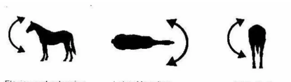
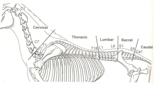
The vertebral column comprises 7 cervical (C), 18 thoracic (T), 5-6 lumbar (L), 5 sacral (S) and approximately 20 caudal (Ca) vertebrae. The vertebrae looks different in the various parts of the vertebral column. Each vertebra can rotate in three planes, which allows flexion/extension, lateral bending and axial rotation of the spine (Fig. 1).
Fig. 1 The three different movements in the vertebral column.
The conformation of the spinous and transverse processes limits the motion range of the different parts of the spine. Atlas (C1) and axis (C2) are highly modified to support and move the head. The cervical part of the spine is the most mobile and the following five vertebrae have small spinous processes and large divided transverse processes, allowing great freedom of movement for the neck. The thoracic vertebrae have large spinous processes that form the withers (T3-T8) and limit the flexion-extension movement. The lumbar vertebrae have limited lateral movement due to long horizontal transverse processes, and sometimes synovial joints develop between the transverse processes of the fourth and fifth lumbar vertebrae. Between L5-6 and the wing of sacrum, the bones are fused. The sacral vertebrae are fused to a single bone (Fig. 2).
Fig. 2 The vertebral column of the horse.
The spine represents a series of curves when viewed from the lateral aspect. The cervical part of the spine forms a ventral curve that does not correspond to the top line of the neck of the horse. The curves formed by the spinous processes of the vertebrae from the withers to the tail correspond to the top line of the horse but not to the curves formed by the bodies of the vertebrae (Sisson & Grossman, 1975).
Ligaments
Several long ligaments connect the vertebrae. Two of these ligaments cover the bottom of the spinal canal (dorsal longitudinal) and the ventral side of the vertebrae (ventral longitudinal). The dorsal longitudinal ligament runs from C2 to the sacrum, and the ventral longitudinal ligament runs ventral to the vertebral bodies from T8/9 to the sacrum. The most well known of the long ligaments is the nuchal ligament that consists of a heavy, elastic structure, running from the back of the skull (occipitalis) to the first sacral vertebrae. From the withers, it is called the supraspinal ligament. This ligament runs over the tips of the spinous processes. It is broad until T12 and then becomes smaller.
The nuchal ligament is connected to the cervical vertebrae by elastic, web-like (laminar) structures. The structure as a whole keeps the head in position and is as such an important element in the functional entity that is formed by the head, the neck and the spinal column. There are also short ligaments between adjacent vertebral bodies.
Muscles
The back of the horse consists of a deep and a superficial muscle layer. The M. multifidus forms part of the deep layer and has been designated as the longest muscle of the body. It consists, in fact, of a long series of muscle bundles, reinforced with ligamentous strips, which run from the sacrum, the sides of the spinous processes of the lumbar vertebrae, the transverse processes, and the thoracic vertebrae to the spinous processes of the vertebrae that are located 2 to 6 positions further forward. The superficial layer is mainly formed by the
M. longissimus dorsi, which in volume is one of the largest muscles of the body. This muscle originates from the spinous processes of the lumbar vertebrae and runs to the first cervical vertebrae. In the lumbar region, there is a union with the fibrous sheet of the medial gluteal muscle, which is one of the most important rotators of the hind limb and the most important determinant of the driving force of the hindquarters. Longissimus dorsi is the most powerful extensor of the back and loins, and flexes the spine laterally, when acting on one side. It should be noted that almost all back muscles are located dorsal of the spinal column and the primary role of the muscles is to control the stiffness rather than to induce movement (Robert et al., 2001). When the back extends, the rectus abdominis is active and when the back is flexed the longissimus dorsi is active (Robert et al., 2001). Although often misunderstood, activity in the epaxial muscles extends the back and makes the curvature of the back hollower. Given the fact that the vast majority of the muscles are located dorsal to the thoracolumbar vertebral column, contraction of this musculature will automatically lead to extension of the back (Fig. 3).
Fig. 3 Vertebral muscles of the horse. Top figure: Transverse section of vertebral muscles at the level of the lumbar vertebrae; a) epaxial muscles (Mm. ileocostalis, longissimus dorsi and spinalis) and b) hypaxial muscles (Mm. psoas major and psoas minor) (Adapted from: Dyce et al. (2002). Middle figure: Longitudinal view of the superficial muscles of the back; a) M. spinalis, b) M. longissimus dorsi and c) M. iliocostalis. Low figure: deep muscles of the back and nuchal ligament; a) nuchal ligament, b) multifidus muscle and c) iliopsoas muscle (M. psoas major and M. iliacus), (Adapted from: Denoix and Pailloux (2001)).
Functional models of the vertebral column and limbs
The spine of the horse is more rigid during locomotion compared to other
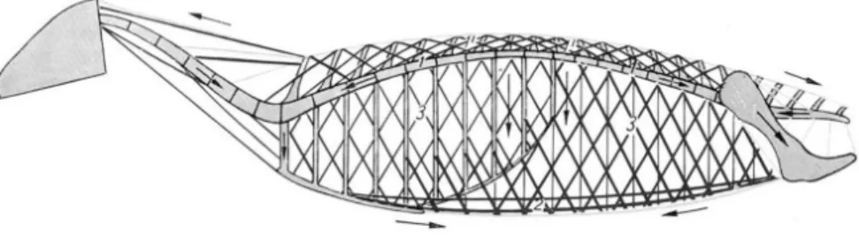
mammals such as cats and dogs. The spine is a complex structure and the two most important functions of the equine spine are 1) to carry part of the body mass and 2) to transmit the forward directed propulsive forces from the hind limbs. Slijper (1946) described the trunk-skeleton as a construction of a bow and string (Fig. 4). The elastic bow, consisting of the vertebral column, the spinal musculature and the pelvis is kept rigid and under tension by a string consisting of the sternum, the ventral abdominal musculature and the linea alba. The bow and string are connected through the ribs and the oblique and transverse abdominal muscles.
Fig. 4 “The bow and string theory” by Slijper (1946) (Modified from: Nickel et al. (1986).
Various factors determine the ultimate loading of the system, i.e. the curvature of the bow. Contraction of the abdominal muscles, especially of the rectus abdominus muscle, tenses the bow (i.e. flex the back). The same will be achieved indirectly by the retraction of the forelimbs or the protraction of the hind limbs. The string is tensed (i.e. the back extended) by protraction of the forelimbs and retraction of the hind limbs, but also by the gravitational force of the abdominal mass that pulls the bow downwards.
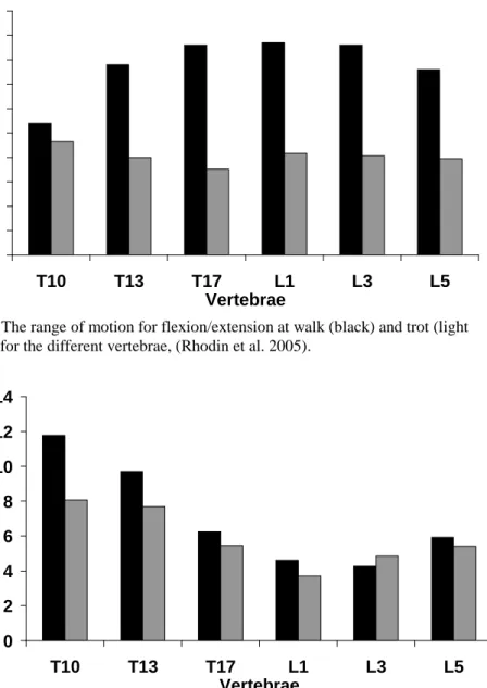
The movement of the back is closely related to the movement of the limbs and consequently there are big differences between gaits. At walk, the range of motion (ROM) for flexion/extension, lateral bending and axial rotation are almost twice as large compared to trot for sound riding horses measured with skin-fixated markers (Fig. 5,6).
0
1
2
3
4
5
6
7
8
9
10
T10
T13
T17
L1
L3
L5
Vertebrae
R a nge of mot io n ( d egrees)Fig. 5 The range of motion for flexion/extension at walk (black) and trot (light grey), for the different vertebrae, (Rhodin et al. 2005).
0
2
4
6
8
10
12
14
T10
T13
T17
L1
L3
L5
Vertebrae
R a nge of mot io n ( d egrees)Fig. 6 The range of motion for lateral bending at walk (black) and trot (light grey), for the different vertebrae, (Rhodin et al. 2005).
During walk, there is an alternation between two or three limbs having contact with the ground simultaneously and the axial rotation and lateral bending move the centre of gravity laterally between the supporting limbs. The muscles of the back restrict the movements induced by the hind limbs and muscle activity is generally low, indicating a passive movement of the back. The trot requires a much higher tension in agonists and antagonist to maintain equilibrium and the activity of the rectus abdominis muscle will prevent excessive extension of the back whereas the longissimus dorsi muscle has the opposite effect. The vertebral column remains as
rigid as possible in order to resist wasteful lateral movements (Robert et al., 1998) and to transmit the forces from the hind limbs into forward movements of the body of the horse. The muscles also limit the twist of the back that arises from the diagonal stance phase. Therefore changes in limb activity will change both the muscle activity and the movement of the back.
Flexion-extension movement has a double sinusoidal motion pattern at the walk and trot, but a single sinusoidal pattern at the canter. Lateral bending and axial rotation have a single periodic motion for all gaits. This has to do with the symmetry of the gait and the effect of hind limb placement on spinal kinematics.
Biomechanical research
Biomechanics is the science of the mechanical behaviour of living organisms and structures. Kinematics deals with the geometry of movement and kinetics with the forces that are responsible for the movements. The first documented biomechanical study on animal locomotion “On the movements of animals” was written by the Greek philosopher Aristotle (384-322BC), who described the quadrupedal
locomotion. A big revolution in equine gait analysis took place in France in the late 19th century. In 1887, the photographer, Eadweard Muybridge, visualised equine locomotion by use of 24 cameras (Muybridge, 1957). Later it is the technical development that has directed the research in equine biomechanics. The next big step for equine locomotion research was the rapid development of computer technology in the late 1970s. This facilitated capture and analysis of the fast movements of the horse when high-speed film was used.
A Swedish research group was the first to use a treadmill for equine locomotion studies (Fredricson et al., 1983). Kinematic studies of the horse back were initiated by Leo Jeffcott, a clinician interested in orthopaedics. He collaborated with the Swedish group performing in vitro studies on the movement of the spine (Jefcott & Dalin, 1980a). He also studied various back-related disorders (Jefcott 1980b).
Knowledge of today
The kinematics of the horse has been studied extensively since the research field was established in the early 1980s. Today we have knowledge about the normal motion pattern of the limbs and back of the horse in the three basic gaits; walk, trot and canter (Back, 2001). The effects of lameness on the motion pattern of the horse has been thoroughly studied as well as the effects of back pain (Buchner, 1995, 1996a, 1996b, 2001; Weishaupt et al., 2004, 2006b;Wennerstrand et al., 2004; Gómez et al. 2007a, 2007b, 2007c). Further, the effects of placing a rider on a horse and the kinetics and kinematics of different equestrian sports activities have been documented (Clayton, 1997, 2001). However, the knowledge of how different training methods and other forms of human interventions affect the riding horse and how a horse should be ridden and trained to avoid injuries, e.g. “Equine ergonomics”, is yet limited (van Weeren, 2005).
There are a few studies that have evaluated the effect of the rider’s level of skill on the horse’ reaction to the rider. Schamhardt et al. (1991) compared ground reaction forces in horses ridden by a novice and an experienced rider and found practically no differences between the two riders. Licka et al. (2004) studied the
hind limb lameness did not change significantly from the in-hand situation when they were ridden by a novice rider but increased when ridden by a dressage rider. Schöllhorn et al. (2006) studied the horse-rider interaction using cluster analysis of kinematic parameters. They found that a professional rider had a higher influence on the head movements of the horse than a recreational rider had. Lagarde et al. (2005) studied the interaction between rider and horse by measuring their ensemble motions at trot and found that an expert rider moved more in phase with the horse than a novice rider, and that the horse at the same time had increased temporal regularity of the oscillations of its trunk when ridden by the expert rider.
Apart from the studies included in this thesis, there are only three more studies published on the effects of different head and neck positions, of which two are based on the same experiment. Roepstorff et al. (2002) and Byström et al. (2006) studied the kinetic and kinematic effects of draw reins on the limb movements. They concluded that the use of draw reins together with normal reins shifted the weight of the horse caudally with an increased flexion of the hock joint angle as a result. Biau et al. (2002) studied the effect of three types of reins on the
locomotion of the horse at walk and trot. The horses were equipped with three types of reins: rubber bands, Chambons and back lifts while they walked and trotted freely in an automatic walker. The gait characteristics were measured with accelerometric technique. They concluded that changes in the head and neck attitude caused by the reins influenced the kinetic variables of the forelimbs, but only few significant effects were found in the hind limbs.
Today there are thus only a handful of studies on which we can base any statements on the influence of the rider’s educational level and different training methods on the riding horse. It is really in the interest of the equestrian sport to increase the knowledge in this field. This thesis aims at providing one more piece.
Training methodology
The position of the head and neck axis has a considerable effect on the back of the horse according to the equestrian literature (Heuschmann, 2007; GNEF, 2002). The back of the horse and its state of relaxation are the keys to success when training a horse, regardless of the equestrian discipline. The effect of different head and neck positions on the motion pattern of the horse is often discussed but there are no quantitative scientific studies supporting these theories.
Relative elevation
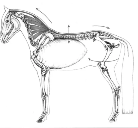
The muscles of the upper neck work freely without tension and the position enables the horse to carry its back freely and without tension. The state of relaxation in the back is mirrored by the purity of rhythm seen in the horse’s gaits. The expression “a carried back” means a back which is working in relaxation (Fig. 7).
Fig. 7 A horse in “relative elevation” with a released back allowing the muscles to work properly will be able to easily lower the croup. Thereby, it will be able to step far forward under its body and the centre of gravity (see arrows). (Adapted from: Heuschmann, 2007).
Absolute elevation
The head and neck axes are positioned higher and compressed beyond the level of the training and muscular development of the horse. When ridden like this, a horse will have difficulty supporting its rider. Initially, the horse tries to lift its trunk, which includes the additional weight of the rider, by tensing its longissimus dorsi muscle, but eventually has to drop its back, with the effect of loosing the connection between its hindquarters and the hand of the rider. The result is unwillingness, tension and resistance which cause poor basic gaits. The rider gets problems sitting comfortably at the trot because of the tension in the back of the horse (Fig. 8).
Fig.8 A horse in “absolute elevation” with a broken neckline, a short neck and a hollow back, with the hind legs strung out or trailing behind. (Adapted from: Heuschmann, 2007).
Hyperflexion
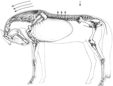
Hyperflexion occurs when the rider wants to raise and “swing” the back of the horse in an overstretched manner. This method puts enormous tension on the upper neck muscles and ligament system and the back through the supraspinous ligament. The back is not relaxed during work. Horses worked regularly in this position develop a straight, flat back line, with inactive trailing hind legs and no noticeable flexion in the haunches during collection or extended movements. The trailing of the hind legs can be explained by the fact that the back fascia connects with the large muscle groups of the hind limbs (Fig. 9).
Fig. 9 A horse in hyperflexion with an overstretched back. Due to the extremely deep position of the head and neck it exhibits a broken neckline, a strongly (incorrectly) lifted back, and a straight croup (overextended sacro-lumbar joint with the hind limbs behind (Adapted from: Heuschmann, 2007)
Aims of the thesis
The objective of the thesis is to evaluate the effect of different head and neck positions on the kinematics of the horse and to discuss findings in a perspective of functional anatomy and riding methodology respectively.
General Hypothesis
All head and neck positions different from the natural will influence the motion pattern of the horse. The more extreme high and low positions the more kinematic changes at both walk and trot will be induced.
Specific aims of the studies
1) To evaluate the effect of 3 different head and neck position on the kinematics of the back in riding horses at walk and trot.
2) To evaluate the effect of long reining and the head and neck position on the kinematics of the back at walk and trot.
3) To quantify the effect of 6 different head and neck positions on thoracolumbar kinematics of the horse at walk and trot.
4) To evaluate the effect of 5 different head and neck positions on the caudal back and limb kinematics in riding horses at walk and trot.
Materials and methods
Horses
The horses used in the studies were privately owned horses in training for competition. The horses were all clinically sound with no resent history of lameness and had no pathological reactions to palpation of the limbs and back. They were fully accustomed to treadmill work beforehand (Buchner et al., 1994).
The horses in study I-II were dressage and show jumping horses, between 5 to 15 years of age and competing at a low level.
In study III-IV the horses were dressage horses at Grand Prix level except one competing at intermediate level. The age was 14.0+4.3 years, the wither height 170+0.07m and body weight 609 +62.3kg.
The horses used their own bridle with a normal snaffle bit and in study IV the horses were ridden by their ordinary rider and with their own fitted saddle. Study I and II were approved by the local committee for the Swedish National Board for Laboratory Animals and study III and IV by the Animal Health and Welfare Commission of the canton of Zürich.
Experimental design and protocol
Paper I
Eight Warmblood riding horses were studied on a treadmill1 at walk (1.7 ms-1) and trot (3.8 ms-1) with the head and neck in 3 different predetermined positions. The three positions were 1) free position; 2) lowering of the neck with the poll close to the level of the withers (low position); 3) elevation of the head and neck with the bridge of the nose slightly in front of the vertical (high position) (Anon 2003). The positions were achieved by use of side reins attached to an anticast roller.
Paper II
Seven Warmblood riding horses were studied when long reined on a treadmill1,2 at walk (1.7 ms-1) and trot (3.3 ms-1) with the head and neck in 3 different pre-determined positions. The three positions were 1) free position; 2) lowering of the neck with the poll close to the level of the withers (low position); 3) elevation of the head and neck with the bridge of the nose slightly in front of the vertical (high position). The positions were achieved either by the long reiner or with side reins attached to an anticast roller.
Paper III
Seven Warmblood dressage horses competing at Grand Prix (n=6) or intermediate (n=1) level were studied walking and trotting on a high-speed treadmill1 with an integrated force measuring system (Weishaupt et al., 2002) with six different head and neck positions. They were achieved using standard side reins, additional side reins and a custom-made over-check as needed to achieve the various head and neck positions. A qualified dressage judge evaluated the different positions being accomplished correctly by the horses. No tension was allowed in the side reins during the measurements. The positions were defined as follows (Fig. 10):
HNP1: Free or natural (voluntarily acquired position, unrestrained with loose reins) HNP2: Neck raised, poll high and bridge of the nose slightly in front of the vertical HNP3: Neck raised, poll high and bridge of the nose slightly behind the vertical HNP4: Neck lowered and flexed, bridge of the nose considerably behind the vertical
HNP5: Neck extremely elevated and bridge of the nose considerably in front of the vertical
HNP6: Neck and head extended forward and downward
Fig. 10 Head and neck positions. Illustration: Matthias Haab.
All horses were studied at their individual preferred speed. The preferred speed was defined as the speed at which the horse moved in a relaxed manner and showed the best performance at each head and neck position according to the dressage judge. As preferred speed changed with the different head/neck positions, reference measurements were made with the head and neck in free position (HNP1) at the same speed as preferred by the horse for the specific head/neck position, in order to have a speed-matched control.
Paper IV
Paper IV is based on the same horses and overall experimental setup as paper III,
HNP1 HNP2
HNP3 HNP4
HNPs were achieved by the rider, and additional reins were not used except for the low position, HNP4. For this position a combination of ordinary reins and draw-reins was necessary to guide the horse to the correct position, but the draw-draw-reins were hanging loose during the measurements (Fig 11).
Fig. 11 Head and neck positions (HNP). Illustration: Matthias Haab.
To achieve HNP6 at the trot, the rider used rising trot and at walk there were only small visual differences between HNP1 and HNP6. Therefore the HNP6 was excluded from further analysis. The correctness of each HNP was judged by an international dressage judge. As in paper III all horses were measured at their individual preference speed. The preference speed was defined as the speed at which the horse moved in a relaxed way and showed the best performance at each of the head and neck positions with reference to the opinion of the rider and the judge. Accordingly, the speed changed with the different head/neck positions. Reference recordings were made with the head and neck in HNP2 at the same speed as for the specific head/neck position to have a speed-matched control.
HNP2 HNP1 HNP4 HNP3 HNP6 HNP5
Measuring and analysis techniques
Measurement
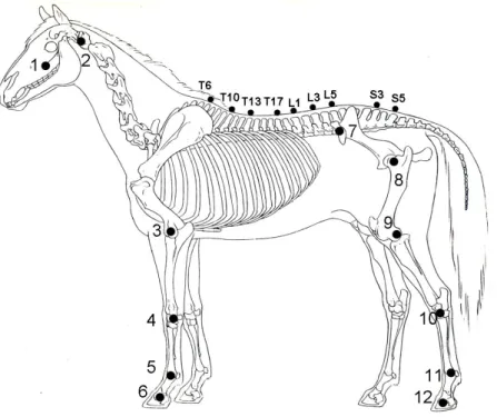
Reflective markers were glued onto the skin above anatomical structures defined through palpation (Fig. 12). A high speed 3D infrared camera system (ProReflex3) was used to capture data. In study I-II, six cameras were positioned around the treadmill and in study III-IV twelve cameras were used. In study III-IV, a treadmill instrumented with a force measuring system was used for simultaneous,
synchronized force measurements (Weishaupt et al. 2002).
Fig. 12 Marker positions with anatomical reference points: 1: Distal part of facial crest, 2: the cranial end of the wing of atlas, 3: the transition between the proximal and the middle thirds of the lateral collateral ligaments of the elbow joint, 4: ulnar carpal bone, 5: the proximal attachment of the lateral collateral ligament of the fetlock joint to the distal end of the third metacarpal bone, 6:lateral hoof wall at the level of the coffin joint, 7:tuber coxae, 8: cranial part of greater trochanter of femur, 9: the proximal attachment of the lateral collateral ligament of the stifle joint to the femur, 10: talus, lateral, 11: the proximal attachment of the lateral collateral ligament of the fetlock joint to the distal end of the third metatarsal bone, 12: lateral hoof wall at the level of the coffin joint. Back markers: Above the thoracic (T), lumbar (L) and sacral (S) spinous processes as numbered in the figure.
Data processing
Qualisys Track Manager software4 was used to capture and reconstruct the 3-dimensional position of each marker. When data were missing for maximal 10 consecutive frames a gap fill function was used. The resulting graphs for the X, Y, Z-positions of the marker in question were checked manually before acceptance. The raw X-, Y-, and Z-coordinates were then exported into Matlab5 for further analysis.
Back movement analysis
For the analysis of the back movements in study I-III data from skin markers placed at the spinal processes of T6, T10, T13, T17, L1, L3, L5, S3, left and right tuber coxae and one hind hoof were used and analyzed by use of a custom written Matlab program, Backkin6. This program has been developed from the results of earlier studies by Faber et al. (2001b) and Johnston et al. (2002).
Faber et al. (2001b) compared the results from measurements based on skin-fixated markers as listed above with the results from measurements with invasive markers (Steinman pins inserted into the same structures) in the same horses at walk (1.6 ms-1) and trot (4.0 ms-1). Data from the invasive markers were analysed by use of a rigid-body model which calculates the 3-dimensional motion of each of the measured vertebrae. This is not possible with skin markers, as this would require three markers per vertebra. Instead it was assumed that the vertebra, for example T10, was oriented in alignment with a straight line between the markers at the preceding and the following of the labelled vertebrae, which then would be T6 and T13. Marker coordinate data were first filtered with an 8th order Butterworth filter with a cut-off frequency set to 40 Hz. The marker data from the two vertebrae was then projected into the saggital and the horizontal planes. The angle between the horizontal axis of the plane and a line between the markers was determined to represent flexion/extension (FE) (in the sagittal plane) and lateral bending (LB) (in the horizontal plane) for each vertebra. Axial rotation of the thoracolumbar vertebrae can not be determined with this method, but the axial rotation of sacrum was calculated by projecting data from the left and right tuber coxae into the frontal (transverse vertical) plane.
Both data sets, e.g. invasive and skin marker based data, were normalized to 101 data points (0-100 % stride) and then averaged over available strides for each horse. The individual stride cycles were determined from the vertical velocity profile of a hind hoof marker. The mean curves for each horse were then compared between the two measurement techniques. Based on the results of this comparison Faber et al. (2001b) concluded that skin-fixated markers can reliably be used to quantify thoracolumbar FE and AR at walk and trot and LB at trot, while at walk LB can reliably be used only in the non-central portions of the thoracolumbar back.
Johnston et al. (2002) used Backkin6 to perform the above described calculations for skin-fixated marker data and evaluated the day-to-day repeatability of the method as well as the comparability between different labs and treadmill types.
In study III data from the treadmill force measurement system (Weishaupt et al., 2002) were used rather than the velocity profile of the hoof marker to determine stride cycles. Apart from this, back movements in study I-III were calculated as described above, using Backkin6. From the stride mean curve a stride average over the 101 points was determined and compared to the angular value for the same
vertebra in a square stance measurement of the same horse. The range of motion (ROM) was determined as the difference between stride maximum and minimum values. In FE positive rotation was defined as clock-wise rotation when seen from the right, e.g. cranial rotation of the vertebra. It is important to remember that this is for each vertebra. However, if the whole back is to flex this requires
simultaneous positive (cranial) rotation of the cranial vertebrae and negative (caudal) rotation of the caudal vertebrae.
Considerable variation of the head and neck position of the horse intuitively seems to cause quite considerable skin displacement in cranio-caudal direction at the withers area. As the Backkin6 method has been validated only for use with the head and neck of the horse in free, unrestrained position. Also, in a previous study using a part of our data for a different purpose it was also found that the cranio-caudal movement of the T6 and T10 was greater than would be likely possible for the underlying bone (Bobbert et al., 2007). Therefore the skin displacement of the T6 and T10 markers was subjected to preliminary study by x-raying a horse with the neck in neutral position at which the markers were placed and with the neck lowered and the neck raised. The findings indicate that the displacement of the markers was not insignificant and must thus be considered in interpreting the results of the measurement comparing different head- and neck positions.
Kinematic analysis in study IV
Left fore- and hind limb joint angles and selected axial parameters were determined as defined by fig. 13.
Fig. 13Angle definitions. Original photo: Martin Larsson Limb length was defined as the global distance from elbow joint to the front hoof and from the greater trochanter at the hip joint to the hind hoof. The protraction-
retraction angle was calculated from elbow joint or L5 and the hooves for the fore and hind limbs respectively to the horizontal plane.
Fig. 14 The angle of the lumbar back was calculated between L5, L3 to the horizontal plane (Fig 14). Due to the ventral curvature of the back the angle is always positive
Fig.15 The three rotations of pelvis: A; roll, B; pitch and C; yaw. Illustration: Jill Rhodin.
To analyze the movement of the pelvis a rigid body model was created based on a previously published algorithm (Söderkvist & Wedin, 1993). In this model, the pelvis was defined by the markers located on the spinal processes of S3, S5 and
the left and right tuber coxae. The rotations of the pelvis around the longitudinal, transverse and vertical axis were described as three angles; roll, pitch and yaw (Fig.15). A square stance measurement of each horse was used to define the zero position.
All variables were normalized to 0-100% of for the whole stride defined by the left front or hind limb using temporal information from the treadmill integrated force measuring system (Weishaupt et al. 2006a). From the stride normalized data, ROM was determined for the angle of the neck in relation to the thorax, vertical excursion of T6 and L5 and pelvic rotations and stride minimums were determined for fore- and hind limb length. The resulting values were averaged over available strides for each horse and measurement after excluding the highest and lowest 5% of the observations, to avoid bias due to outliers.
Use of skin fixated markers to measure movements of the skeleton always includes the risk of skin displacement errors. Distal to the elbow in the forelimb and the stifle in the hind limb, the skin movement artefacts are small enough to be neglected (van Weeren et al., 1992). I am aware of the problem with skin displacement for the proximal limb markers. However, these displacements are likely not effected by the head and neck position and as the different positions were compared pair-wise, with each horse acting as its own control, the skin displacement should not affect the differences observed between different head and neck positions.
Statistics
In the studies included in this thesis the number of horses ranged from seven to eight. With this low number of horses it is not possible to test data for normality in a reliable way. Therefore a paired non-parametric (Wilcoxon signed rank) test was used. Differences were considered to be significant at p<0.05.
Results
Paper I) The head and neck positions influenced the movements of the back, especially at the walk. When the head was fixed in a high position at the walk, the flexion-extension movement and lateral bending of the lumbar back, as well as the axial rotation, were significantly reduced when compared to movements with the head free or in a low position. At walk, head and neck position also significantly influenced stride length, which was shortest with the head in a high position. At trot, the stride length was independent of head and neck position and there were few kinematic changes between the different positions.
Paper II) The most pronounced reduction of the flexion-extension movement of the caudal back was found with the head in a high position at the walk. At the trot the effect of long reining on the movement of the back was less pronounced. The movement of the back and the displacement of the angular motion pattern at the walk differed significantly between long reins and side reins.
Paper III) The head and neck positions only affected vertebral angular motion patterns in the sagittal plane (flexion-extension). There was no influence on lateral bending or axial rotation. The positions in which the neck was extended (HNP2, 3, 5) increased extension in the anterior thoracic region, but increased flexion in the posterior thoracic and lumbar region. For HNP4 the pattern was opposite. Range of motion (ROM) of vertebrae was reduced at walk in the lumbar region in HNP2 and HNP5, and at trot also in HNP3. Restriction was more evident at trot than at walk and most evident in HNP5. In this position there was an increase in ROM of lateral bending in the thoracic region at walk and of axial rotation at trot. In HNP4 there was an overall increase in flexion-extension ROM, at walk mainly thoracic, at trot also lumbar. HNP5 was the only position that negatively affected intra-vertebral pattern symmetry and reduced hind limb protraction.
Paper IV) The unrestrained horse seems to rely more on the forelimbs for vertical support and use the hind limbs in a more horizontal direction. At walk the stride length and the movement of the pelvis increased compared to HNP2, which can be useful for training purposes, while at trot the gait economy is likely improved with a free head and neck position.
Extreme elevation of the head and neck increased vertical limb movement with increased limb flexion both during stance and during swing. Pelvic rotations were increased at trot, while decreased at walk.
More slight differences in head and neck position clearly influenced the movement pattern of the horse at walk, but had few effects at trot.
Additional analysis of data from study IV
To compare the kinetic and kinematic results, the angular motion pattern of the fetlock joint, tarsal joint and vertical force of left hind during stance, were compared between HNP5 and HNP2. The effect of speed was compared by using the lowest and highest speed for HNP2 for the same variables at both walk and trot.
Fig. 16 The tarsal joint, fetlock joint and vertical force for left hind during the stance phase normalized to 100%. A HNP5 walk, B HNP2 different speeds walk. Blue line for HNP2 control and speed (mean 1.43+.5.27 ms-1). Green line for HNP5 and speed (mean 1.70+.4.89 ms-1). Red line indicate significant differences (p<0.05) between the two situations compared.
B
walk
Fig. 17 The tarsal joint, fetlock joint and vertical force for left hind during the stance phase normalized to 100%. C HNP5 trot and D. HNP2 different speeds trot. Blue line for HNP2 control and speed (mean 2.85+.7.88 ms-1) Green line for HNP5 and speed (mean 3.24+.4.00 ms-1). Red line indicate significant differences
(p<0.05) between the two situations compared.
C
trot
HNP1 walk (A) HNP5 walk (B)
HNP 1 trot (C) HNP 5 trot (D)
Fig. 18 Stick figures illustrating the hind limb position during first contact (FC), midstance and toe off (TO) for HNP1 walk (A), HNP5 walk (B) HNP1 trot and (C) HNP5 trot (D) (green line) compared to HNP2 (blue line) at walk and trot.
Markers from proximal to distal; greater trochanter of the hip joint, stifle joint, hock joint, fetlock joint and hind hoof.
Evaluation of skin displacement.
To get an approximate estimation of the amount of skin displacement for
the T6, T10 and T13 marker, a radiographic study was performed. One
horse was radiographed with the head and neck in three different positions.
Three radiodense markers were placed on the skin above the spinous
processes of T6, T10 and T13. A large skin displacement was seen for the
T6 marker, and a lesser for the T10 marker (Fig. 19), whereas T13 was not
affected (not shown in the pictures).
Fig. 19
. A horse with a high (A), neutral (N) and low (L) neck position.
Discussion
Data collection and processing
This thesis considers the effect of human manipulation on the biomechanics of riding horses. Horses are frequently used for sport and leisure and are trained to
A
B
perform at a level which sometimes is far beyond what a horse would do by its own free will. Even though a free roaming horse at times may clear an obstacle, run fast, or show an impressive posture, we can not refer to equestrian sports as natural for the horse. Human intervention on the horse may be harmful (Murray et al. 2006), but there are few scientific studies on the physical and mental effects of this rider-horse interactions. Such studies are difficult to perform, which is illustrated by the low number of studies published.
In order to be valid, studies on the effects of equestrian sports require research horses, representative of the population on which the results should be applied. All horses used in the studies included in this thesis were privately owned riding horses and the majority were actively participating in equestrian sports competitions at the time of study. It is clearly an advantage, that these horses are more likely to be representative of the horses we aimed to study compared to using horses only aimed for research purposes. However, this limited our choice of measurement techniques to non-invasive, harmless alternatives. At present, researchers are generally limited to conduct measurements on a treadmill, due to the properties of the technical equipment used. We can not be completely sure that results obtained using the treadmill are valid in the over-ground situation. A previous study comparing trot on treadmill vs. over-ground found that forelimb stance duration increased and diagonal advanced placement of the hind limb decreased, e.g. the forelimb was placed earlier than the hind limb (Buchner et al., 1994). However, as all our measurements were performed on a treadmill, changes introduced by the treadmill per se must not necessarily have influenced our results comparing various head and neck positions. Further, the treadmill has the advantage to enable speed control, which is an important factor in locomotion research.
Skin displacement
For the analysis of the back movements in study I-III, a kinematic method based on skin-fixated markers were used (Faber et al., 2001b). With this method vertebral orientation is determined from sagittal and horizontal plane projection angles for flexion-extension and lateral bending, respectively. The axial rotation of the pelvis is defined from a transverse plane projection angle between the left and right tuber coxae. As these angles are determined with respect to the horizontal axis of the plane rather than the angle of the preceding vertebra they will be influenced not only by the degree of flexion and extension between the vertebrae of the back, but also by changes in the trunk angle. Faber et al. (2001b) validated the method and found that the results for lateral bending of the mid-thoracic back differed considerably from the movement measured with invasive markers in the same vertebrae. Also, the validation did not include manipulation of the head and neck position. From preliminary studies we suspect that significant skin
displacement related positional changes may have occurred for the marker at T6 between the various neck positions evaluated (Fig. 17). When an un-proportionally large change in flexion-extension angle of T10 is found compared to T13 however, this results must be questioned. These limitations must be kept in mind, when interpreting the results of study I-III.
Statistics
When analyzing large numbers of variables and particularly when comparing each variable at multiple points during a stride, a number of random significances will inevitably be encountered. If 1000 comparisons are made at the 5 % level of significance there would be about 50 significant associations in the data all representing false positive results. These associations occur randomly and are denoted type 1 errors. To reduce this problem, one possibility is to lower the p-value at which a difference is considered significant. However, with the low number of individuals used in the studies included in this thesis, changing the level of significance does not actually solve the problem. Rather it merely produces a smaller type 1 error at the expense of a greater type 2 error e.g. true differences between groups or situations may not be observed. This can, at least in some situations, be just as serious as the type 1 error. Therefore, adjustments of the level of significance should not be made, even if a large number of comparisons are reported, provided that it is clear how many comparisons have been made and that all non-significant results have been reported along with the significant results (Rothman 1986).
The influence of the head and neck position on the movements of the back
To the authors knowledge study I was the first to evaluate the effect of different head and neck positions on the back movements in riding horses. It was found that the head and neck position influenced the movements of the back at both walk and trot, but the effects were more pronounced at walk. The high position significantly reduced the ROM for flexion-extension and lateral bending of the lumbar back and axial rotation of the pelvis at walk, compared both to the free and the lowpositions. This was accompanied with a decreased ROM for the pro- and retraction of the hind limb, which was lowest in the high position. Previous studies of the kinematics of the thoracolumbar back at walk and trot have identified the hind limb movements as important for the movements of the back (Faber et al., 2000, 2001a). It is therefore likely that the concurrent decreases in back movement and hind limb pro- and retraction are coupled, but it is more difficult to speculate on the causal relationship. The findings in study I was in contrast to the opinions of some riders and trainers, who state that they use a low head and neck position to engage the back of the horse and increase the movements of the back (Janssen, 2003).
Further, placing the head and neck of the horse in a defined position by use of side reins potentially yields a different response compared to when the same head and neck position is achieved by a rider using the reins in conjunction with other aids. Unfortunately, however, only the caudal back and pelvis can be studied in the ridden horse. One way to simulate the ridden situation in the unmounted horses is long reining. A highly experienced long reiner participated in a second study comparing the same head and neck positions, achieved with side reins or by long reining. The main differences with long reining, compared to side reins, are that the horses could be encouraged from behind and that the aids given through the reins could be adjusted in a similar way as in the ridden situation. Despite this, few significant differences were found between long reining and side reins. The results for the different positions in study II, free compared to low and high, were generally similar to the findings of study I. Long reining in the high position
increased the flexion-extension angle for the thoracolumbar transition (T17, L1) significantly at the walk, compared to side reins. The conclusion is that the cranial rotation of L1 causes the back in this area to be more extended on average during the stride, however, the ROM for flexion-extension was unchanged. Although the changes were small, this was an unexpected result, since one of the goals with long reining a horse “on the bit” is to increase hind limb activity and cause an increased dorsal flexion of the back. The low position increased the flexion of the back at L1 compared to the high position and at L3 compared to the free position.
In study III the head and neck positions were made more diverse, in order to possibly further define the specific effects of each position. In this study, an extremely elevated head and neck position, HNP5, decreased ROM both of the hind limb pro- and retraction, lumbar back (L3, L5) flexion-extension and pelvic axial rotation. In HNP2, the second highest position, only ROM at L5 was decreased and the decrease in ROM of protraction-retraction of the hind limb was not significant. This again, confirms a coupling between stride length and back movements at walk. In the three other positions evaluated, HNP3, HNP4 and HNP6, the ROM for protraction-retraction of the hind limb was unchanged and in addition, no decrease in ROM of the back movement were observed compared to free position, despite of the restrained head and neck.
Compared to HNP1 at trot, the three higher positions, HNP2, HNP3 and HNP5, all decreased back flexion-extension ROM in the caudal thoracic and/or cranial lumbar back and the low positions, HNP4 and HNP6, increased ROM at T10, L1, L3 and L5 and T10 and T13, respectively. The latter finding is thus in alignment with the subjective impression by some riders and trainers that a low head and neck position increases back movements. The ROM of hind limb pro- and retraction were not significantly different in of the evaluated head and neck positions compared to HNP1. This indicates that stride length and back movements are less closely coupled at trot, the head and neck position by it self seems to have more influence. Comparing the mean angular motion patterns, greater differences were found between the various head and neck positions at walk than at trot.
From the kinematic evaluation of the same head and neck positions in the ridden situation, it became clear that not only the movement pattern of the back, but also the horse as a whole was more sensitive to the head and neck position at walk than at trot. As far as the variables measured could be compared, the different head and neck positions seem to have produced similar changes in the back movement pattern in both the ridden and unmounted situations, but a direct comparison is difficult to make due to differences in data analysis and due to the use of different HNP controls, free position (HNP1) for the unmounted measurements and HNP2 in the ridden situation.
Movement pattern of the gait is much more sensitive to a restrained head and neck position at walk than at trot. During walk, two or three limbs have contact with the ground in an alternating manner, whereas at trot there is a diagonal stance phase followed by a suspension phase. These differences are reflected in the function and the motion pattern of the thoracolumbar spine. The trot requires much more muscular activity to maintain equilibrium and the vertebral column is made as rigid as possible in order to resist wasteful lateral movements (Robert et al., 1998), to limit the twist of the back that arises from the diagonal stance phase and to transmit the forces from the hind limbs to the forequarter. The range of motion
to trot and the ROM for lateral bending is 50% greater (Faber et al., 2000, 2001a). It can be hypothesised that it is much easier for a rider or other intervention to increase tension in muscles which are relatively relaxed and to reduce the movements of structures with a large ROM. These findings are also in agreement with the common opinion among riders and trainers that the quality of the walk is more sensitive to mental tension in the horse and that the walk is more easily destroyed by an unskilled rider compared to the trot. The walk is therefore also more interesting in studies of the rider-horse interaction.
However, we can not tell from our studies whether the observed kinematic effects are beneficial or harmful to the horse. The difference between training effect and injury is likely a matter of dosage. Also, we can not say that the effects of changing head and neck position at trot, though to a great extent statistically insignificant in our studies, are insignificant to the horse. The balance between training effect and injury could be very delicate especially when training on an advanced level with high intensity, longer periods and/or specialised training regimes. Also many things not studied in these experiments as fatigue, gait characteristics, speed, conformation, shoeing and trimming and so on could influence the balance. For the future, it is necessary to further study the influence of different head and neck position and riding techniques by quantifying the influence of the rider and correlate this to the response of the horse, for example by measuring saddle pressure, rein forces and kinematics of the rider. It is also necessary to perform long time studies of different training methods in order to clarify if and in what way they may be beneficial, or potentially harmful to the horse.
The concept of collection
Collection is a central theme in equestrian dressage. Despite this, there is no generally accepted definition of collection. Descriptions in the equestrian literature can differ quite considerably. However, some common points are frequently stressed and the hind limbs are always assigned an important roll.
One description of the collected horse is that the “increased weight bearing of the hind limbs, which in turn lightens the forehand, gives more freedom to the movements of the forelimbs” (GNEF 2002). Weishaupt et al. (2006a) found that extreme elevation of the head and neck (HNP5) resulted in a higher degree of “collection” as the relative weight bearing of the hind limbs increased. At walk, a more normal riding head and neck position (HNP2) also caused a shift of weight from fore to hind compared to when the horse was walking with the reins loose (HNP1), but a similar tendency at trot was not significant (Weishaput et al., 2006a). This could indicate that HNP2 was more collected than walk and trot on loose reins. Most riders and dressage trainers would likely agree with the latter statement but would not accept HNP5 to display a higher degree of collection than HNP2. Consequently, perhaps increased fractional weight bearing of the hind limbs alone is not enough to define collection.
Another statement describing collection is if, “the hind limbs (the hock and stifle joints) bend more during stance” (GNEF, 2002). At trot, the only position which increased flexion of the stifle and hock joints was HNP5. At walk hock and stifle flexion was increased in HNP5 and HNP3, while decreased flexion was found in HNP1. However, these changes can be secondary to other changes, such as hind