DOI: 10.1097/01.gme.0000222475.93345.b3 *2006 by The North American Menopause Society 5Text printed on acid-free paper.
P
OSITION
S
TATEMENT
Management of osteoporosis in
postmenopausal women: 2006 position statement
of The North American Menopause Society
ABSTRACT
Objective: To update the evidence-based position statement published by The North
American Menopause Society (NAMS) in 2002 regarding the management of osteoporosis in postmenopausal women.
Design:NAMS followed the general principles established for evidence-based guidelines to
create this updated document. A panel of clinicians and researchers expert in the field of metabolic bone diseases and/or women`s health was enlisted to review the 2002 NAMS position statement, compile supporting statements, and reach consensus on recommendations. The panel`s recommendations were reviewed and approved by the NAMS Board of Trustees.
Results: Osteoporosis, whose prevalence is especially high among elderly postmenopausal
women, increases the risk of fractures. Hip and spine fractures are associated with particularly high morbidity and mortality in this population. Given the health implications of osteoporotic fractures, the primary goal of osteoporosis therapy is to prevent fractures, which is accomplished by slowing or stopping bone loss, maintaining bone strength, and minimizing or eliminating factors that may contribute to fractures. The evaluation of postmenopausal women for osteoporosis risk requires a medical history, physical examination, and diagnostic tests. Major risk factors for postmenopausal osteoporosis (as defined by bone mineral density) include advanced age, genetics, lifestyle factors (such as low calcium and vitamin D intake, smoking), thinness, and menopause status. The most common risk factors for osteoporotic fracture are advanced age, low bone mineral density, and previous fracture as an adult. Management focuses first on nonpharmacologic measures, such as a balanced diet, adequate calcium and vitamin D intake, adequate exercise, smoking cessation, avoidance of excessive alcohol intake, and fall prevention. If pharmacologic therapy is indicated, government-approved options are bisphos-phonates, a selective estrogen-receptor modulator, parathyroid hormone, estrogens, and calcitonin.
Conclusions:Management strategies for postmenopausal women involve identifying those at
risk of low bone density and fracture, followed by instituting measures that focus on reducing modifiable risk factors through lifestyle changes and, if indicated, pharmacologic therapy.
Key Words:Menopause Y Osteoporosis Y Fractures Y Bone mineral density Y Estrogen
therapy Y Hormone therapy Y Bisphosphonate Y Selective estrogen-receptor modulator Y
CalcitoninYParathyroid hormone YCalciumYVitamin D YNAMS.
Received February 27, 2006.
The Board of Trustees of The North American Menopause Society (NAMS) developed this manuscript with assistance from an Editorial Board composed of Bruce Ettinger, MD; Steven T. Harris, MD, FACP; David Kendler, MD; Bruce Kessel, MD; and Michael R. McClung, MD. It was edited, modified, and subsequently approved by the NAMS Board of Trustees on February 24, 2006.
The development of this position statement was supported by unrestricted educational grants from the Novartis Pharmaceuticals Corporation. Address correspondence to: NAMS, P.O. Box 94527, Cleveland, OH 44101. E-mail: [email protected].
O
steoporosis becomes a serious health threat for aging postmenopausal women by predisposing them to an increased risk of fracture. Osteoporotic fractures are associated with substantial morbidity and mortality in postmenopausal women, especially older women.In response to the need to define standards of clinical practice in North America as they relate to menopause-associated health conditions, The North American Menopause Society (NAMS) has created this evidence-based position statement. The objective of this position statement is to provide guidance on the diagnosis, prevention, and treatment of osteopo-rosis in postmenopausal women to physicians, physi-cian assistants, nurse practitioners, nurses, and other healthcare professionals caring for postmenopausal women, especially those in the clinical practice fields of obstetrics and gynecology, internal medicine, family medicine, and geriatrics.
This position statement is an update of the NAMS position statement published in 2002.1Since then, the publication of additional scientific evidence has created a need to update the position statement.
For this revision, NAMS conducted a search of the medical literature published since the previous posi-tion statement was submitted for publicaposi-tion in November 2001. A search was made for clinical trials, meta-analyses, and clinical practice guidelines published in English and related to osteoporosis in postmenopausal women using the database MED-LINE. The Medical Subject Headings (MeSH) used for the search were postmenopausal osteoporosis and bone loss with subheadings for epidemiology, eti-ology, diagnosis, prevention and control, and therapy. The National Guideline Clearinghouse was searched for relevant clinical practice guidelines and the Cochrane Library was searched for relevant system-atic reviews. Priority was given to evidence from randomized controlled clinical trials and meta-analyses of such trials, followed by evidence from controlled observational studies, using criteria described else-where.2<4 Conclusions from other evidence-based guidelines also were reviewed. Because standards of care and available treatment options differ throughout the world, the focus is limited to therapies available in North America.
To help with this revision, NAMS enlisted a five-person Editorial Board composed of endocrinologists and gynecologists from both clinical practice and research with expertise in metabolic bone diseases and/or women`s health. The Editorial Board reviewed the previous position statement and incorporated data
published since that statement, compiled supporting statements, and made recommendations. Where the evidence was contradictory or inadequate to form a conclusion, a consensus-based opinion was estab-lished. (Practice parameter standards related to NAMS position statements have been described in an editorial.5) The NAMS Board of Trustees was responsible for the final review and approval of this document. Updates to this revised position statement will be published as developments occur in scientific research that substantially alter the conclusions.
BACKGROUND
OsteoporosisVthe most common bone disorder affecting humansVis a skeletal disorder charac-terized by compromised bone strength predisposing a person to an increased risk of fracture.6 Bone strength (and, hence, fracture risk) is dependent on both bone quality and bone mineral density (BMD).6 Expressed as grams of mineral per area or volume, BMD at any given age is a function of peak bone mass (reached around age 30 years) and how much bone is subsequently lost. Qualities of bone other than BMD (including degree of mineralization, hydroxyapatite crystal size, collagen structure, heter-ogeneity of bone microstructure, connectivity of trabeculae, and microdamage) are difficult or impos-sible to measure in clinical practice.
To standardize values from different bone densi-tometry tests, results are reported as either a Z-score or a T-score, with both expressed as standard deviation (SD) units.
&
T-score is calculated by comparing current BMD to the mean peak BMD of a normal, young adult population of the same gender. For women, the reference database is white (nonYrace-adjusted) women aged 20 to 29 years. Use of T-scores is the preferred choice for postmenopausal women.&
Z-score is based on the difference between the woman`s BMD and the mean BMD of a reference population of the same gender, age, and ethnicity. NAMS supports the World Health Organization (WHO) definition7 of osteoporosis in a postmeno-pausal woman as a BMD T-score less than or equal toj2.5 at the total hip, femoral neck, or lumbar spine (posterior-anterior, not lateral) (see below). If ana-tomic factors such as obesity or arthritis make measurements invalid, the distal one-third radius bone density may be considered a diagnostic site. However, the relationship between the T-score at this site and fracture risk has not been systematically examined.
In addition to diagnosis through densitometry, os-teoporosis can be diagnosed clinically, regardless of the T-score. Presence of a fragility fracture constitutes the clinical diagnosis of osteoporosis.
Peak bone mass is achieved during a woman`s third decade of life.8The process of bone loss begins at that time and accelerates at menopause. By age 80, many women have lost, on average, approximately 30% of their peak bone mass.9However, osteoporosis is not always the result of bone loss. A woman who does not achieve an adequate peak bone mass as a young adult may have low bone mineralization without substantial bone loss as she ages.
Osteoporosis has no warning signs. Often, the first indication of the disease is a fracture. Nearly all nonvertebral fractures are caused by a fall; however, vertebral fractures often occur without a fall. Wrist fracture, which tends to occur at a younger age than vertebral or hip fracture, may also be an early clini-cal expression of osteoporosis.10
Osteoporosis is categorized as either primary or secondary. Primary osteoporosis is usually due to bone loss that occurs with aging. Secondary osteoporosis is a result of medications (eg, glucocorticoids), certain medi-cal conditions (eg, hypogonadism), or diseases (eg, malabsorption) that adversely affect skeletal health.
The primary clinical goal of osteoporosis management is to reduce fracture risk. This may be accomplished by slowing or stopping bone loss, increasing bone mass or improving bone architecture, maintaining or increasing bone strength, and minimizing factors that contribute to falls. Management strategies include general preventive health measures and pharmacologic interventions.
Prevalence
Most cases of osteoporosis occur in postmeno-pausal women, and the prevalence of the disorder as defined by low BMD increases with age. Data from the Third National Health and Nutrition Examination Survey (NHANES III) indicate that 13% to 18% of white American women aged 50 or older have osteoporosis of the hip, which the survey defined as
femoral BMD greater than or equal to 2.5 SD below the mean of young, healthy white women (ie, T-score ofj2.5).11Another 37% to 50% have osteopenia (or low bone mass) of the hip, defined as a T-score between 1 and 2.5 SD below the mean.11Prevalence increases from 4% in women 50 to 59 years old to 52% in women 80 and older.9
Osteoporosis as defined by low BMD is a common contributor to fractures, responsible for an estimated 90% of all hip and spine fractures in white American women aged 65 to 84 years.12 However, most post-menopausal women with fractures do not have bone density values consistent with osteoporosis based on the WHO criterion.13 In the Study of Osteoporotic Fractures,14 the fracture risk attributable to osteopo-rosis (total hip BMD ofj2.5 or less) was 28% for hip, 25% for spine, and 13% for all fractures. For BMD of
j1.5 or less, the risks were 51%, 38%, and 25%, respectively. In a 2-year follow-up of women older than age 65, 49% of hip fractures occurred in women with total hip BMD T-scores better than j2.5; 28% occurred in women with T-scores better thanj2.0.15
For an American woman at age 50 years, the risk of suffering an osteoporotic fracture in her remaining lifetime has been estimated at 40%,16with two thirds of the fractures occurring after age 75.17 The estimated remaining lifetime risks after age 50 years for hip, vertebral, and forearm fracture are 17.5%, 15.6%, and 16.0%, respectively.16
In the United States, the rates of osteoporosis and fracture vary with ethnicity. In one large study of postmenopausal women from five ethnic groups (white Americans, African Americans, Asian Ameri-cans, Hispanic AmeriAmeri-cans, and Native Americans),18 African Americans had the highest BMD, while Asian Americans had the lowest; only the BMD differences for African Americans were not explained by differences in weight. After adjusting for weight, BMD, and other covariates, white Americans and Hispanic Americans had the highest risk of osteo-porotic fracture, followed by Native Americans, African Americans, and Asian Americans. The age-adjusted lifetime risks of hip fracture in US women are 17% for white Americans, 14% for Hispanic Americans, and 6% for African Americans.11 These differences, however, may be related more to body size than to race.12,19
Morbidity and mortality
Hip fractures, which occur on average at age 82 years, elicit a particularly devastating toll, resulting in higher cost, disability, and mortality than all other
BMD-based definitions of bone density
Normal: T-score above (ie, better than) j1.0 Low bone
mass:*
T-score betweenj1.0 andj2.5 Osteoporosis: T-score below (ie, worse than) or
equal toj2.5 *osteopenia
osteoporotic fracture types combined. Hip fractures cause up to a 25% increase in mortality within 1 year of the incident. Approximately 25% of women require long-term care after a hip fracture, and 50% will have some long-term loss of mobility.20
Fractures at other sites can also result in serious morbidity. Vertebral fractures occur, on average, in a woman`s mid-70s. Multiple or severe vertebral frac-tures may cause substantial pain as well as loss of height and exaggerated thoracic kyphosis. Spinal pain and deformity can greatly restrict normal movement, including bending and reaching. Importantly, existing vertebral fractures greatly increase (fivefold to seven-fold) the risk of subsequent vertebral fracture.21,22 Thoracic fractures may restrict lung function and cause digestive problems.23In the Fracture Intervention Trial,24 after an average of 3.8 years of follow-up, the relative risk of mortality was 6.7 (95% CI, 3.08-14.52) for hip fracture and 8.64 (95% CI, 4.45-16.74) for vertebral fracture.
Osteoporotic fractures take a psychological toll as well.25 Hip and vertebral fractures and the resultant pain, loss of mobility, changed body image, and loss of independence can have a significant impact on self-esteem and mood.
PATHOPHYSIOLOGY
Bone remodeling is the process of bone resorption and bone formation. At the cellular level, osteoclasts promote bone resorption by stimulating the produc-tion of acid and enzymes that dissolve bone mineral and proteins. Osteoblasts promote bone formation by creating a protein matrix consisting primarily of collagen, which is soon calcified, resulting in miner-alized bone.
In normal bone remodeling, bone resorption is balanced by bone formation. Bone loss occurs when there is an imbalance between bone resorption and bone formation, resulting in a decrease in bone mass and an increase in the risk of fracture.
Menopause is associated with a few years of rapid bone loss attributed to lower circulating levels of 17A -estradiol, related primarily to the loss of estrogen-mediated inhibition of bone resorption without a fully compensatory increase in bone formation26 However, there is only a weak association between serum estradiol levels and rates of bone turnover in post-menopausal women.
RISK FACTORS
In determining risk factors, it is important to dis-tinguish between risk factors forosteoporosis as defined
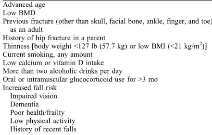
by BMD(both primary and secondary causes) and risk factors for osteoporotic fracture. For BMD-defined osteoporosis, major risk factors in postmenopausal women are advanced age, genetics, lifestyle factors (eg, low calcium and vitamin D intake, smoking), thinness, and menopause status. Risk factors for osteoporotic fracture are listed in Table 1; the most common are advanced age, low BMD, and previous fracture (other than skull, facial bone, ankle, finger, and toe) as an adult.
Risk factors for BMD-defined osteoporosis and osteoporotic fracture overlap, given that BMD is a risk factor for fracture. Importantly, however, many fracture risk factors are not related to BMD.
BMD and fracture risk
Bone density is an important determinant of fracture risk, especially in women aged 65 and older.27,28
In general, lower BMD scores indicate more severe osteoporosis and higher risk of fracture. A decrease of 1 SD in BMD represents a 10% to 12% decrease in BMD and an increase in fracture risk by a factor of 1.5 to 2.6.29,30 BMD and fracture risk are most closely related when bone density is used to predict the fracture risk at that same site. Risks for spine fracture and hip fracture increase 2.3-fold and 2.6-fold, respectively, for each decrease of 1 SD in age-adjusted BMD at spine and hip, respectively.30
Fracture risk, however, depends largely on factors other than BMD. Furthermore, a reliable marker to predict fracture risk or determine fracture risk reduction from therapy is not currently available. The use of BMD scores to assess fracture risk can be markedly improved by combining BMD with infor-mation about other risk determinants, particularly the
TABLE 1.Risk factors for osteoporotic fracture Advanced age
Low BMD
Previous fracture (other than skull, facial bone, ankle, finger, and toe) as an adult
History of hip fracture in a parent
Thinness [body weightG127 lb (57.7 kg) or low BMI (G21 kg/m2)] Current smoking, any amount
Low calcium or vitamin D intake More than two alcoholic drinks per day
Oral or intramuscular glucocorticoid use for93 mo Increased fall risk
Impaired vision Dementia Poor health/frailty Low physical activity History of recent falls
woman`s age and fracture history. The WHO is currently developing a model to estimate the 10-year absolute fracture risk based on known risk factors. It will offer substantial benefits to healthcare providers compared with current methods.
Treatment-induced changes in BMD do not always correlate well with reductions in vertebral fracture risk.31<34 In addition, fracture risk reductions in response to antiresorptive therapy occur much more rapidly than discernible BMD changes. For example, significant fracture risk reduction has been reported after 6 months of risedronate therapy,35 although minimal BMD increases were observed at that time.36
Advanced age
As women age, their risk of fracture increases. In general, the risk of osteoporotic fracture doubles every 7 or 8 years after age 50. The median age for hip fracture is 82 years. The median age for vertebral fracture is thought to occur in a woman`s 70s.12
Older women are at substantially greater risk of fracture at any given BMD value.31 For example, at the same T-score of j2.5, a 75-year-old woman has about 8 to 10 times the 10-year hip fracture risk of a 45-year-old woman.
Fracture history
In two analyses of studies, a peri- or postmeno-pausal woman who has had a fracture has approx-imately a 2.0 increased risk of sustaining another fracture; adjustment for BMD did not significantly affect the risk.22,37 A study of older women (mean age, 74 years) with recent vertebral fracture found that approximately 20% of these women experienced another vertebral fracture within 1 year of an incident vertebral fracture.21 However, the risk of recurrent fracture was significantly affected by the number of existing fracturesVwomen with two or more verte-bral fractures had a significantly increased risk (relative risk, 11.6) of another vertebral fracture within 1 year.
Genetics
The greatest influence on a woman`s peak bone mass (ie, the maximal BMD gained during the skeletal development and maturation phase) is heredity. Studies have suggested that up to 80% of the variability in peak bone density might be attributable to genetic factors.38,39Female children of women who have osteoporotic fractures have lower bone density than would be expected for their age.40,41 First-degree relatives (ie, mother, sister) of women with
osteoporosis also tend to have lower bone density than those with no family history of osteoporosis.42
A history of fracture in a first-degree relative also significantly increases the fracture risk. In a meta-analysis,43 a family history of fracture was found to be associated with significant increases in any osteoporotic fracture. Hip fracture risks were nearly 50% higherV127% higher if a hip fracture had occurred in a parent.
Lifestyle factors
Several lifestyle factors are associated with the risk of low BMD and fracture. These include nutrition, physical activity, cigarette smoking, and heavy alcohol consumption.
Nutrition
A balanced diet plays an important role in bone development and maintenance of bone health throughout life. Both calcium and vitamin D have well-known roles in bone metabolism. Adequate intake of calcium and vitamin D is required throughout life for a woman to achieve her genetically determined peak bone mass and to maintain optimal bone mass and strength after peak bone mass is attained.44
Low vitamin D intake has been linked to impaired muscle strength, increased fall risk, and increased fracture risk along with increased rates of bone loss.45 Furthermore, treatment with vitamin D has been found to reduce fracture risk in elderly postmeno-pausal women,46<49 although not in all studies.50,51 Elderly postmenopausal women have an increased risk of hip fracture associated with low dietary calcium intake.52
Physical activity
There is general agreement that weight-bearing exercises confer a positive effect on the musculo-skeletal system and that weight-bearing exercises (eg, walking, running, step aerobics, gymnastics) provide the greatest osteogenic stimulus.53<55 The effects of exercise on bone mass could be caused by osteo-blast activity.56
Regular exercise has been associated with reduced fracture risk.6Exercise also appears to reduce the risk of falls by increasing muscle mass, strength, and balance, although it is unclear whether exercise affects the risk of fracture from falls that do occur.6
Long-term immobilization, such as prolonged bed rest, has been associated with rapid and significant bone loss.57,58However, no evidence indicates that a sedentary lifestyle increases the risk of bone loss.
Cigarette smoking
Compared with nonsmokers, women smokers tend to lose bone more rapidly, have lower bone mass, and reach menopause 2 years earlier, on average.59<61In addition, some data show that postmenopausal women who currently smoke have significantly higher fracture rates than nonsmokers.62 The risk imparted by smoking remains significant even after adjust-ing for BMD.63
The mechanisms by which smoking might adversely affect bone mass are not known, although evidence suggests that cigarette smokers may have impaired calcium absorption59,64,65 and lower 17A -estradiol levels.66
Alcohol consumption
Heavy alcohol consumption [defined in the Fra-mingham Study as Q7 oz/wk (200 mL/wk)]67 has been shown to increase the risk of falls and hip fracture. A recent meta-analysis68 showed that con-suming as few as two drinks per day significantly increases the fracture risk. Very heavy alcohol consumption also has detrimental effects on BMD. However, moderate alcohol consumption [1-2 oz/wk (30-60 mL/wk)] in women 65 years of age and older is associated with higher BMD69 and decreased risk of hip fracture.70
Thinness
Being thinVoften cited as body weight less than 127 lb (57.7 kg), the lower quartile of weight for US women older than age 65, or a body mass index (BMI) less than 21 kg/m2Vis a risk factor for low BMD.71 Thinness has also been associated with increased fracture risk, especially in older women.72 In a meta-analysis of population-based cohort studies,73 a BMI of 20 kg/m2 conferred nearly a twofold increased risk of fracture compared with a BMI at the upper range of normal (Q25 kg/m2). Being thin seems to have its primary effect on fracture risk by its association with low bone density.
Menopause status
The increased rate of bone resorption immedi-ately after menopause clearly indicates a hormonal influence on bone density in women. The most likely explanation for this increased resorption is the drop in ovarian estrogen production that accompa-nies menopause.
Bone loss begins to accelerate approximately 2 to 3 years before the last menses, and this acceleration
ends 3 to 4 years after menopause. For an interval of a few years around menopause, women lose 2% of bone annually. Afterward, bone loss slows to about 1% to 1.5% per year.74,75A prospective, longitudinal study of white women reported BMD losses during this 5- to 7-year interval of 10.5% for the spine, 5.3% for the femoral neck, and 7.7% for the total body.74 Although some of the decline can be attributed to age-related factors, lower estrogen levels were impli-cated as the cause for approximately two thirds of the bone loss. Lower estrogen levels have also been significantly associated with increased fracture risk in older women (mean age, 75 years).76
Women experiencing menopause at or before age 40Veither spontaneously or induced (eg, through bilateral oophorectomy, chemotherapy, pelvic radia-tion therapy)Vare at greater risk of low BMD than other women of the same age who have not reached menopause.77However, by age 70, when fractures are more likely to occur, these women have the same risk of low BMD or fracture as women who reached menopause at the average age.78,79
Secondary causes of bone loss
Various medications, disease states, and genetic disorders are associated with bone loss (Table 2). Oral glucocorticoid use causes the most common form of drug-related osteoporosis. Evidence suggests that high-dose inhaled glucocorticoids may cause bone loss.80,81 Other studies82,83 suggest that no effect occurs with the approved doses of inhaled steroids. In a meta-analysis of seven population-based cohort studies (N = 42,500 men and women),84current and previous oral glucocorticoid use was found to be significantly associated with increased risk of osteo-porotic fracture. This risk appears within 3 months of beginning corticosteroid use.
Current use of two drugs prescribed for premeno-pausal womenVgonadotropin-releasing hormone (GnRH) and intramuscular medroxyprogesterone acetate (MPA)Vhas been associated with bone loss. Use of GnRH contributes to bone loss by creating iatrogenic hypogonadism.85 Bone loss with short-term use of GnRH agonist therapy is reversible. Bone loss with long-term use can be ameliorated by
Badding back^ low-dose estrogen therapy. Use of depot MPA (150 mg/3 months) as a contraceptive has been associated with bone loss.86,87 This bone loss, which has never been linked to the occurrence of osteoporotic fracture, has been shown to be reversible in some studies; however, other studies have indi-cated that BMD only partially recovers.88
Medical conditions associated with bone loss include excess urinary calcium excretion, which may be caused by a renal calcium leak or hyper-thyroidism. Vitamin D deficiency, an especially common condition in the older women, is a correct-able cause of secondary hyperparathyroidism and accelerated bone loss. Other conditions that can have a detrimental effect on bone include multiple mye-loma, endocrine disorders such as hyperparathy-roidism and Cushing`s syndrome, and disorders of collagen structures. Renal failure can cause either increased bone resorption (secondary/tertiary hyper-parathyroidism) or decreased bone formation, leading to renal osteodystrophy.
EVALUATION
All postmenopausal women should be assessed for risk factors associated with osteoporosis and fracture. This assessment requires a history, physical examination, and any necessary diagnostic tests. The
goals of this evaluation are to identify risk factors for fractures, including whether osteoporosis is present, and, if so, assessing its severity, ruling out secondary causes for osteoporosis, and identifying modifiable risk factors for falls and injuries.
History and physical examination
The medical history and physical examination should focus on the detection of clinical risk factors for osteoporosis and fracture. This includes a personal history of fracture as well as a history of hip fracture in a parent. Most of these risks can be uncovered with a simple questionnaire. Although risk factors may help identify contributing causes of osteoporosis or help guide therapeutic recommendations, they cannot be used to diagnose osteoporosis.
Loss of height may be a sign of vertebral fracture. After achieving maximal height, women (and men) can lose up to 1.0 to 1.5 inches (2-3 cm) of height as part of the normal aging process, primarily as a result of shrinkage of intervertebral disks. Height loss greater than 1.5 inches (3 cm) increases the likelihood that a vertebral fracture is present.89Height should be measured annually with an accurate method, such as a wall-mounted ruler or a stadio-meter. Loss of 1.5 inches (3 cm) or more calls for evaluation by a lateral thoracolumbar radiograph to identify silent vertebral fractures.
Weight also should be recorded to identify those with a body weight of 127 lb (57.7 kg) or lower and to calculate BMI.
The examination should include an assessment for acute or chronic back pain, especially in the middle back, which may indicate the presence of vertebral fractures. The midback vertebrae T11Y12 and L1 are the most common fracture sites, followed by T6 through T9.90<92 Multiple, severe vertebral compres-sion fractures ultimately result in kyphosis (abnormal curvature of the thoracic spine), the most obvious sign of osteoporosis.
Because back pain, height loss, and kyphosis can occur without osteoporosis, and two thirds of verte-bral fractures are asymptomatic,93,94 vertebral frac-ture must be confirmed, usually by lateral spine radiographs. In addition, some dual energy x-ray absorptiometry (DXA) techniques (eg, instant verte-bral assessment, morphometric x-ray absorptiometry) allow vertebral fracture assessment and, hence, can be used to visualize a fracture at the same time that BMD is being measured.95,96 Height loss of more than 20% (or 4 mm) of the anterior, mid, or posterior
TABLE 2. Secondary causes of bone loss Medications
Oral or intramuscular use of glucocorticoids for93 mo Excessive thyroxine doses
Aromatase inhibitors
Long-term use of certain anticonvulsants (eg, phenytoin) Heparin
Cytotoxic agents
Gonadotropin-releasing hormone agonists or analogues Intramuscular medroxyprogesterone contraceptive Immunosuppressives (eg, cyclosporine)
Genetic disorders Osteogenesis imperfecta Thalassemia
Hypophosphatasia Hemochromatosis Disorders of calcium balance
Hypercalciuria Vitamin D deficiency Endocrinopathies
Cortisol excess Cushing`s syndrome
Gonadal insufficiency (primary and secondary) Hyperthyroidism
Type 1 diabetes mellitus Primary hyperparathyroidism Gastrointestinal diseases
Chronic liver disease (eg, primary biliary cirrhosis)
Malabsorption syndromes (eg, celiac disease, Crohn`s disease) Total gastrectomy
Billroth I gastroenterostomy Other disorders and conditions
Multiple myeloma Lymphoma and leukemia Systemic mastocytosis
Nutritional disorders (eg, anorexia nervosa) Rheumatoid arthritis
dimension of a vertebra on spinal radiograph is also indicative of vertebral fracture.97,98
After menopause, a woman`s risk of falls should be assessed at least annually. Clinical factors related to an increased risk of falls include the following:
&
A history of falls, fainting, or loss of consciousness&
Muscle weakness or coordination&
Dizziness or balance problems&
Difficulty standing or walking&
Arthritis&
Impaired visionThe risk of falls is also increased by use of medications that affect balance and coordination (eg, sedatives, narcotic analgesics, anticholinergics, anti-hypertensives) or by use of multiple medications.99
The greater the number of risk factors, the greater the risk is of falling. In one study, having four or more of these risk factors increased the risk of falls by nearly 80%.100
Safety hazards in the home and work environ-ment, such as obstacles and poor lighting, also contribute to the risk of falls. These hazards can be assessed by questioning the woman or through a home and/or workplace visit by an occupational therapist or other healthcare professional knowledge-able in fall prevention.
Bone mineral density measurement
BMD testing is the technical standard for diagnos-ing osteoporosis. Determinants of bone strength other than bone density cannot be measured in the clinical setting.
Indications for BMD testing
Testing of BMD should be performed based on a woman`s risk profile. Testing is not indicated unless the results will influence a treatment or management decision. Other factors such as availability of BMD testing equipment and reimbursement by insurance also affect the decision to measure BMD.
NAMS recommends that BMD be measured in the following populations:
&
Postmenopausal women with medical causes of bone loss, regardless of age&
Postmenopausal women at least 65 years of age, regardless of additional risk factorsTesting should be considered for healthy post-menopausal women younger than age 65 when one or more of the following risk factors for fracture have
been identified (the greater the number of risk factors, the greater is the need for testing):
&
Fracture (other than skull, facial bone, ankle, finger, and toe) after menopause&
Thinness [body weightG127 lb (57.7 kg) or BMIG21 kg/m2]
&
History of hip fracture in a parent&
Current smokerBMD testing options
Several tests to measure BMD are available. DXA is the preferred technique for measuring central (eg, spine, hip) BMD and for diagnosing osteoporosis because it measures BMD at the important sites of osteoporotic fractures.101
When BMD testing is indicated, NAMS recom-mends measuring the total hip, femoral neck, and posterior-anterior lumbar spine, and using the lowest of the three BMD scores. In some older patients (older than 60 years), there can be artifacts of the spine that make measurements unreliable. The spine, however, is a useful site for BMD measurement in early postmenopausal women because they tend to lose bone faster in the spine than in the hip.
Although tests at peripheral sites (eg, wrist, calcaneus) can identify women with low bone mass, they are not as useful as central-site tests because the prediction of risk with the results is not well determined. WHO diagnostic criteria cannot be ap-plied to peripheral sites with the exception of the distal radius, although BMD measurement has been predictive of fracture risk.102 Peripheral site mea-surements should be limited to the assessment of fracture risk when DXA is not available. They cannot be used to diagnose osteoporosis or to follow re-sponse to therapy.103
Follow-up BMD testing
In most cases, repeat DXA testing in untreated postmenopausal women is not useful until 3 to 5 years have passed, given the rate of bone loss of 1% to 1.5% per year. Postmenopausal women, after substantial BMD losses in early postmenopause, generally lose about 0.5 T-score units every 5 years.74,104
For women receiving osteoporosis therapy, BMD monitoring may not provide clinically useful infor-mation until after 2 years of treatment. The lack of an increase in BMD is not evidence of treatment failure. In two randomized controlled trials, most women who appeared to have lost more than 4% of BMD during the first year of treatment with either alendronate or
raloxifene showed substantial gains the second and third years while remaining on the same therapy.105,106This variability was seen despite excel-lent quality assurance programs and is a consequence of the imprecision of DXA testing.
A statistically insignificant decrease in BMD on treatment may be related to imprecision in the DXA measurement rather than to treatment failure. How-ever, a statistically significant decrease in BMD (usually >4%-5%) would warrant further consider-ation of secondary causes of bone loss and evaluconsider-ation of adherence to therapy.
Bone turnover markers
Biochemical markers of bone turnover cannot diagnose osteoporosis and have varying ability to predict fracture risk. Nevertheless, these tests have been studied as a means to assess therapeutic response earlier than through BMD changes, some-times within a few months as opposed to the 1 to 3 years required with BMD.107<109 However, bone turnover markers vary from day to day, are affected by food intake and time of day, and lack assay standardization, limiting their clinical utility.
The value of bone turnover markers in routine clinical practice has not been established. Although some clinicians have found that these data can encourage adherence to therapy, several trials have found no difference in adherence when marker values are communicated to women.110,111
Tests for secondary causes
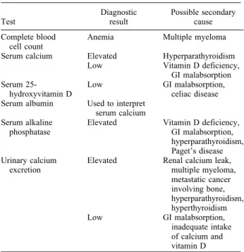
Once osteoporosis is diagnosed, any secondary causes should be identified. Various laboratory tests can be useful (Table 3). Routine tests include a complete blood cell count plus serum levels of calcium, 25-hydroxyvitamin D, alkaline phosphatase, and albumin, as well as urinary calcium excretion to identify calcium malabsorption or renal calcium leak. If the clinical history, physical examination, or routine laboratory tests indicate a need, special tests that may be appropriate include measurement of thyroid-stimulating hormone, urinary cortisol, serum protein electrophoresis, and parathyroid hormone.
MANAGEMENT: LIFESTYLE APPROACHES
All postmenopausal women, regardless of their osteoporosis risk factors, should be encouraged to take steps to prevent bone loss and fractures, such as eating a balanced diet, obtaining adequate calcium
and vitamin D, participating in appropriate exercise, not smoking, avoiding excessive alcohol consump-tion, and instituting measures to prevent falls. These steps offer health benefits beyond their effects on osteoporosis.
Nutrition
A balanced diet is important for bone development as well as for general health. Some populations, such as elderly women (older than age 65) with reduced appetites or women who diet frequently or have eating disorders, may not consume adequate vitamins and minerals to maintain optimal bone mass. Elderly women who lose weight, purposely or not, run the risk of accelerated bone loss and a higher risk of hip fracture.112 In general, women should be advised to maximize consumption of fruits and vegetables and minimize consumption of fats.
For women older than age 75, data from the Framingham Osteoporosis Study, a longitudinal cohort study, suggest that adequate protein intake may help minimize bone loss.113,114 Protein sup-plements (20 g/day) in older patients (mean age, 82 years) who have sustained a hip fracture have been shown to significantly shorten the hospital stay (median stay, 69 days vs 102 days for placebo recipients) after
TABLE 3. Routine laboratory tests for osteoporosis evaluation Test Diagnostic result Possible secondary cause Complete blood cell count
Anemia Multiple myeloma Serum calcium Elevated Hyperparathyroidism
Low Vitamin D deficiency, GI malabsorption Serum
25-hydroxyvitamin D
Low GI malabsorption, celiac disease Serum albumin Used to interpret
serum calcium Serum alkaline
phosphatase
Elevated Vitamin D deficiency, GI malabsorption, hyperparathyroidism, Paget`s disease Urinary calcium
excretion
Elevated Renal calcium leak, multiple myeloma, metastatic cancer involving bone, hyperparathyroidism, hyperthyroidism Low GI malabsorption, inadequate intake of calcium and vitamin D GI, gastrointestinal.
hip fracture and improve the clinical outcomes while in the hospital.115 Compared with the controls, pro-tein recipients also had significantly lower rates of complications and mortality 7 months after their hip fracture.
An adequate intake of both calcium and vitamin D is important for bone health, and it is recognized as an important component of any osteoporosis pre-scription-drug regimen. For example, a review of 31 clinical trials evaluating estrogen therapy with and without calcium supplements found annual BMD gains at the hip were significantly greater for those receiving estrogen plus calcium (2.4%) compared with estrogen alone (0.9%).116
Calcium, either alone or with vitamin D, is not as effective as pharmacotherapy with either estrogen alone (ET) or estrogen plus progestogen (EPT), a selective estrogen-receptor modulator (SERM), or a bisphosphonate. Nevertheless, calcium and vitamin D are both important components of osteoporosis therapy in combination with all antiresorptive agents.
Calcium
Evidence has established the role of adequate calcium intake on bone health, primarily in increasing BMD and improving the efficacy of therapeutic agents. Calcium has not been shown to have a positive effect on fracture risk;50,117 however, in the Women`s Health Initiative (WHI) trial,117 hip frac-tures were significantly reduced in older women who were adherent to the calcium regimen.
Calcium requirements increase with advancing age, particularly after menopause, owing in part to both reduced intestinal calcium absorption and renal calcium conservation. The primary factor influencing the amount of calcium absorbed is the amount of calcium ingested.
Most experts support the published recommenda-tions for daily calcium consumption from either the
National Institutes of Health (revised in 1994)118 or the National Academy of Sciences (revised in 1997).119 Recommendations related to peri- and postmenopausal women are presented in Table 4.
No single laboratory test can accurately detect calcium deficiency. However, a 24-hour urine cal-cium level of less than 50 mg suggests either insufficient intake or poor absorption. In general, postmenopausal women in the United States and Canada have dietary calcium intakes that are low, with median intakes of approximately 600 mg/ day.120,121 Specific populations of postmenopausal women at increased risk of inadequate calcium intake include women who are older, are lactose intolerant, follow a vegetarian diet, or have poor eating habits.
Dietary sources should be the primary source of calcium intake because of the other essential nutrients found in high-calcium food. Dairy products are the best sources of calcium based on their high elemental calcium content, high absorption rate, and low cost relative to total nutritional value. To achieve maximal calcium absorption from food sources, food selection decisions should reflect the food`s calcium bioavail-ability and the presence in the meal of other foods that may inhibit calcium absorption (eg, oxalic acid-containing foods, such as spinach, and phytate-rich grains, such as wheat bran).122
Calcium supplements and calcium-fortified foods are alternative sources for women unable to consume enough dietary calcium; most women need an addi-tional 600 to 900 mg/day (two to three dairy portions) over their usual daily intake to reach recommended levels. Calcium citrate supplements are well absorbed when taken with meals or on an empty stomach; calcium carbonate is better absorbed when taken with food. It is best to take calcium in divided doses for better absorption, although single doses of up to 1,000 mg can be taken.
Total calcium intakes of up to 1,500 mg/day do not appear to increase the risk of developing renal calculi and may actually reduce it.123 Calcium supplements are contraindicated in a woman with a calcium-containing renal calculus until her urinary biochemi-cal profile has been assessed. Larger amounts of calcium (>2,500 mg/day) should be avoided.
Calcium intervention trials have not reported any serious adverse events. Nevertheless, some women have difficulty swallowing the large tablet or have gastrointestinal (GI) adverse effects (ie, gaseousness, constipation). Tolerability can be addressed by switching the type of calcium or reducing the dose. GI adverse effects are often related to a woman`s
TABLE 4. Recommended daily elemental calcium intake in peri- and postmenopausal women
National Academy of Sciences
Age 31-50 1,000 mg
Age 51 and older 1,200 mg
National Institutes of Health
Premenopausal women aged 25-50 1,000 mg Postmenopausal women younger than age 65 and
using estrogen therapy
1,000 mg Postmenopausal women not using estrogen therapy 1,500 mg All women aged 65 and older 1,500 mg Adapted from the National Institutes of Health118 and the National
taking more calcium than required, not dividing doses, or perhaps confusing supplemental intake with recommended total daily intake.
Vitamin D
The nutrient vitamin D is essential for the intestinal absorption of calcium. The current National Academy of Sciences recommended dietary intake for vitamin D is 400 IU/day for women aged 51 to 70 years and 600 IU/day for women older than age 70.119 In addition, NAMS recommends intake of 700 to 800 IU/day for women at risk of deficiency because of inadequate sunlight exposure, such as older, frail, chronically ill, housebound, or institutionalized women or those who live in northern latitudes.124 Doses as high as 2,000 IU/day are safe.119 Much higher doses may introduce risks such as hyper-calciuria and hypercalcemia.
Sources of vitamin D include sunlight, vitamin DY
fortified dairy products, fatty fish, and supplements. Daily requirements can usually be met with a multi-vitamin supplement (typically containing 400 IU vitamin D), vitamin DYfortified foods (eg, milk, breakfast cereals providing 100 IU per serving), or calcium supplements containing vitamin D (usually 200 IU per tablet). Many women over the age of 65 who have little or no sun exposure and rely on multivitamins alone for vitamin D intake may have suboptimal vitamin D levels.125Currently, there is no worldwide consensus on criteria for acceptable serum 25-hydroxyvitamin D values, but if parathyroid hor-mone concentration were used as an index of calcium absorption, as some suggest, the lower end of the normal 25-hydroxyvitamin D concentration would be in the range of 30 ng/mL (70-80 nmol/L).126Because of the long half-life of vitamin D, taking vitamin D at the same time as calcium is not necessary, although it can be a convenient way to obtain adequate intake of both nutrients.
Regarding the effect of vitamin D on fracture risk, several large randomized controlled studies of vita-min D (400 and 800 IU/day) plus 1,000 mg calcium50,51,117 have shown that the nutrients do not have a significant effect on fracture risk. However, a meta-analysis49of randomized clinical trials in post-menopausal women (mean ages, 71-85 years) found that a vitamin D dose of 700 to 800 IU/day was associated with significant reductions in the risk of both hip and nonvertebral fractures. No significant changes were found in either fracture outcome in trials that used only 400 IU/day.
Studies have found that vitamin D (600-700 IU/day) with supplemental calcium can reduce the rate of post-menopausal bone loss, especially in older women.127 More recent results from the WHI117 found calcium (1,000 mg/day) plus vitamin D (400 IU/day) recipients had a small but significant 1% improvement in hip BMD. Vitamin D supplementation also has been found to improve muscle strength128 and balance,129 and reduce the risk of falling.130
Vitamin K
Supplementation with vitamin K (1 mg/day) appears to be associated with beneficial effects on bone turnover and bone density. Taking vitamin K as part of a daily multivitamin supplement may contribute to reducing postmenopausal bone loss, especially in the hip,131 but further validation is needed to define its contribution. Vitamin K supplements are contraindicated in women taking warfarin.
Magnesium
Another nutrient, magnesium, is sometimes men-tioned as a necessary supplement for the protection of bone health and/or for absorption of calcium. However, in most trials focused on BMD or osteoporotic fracture, benefits of calcium were observed without magnesium supplements. Moreover, a study with calcium absorption as the end point found that adding 789 to 826 mg/day of magnesium, more than double the daily average mag-nesium intake (280 mg) for postmenopausal women, had no effect on calcium absorption.132Nevertheless, in women with excessive magnesium loss, usually due to GI disease (eg, diarrhea, vomiting), magnesium sup-plementation would be appropriate.133,134
Isoflavones
Clinical trial data do not support the use of isoflavones (a class of phytoestrogens found in rich supply in soybeans and soy products as well as in red clover) to prevent or treat osteoporosis. Although some data suggest that isoflavones may favorably affect bone health,135accumulating data from several more recent studies indicate a lack of bone benefits from isoflavones, regardless of the source (ie, extracted from red clover or soy or consumed in soy foods).136<138Ipriflavone, a synthetic isoflavone avail-able without a prescription in the United States and Canada, has not demonstrated a positive effect on bone density, bone turnover markers, or fracture risk in women with osteoporosis.139
Exercise
Local increases in bone mass occur in response to activities that cause significant stress to bone. Active exercises that involve weight training can increase bone mass if they increase muscle mass and strength. Applying passive stress tests to bone also shows promise, with the most positive results coming from use of high-frequency, whole-body vibration systems.140,141
In early postmenopausal women, strength training provides small but significant benefits on bone mass.142 A meta-analysis143 found that women who exercised increased spinal BMD by approximately 2%. For women who use menopausal estrogen-containing therapy, strength training provides addi-tional BMD benefits over hormone therapy alone.144 Most strength training studies have used progressive resistance obtained with machines designed for this purpose (eg, Nautilus). However, strength training can be performed as few times as twice a week and need not involve expensive equipment. Exercise for women with established osteoporosis should not include heavy weight-bearing exercises or activity so vigorous that it may trigger a fracture.
For elderly women with osteoporosis, physical activity plays an important role in reducing the risk of falls. Among women aged 75 and older, muscle-strengthening and balance exercises have been shown to reduce the risk of falls and fall-related injuries by 75%.145
Fall prevention
Falls are the precipitating factor in nearly 90% of all fractures.146 In the United States and Canada, approximately one third of women older than age 60 fall at least once a year.100,147 In nearly one half of these cases, it is a recurrent fall. The incidence of falls increases with age, rising to a 50% annual rate in people older than age 80. Elderly women have a significantly higher risk of falls than do men of the same age. As a result, prevention of falls that can cause fractures should be an aspect of routine care for all postmenopausal women.
Several healthcare interventions have proven effec-tive in reducing the risk of falls. These focus primarily on exercises to improve balance and muscle strength, adjusting medication use (especially psycho-tropic drugs), and reducing fall hazards in the home.148 Tapering or discontinuing use of benzo-diazepines, neuroleptic agents, and antidepressants has been found to reduce the risk of falling by more
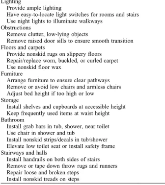
than 60%.149 Implementing relatively inexpensive measures to eliminate safety hazards in the home may also reduce this risk (Table 5), but home hazard intervention studies have failed to show significant reductions in fracture.148
Hip protectors worn during the day have been shown to reduce the likelihood of hip and pelvis fractures from falls among elderly postmenopausal women (aged 75 years and older) with a history of frequent falls.150 However, a Cochrane review151 found the overall evidence inconclusive regarding efficacy in reducing hip fractures. Furthermore, the adherence rates in studies were low, averaging approximately 50%, primarily due to the inconvenience of wearing the protective garment day and night.
Smoking cessation
Because smoking can lead not only to lower BMD but also to a wide range of health problems, including increased fracture risk, smoking cessation should be encouraged for all smokers. A wide array of smoking cessation aids are available, including prescription products (with and without nicotine) and behavior-modification programs.
Alcohol avoidance
The level of alcohol consumption associated with an increased risk of falls is more than seven drinks a
TABLE 5. Recommendations for fall prevention Lighting
Provide ample lighting
Have easy-to-locate light switches for rooms and stairs Use night lights to illuminate walkways
Obstructions
Remove clutter, low-lying objects
Remove raised door sills to ensure smooth transition Floors and carpets
Provide nonskid rugs on slippery floors Repair/replace worn, buckled, or curled carpet Use nonskid floor wax
Furniture
Arrange furniture to ensure clear pathways Remove or avoid low chairs and armless chairs Adjust bed height if too high or low
Storage
Install shelves and cupboards at accessible height Keep frequently used items at waist height Bathroom
Install grab bars in tub, shower, near toilet Use chair in shower and tub
Install nonskid strips/decals in tub/shower Elevate low toilet seat or install safety frame Stairways and halls
Install handrails on both sides of stairs Remove or tape down throw rugs and runners Repair loose and broken steps
week, as established by the Framingham Heart Study.70 Postmenopausal women who drink should be advised to drink moderately and not to exceed seven drinks a week. One drink is considered to be one 12-oz (360 mL) beer, 4 oz (120 mL) of wine, or 1 oz (30 mL) of liquor.
MANAGEMENT:
PHARMACOLOGIC APPROACHES
A management strategy focused on lifestyle approaches may be all that is needed for postmeno-pausal women who are at low risk of osteoporotic fracture. NAMS recommends adding osteoporosis drug therapy in the following populations:
&
All postmenopausal women who have had an osteoporotic vertebral fracture&
All postmenopausal women who have BMD values consistent with osteoporosis (ie, T-scores equal to or worse thanj2.5)&
All postmenopausal women who have T-scores from j2.0 to j2.5 and at least one of the following risk factors for fracture: thinness [body weightG127 lb (57.7 kg) or low BMI (G21 kg/m2)], history of fragility fracture since menopause, or history of hip fracture in a parentThe diagnostic categorization should be based on the lowest of the BMD values among the three measured sites of total hip, femoral neck, and posterior-anterior lumbar spine. Current treatment guidelines are based on specific bone density thresh-olds and risk factors. Available in the near future will be an improved method from the WHO that uses algorithms that incorporate estimates of absolute fracture risk.
Several pharmacologic options are available for osteoporosis therapy, including bisphosphonates, the SERM raloxifene, parathyroid hormone, estrogens, and calcitonin. No studies have compared these therapies for antifracture efficacy.
Adherence to therapy is poor. In studies of 6 months to 1 year, adherence rates for prescription drugs ranged from below 25% to 81%, depending on the therapy.152<154 Ensuring adherence to the treatment plan is perhaps the most important follow-up measure for clinicians.
Bisphosphonates
This class of drugs works by inhibiting the activity of osteoclasts and shortening their life span, thereby
reducing bone resorption.155Bisphosphonates do not have known beneficial effects on the body other than on bone. The most common adverse effect of bisphosphonate therapy is esophageal and gastric irritation, particularly affecting individuals who dose inappropriately. Before starting bisphosphonate therapy, serum creatinine should be used to estimate the glomerular filtration rate; treatment may be initiated only if the rate is 30 mL/min or greater.
Clinical trials have demonstrated that bisphospho-nates significantly increase BMD at the spine and hip in a dose-dependent manner in both younger and older postmenopausal women. In women with osteo-porosis, bisphosphonates have reduced the risk of vertebral fractures by 40% to 50% and reduced the incidence of nonvertebral fracture, including hip fracture, by about half this amount.106,155
All the bisphosphonates approved for osteoporosis therapy in both the United States (alendronate, ibandronate, and risedronate) and Canada (alendro-nate, etidro(alendro-nate, and risedronate) are available in oral formulations for daily and intermittent dosing regi-mens. Weekly oral dosing regimens of alendronate and risedronate and monthly oral dosing regimens and IV dosing of ibandronate have been approved based on clinical trials that showed BMD responses equivalent to those observed with daily treat-ment.156<159 All fracture data are from trials with daily dosing; the bridging studies beyond daily dosing were not designed with fracture end points.
Alendronate
This bisphosphonate, marketed as Fosamax, is approved in both the United States (as an oral tablet and liquid) and Canada (oral tablet only) for post-menopausal osteoporosis prevention (5 mg/day or 35 mg/wk) and treatment (10 mg/day or 70 mg/wk). Alendronate is also available in a single weekly oral tablet of 70 mg with 2,800 IU of vitamin D (Fosamax Plus D).
For women in early postmenopause, 2 to 6 years of treatment with alendronate (5 mg or more daily) has been shown to significantly increase BMD at the spine and hip by approximately 1% to 4% from baseline, whereas BMD in placebo recipients decreased by 2% to 4% during that time.160,161 In older women with osteoporosis,162 therapy with 10 mg daily significantly increased BMD in the spine (8.8%) and the femoral neck (5.9%) after 3 years, compared with placebo. In 7- and 10-year trials in women with low bone density,163,164 alendronate therapy resulted in increases from baseline of 5% to
10% at the spine and hip in postmenopausal women who had low BMD or established osteoporosis. Because placebo groups were not followed for the duration of the studies,163,164 the antifracture effects of long-term alendronate therapy could not be adequately evaluated. However, there was no appa-rent increase in fracture risk over time.
The efficacy of alendronate in decreasing fracture risk has been demonstrated only in postmenopausal women with osteoporosis. Similar to other bisphos-phonates, alendronate has shown lesser effects in women without osteoporosis.
In the Fracture Intervention Trial (FIT),165 daily alendronate therapy for 2.9 years significantly reduced the risk of vertebral fracture by 47% and of hip fracture by 51% in women with low BMD and previous vertebral fracture. The incidence of clinical vertebral fractures was reduced by 59% within the first year.166In a composite analysis of the two arms of the FIT study,1663 years of alendronate therapy in a subgroup of women with osteoporosis (ie, vertebral fracture or T-score equal to or worse than j2.5) significantly reduced the risk of nonspine fracture by 27% and new spine fracture by 50%.
Risedronate
This bisphosphonate, marketed as Actonel, is approved for the prevention and treatment of post-menopausal osteoporosis in oral tablet doses of 5 mg daily or 35 mg once weekly. Recently available in the United States is a packet containing both risedronate and calcium (marketed as Actonel with Calcium) that provides 4 weeks of risedronate therapy (35 mg/wk) and calcium carbonate (500 mg for the no-risedronate days).
In a randomized clinical trial of early postmeno-pausal women (age range, 40-61 years; mean age, 51-52 years) with normal bone density, risedronate doses of 5 mg/day for 2 years produced significant BMD increases of 5.7% in the lumbar spine and 5.4% in the hip greater than with placebo.167 In a randomized controlled trial in older postmenopausal women (mean age, 68-69 years),36 3 years of risedronate therapy (5 mg/day) resulted in significant BMD increases of 4.3% in the spine and 2.8% in the femoral neck compared with placebo. Therapy for 7 years resulted in progressive increases in BMD of 11.5% from baseline (with no placebo group after 5 years).168
Several randomized controlled trials have found fracture risk reductions with risedronate. In two trials of postmenopausal women with osteoporosis,36,169
1 to 3 years of treatment with 5 mg/day of risedronate significantly reduced the risk of vertebral fracture (41% - 49%) compared with placebo. Within the first year of therapy, the relative risk of vertebral fracture was reduced by 61% to 65%. After 3 years of therapy, vertebral fracture risk reductions were still statistically significant relative to placebo. In one of these trials,36the risk of nonvertebral fracture was significantly reduced by 39%. In the other trial,169 nonvertebral fracture risk was reduced by 33%, although this was not statistically significant versus placebo.
In the Hip Intervention Program Study Group,170a randomized controlled trial of 5,445 postmenopausal women aged 70 to 79 years, daily risedronate therapy significantly reduced the relative risk of hip fracture by 40% in women with BMD values consistent with osteoporosis. It reduced the risk of hip fracture by 60% in the group with previous vertebral fractures. However, therapy did not significantly lower the hip fracture risk in women 80 years of age and older who had risk factors for falling but who did not have BMD testing performed to confirm osteoporosis.
In a randomized controlled trial of 265 postmeno-pausal women (mean age, 72 years), the incidence of vertebral fractures in women treated with risedronate 5 mg/day was significantly reduced during years 4 and 5 compared with placebo,171 and appeared to remain reduced through 7 years of treatment (no placebo group after 5 years).168 No new adverse events were observed in these trials.
Ibandronate
Ibandronate, marketed as Boniva, is approved at an oral tablet daily dose of 2.5 mg as well as in a once-monthly oral tablet dose of 150 mg for the prevention and treatment of postmenopausal osteo-porosis. It is also approved in an IV formulation at a dose of 3 mg every 3 months (administered by a healthcare professional) for the treatment of post-menopausal osteoporosis.
In early postmenopausal women (mean ages, 57.6-58.8 years) without osteoporosis, those receiv-ing oral ibandronate at 2.5 mg/day had significant BMD increases of 1.9% in the lumbar spine (vs
Y1.9% for placebo) and 1.2% in the total hip (vs
Y0.6% for placebo) after 2 years.172In older women (mean age, 69 years) with low spinal BMD and prevalent vertebral fractures, oral ibandronate at 2.5 mg/day significantly increased BMD compared with placebo in the spine (5.2%) and femoral neck (4.1%) after 3 years.173 Daily oral ibandronate
therapy reduced morphometric vertebral fractures by 52% over the 3 years, but there was no significant effect on nonvertebral fracture risk in the overall study population. In a post hoc analysis, a 69% reduction of nonvertebral fracture risk was described, but only in the subgroup of study patients with baseline femoral neck T-scores below j3.
Etidronate
The bisphosphonate etidronate, marketed as Didronel oral tablets, is approved in Canada for osteoporosis prevention and treatment in postmenopausal women. In the United States, it is approved only for treatment of Paget`s disease, not for osteoporosis therapy.
A meta-analysis174 of 13 trials investigating inter-mittent cyclic etidronate therapy for postmenopausal osteoporosis found that, relative to control groups, 1 to 3 years of therapy increased BMD by 4.1% in the lumbar spine and 2.3% in the femoral neck. This analysis concluded that etidronate significantly reduced the risk of vertebral fracture (37%) but not the risk of nonvertebral fracture.
For osteoporosis therapy, etidronate is typically administered at 400 mg/day for 14 days every 3 months, with calcium taken between the cycles. A cyclic regimen is used because daily high-dose use may interfere with bone mineralization.175 This is not the schedule for Paget`s disease.
Adverse events with bisphosphonate therapy
Bisphosphonates may cause upper GI disorders such as dysphagia, esophagitis, and esophageal and gastric ulcer, a contraindication in those with esoph-ageal abnormalities that delay esophesoph-ageal emptying or in those who are unable to stand or sit upright for at least 30 to 60 minutes after ingestion. Studies are not adequate to determine upper GI adverse effect differences among oral bisphosphonates, although once-quarterly IV ibandronate labeling does not carry the warnings of the tablet formulations regarding upper GI adverse events.
IV ibandronate labeling includes an enhanced pre-caution on hypocalcemia and renal impairment; how-ever, no cases of acute renal failure have been observed in clinical trials. Patients who receive IV ibandronate should have serum creatinine measured before each dose administration.
Bisphosphonates are poorly absorbed; typically, approximately 0.5% of an oral dose is absorbed, even when taken on an empty stomach with plain water.
Therefore, oral bisphosphonates must be taken the first thing in the morning when the stomach is empty. Food, drink, and medications (including supplements) must be avoided for 30 minutes (alendronate and risedronate) to 60 minutes (ibandronate) after dosing; etidronate labeling recommends waiting 2 hours.
A transient flu-like illness, often called an acute-phase reaction, occurs infrequently with large doses of oral or IV bisphosphonates. This has been observed infrequently after monthly oral or quarterly IV dosing with ibandronate. Symptoms are generally mild, most often occur with the first, but not subsequent, doses and are treated symptomatically.
A theoretical concern exists regarding possible oversuppression of bone turnover with long-term bisphosphonate therapy, resulting in a more brittle skeleton. Individual cases with poor fracture healing after alendronate therapy have been described,176but most of those patients were receiving combined alendronate-estrogen therapy or had serious under-lying medical problems.
Jaw lesions, usually after dental extraction (often described as osteonecrosis of the jaw), have been observed with bisphosphonate use, most often in patients treated with large intravenous doses for cancer-related bone diseases.177,178 The large dose amount, not the duration of therapy (12-25 months), was linked to the osteonecrosis. In 2- to 10-year randomized clinical trials of alendronate or risedro-nate using smaller oral doses appropriate for osteopo-rosis, no osteonecrosis of the jaw has been observed, although these lesions have been anecdotally reported.179,180
Long-term safety of bisphosphonate therapy
Randomized clinical trials of more than 5 years`
duration with alendronate or risedronate161,163,164,168 have demonstrated persistent reduction of bone turn-over without evidence of unexpected adverse effects or abnormal bone histomorphometry. No data are available on effects of long-term (>3 years) iban-dronate or etiiban-dronate therapy. Current evidence does not support recommendations regarding the optimal duration of bisphosphonate therapy.
Discontinuation of bisphosphonate therapy
Following discontinuation of alendronate after 4 to 5 years of therapy, bone turnover remains relatively suppressed with BMD remaining stable or decreasing slowly.163,164,181 Bone turnover markers remain
suppressed but return to pretreatment levels over time. Whether the fracture protection afforded by alendronate therapy persists after discontinuation is not known, although there is no apparent abrupt increase in fracture rate upon treatment cessation.
Discontinuation of risedronate therapy after 2 years in young postmenopausal women (mean ages, 51-52 years) has been shown to result in significant bone loss at both the spine and hip during the first year after treatment is stopped.167The effects of stopping therapy in older women or after longer treatment intervals are not known.
No data are available regarding discontinuation of ibandronate or etidronate therapy.
SERMs
The SERMs are nonsteroidal agents of various chemical structures that act as estrogen receptor agonists and/or antagonists. The SERM raloxifene (marketed as Evista oral tablets) is government approved for the prevention and treatment of osteo-porosis at a dose of 60 mg/day. No other SERM is approved for osteoporosis therapy, although several are in clinical development.
Raloxifene has beneficial effects on BMD, and it decreases bone turnover as assessed by biochemical markers. In a 2-year randomized controlled trial of 601 postmenopausal women without osteoporosis (mean age, 55 years), raloxifene at a dose of 60 mg/day significantly improved BMD at the lumbar spine (1.6%) and femoral neck (1.2%) compared with placebo (decreases of 0.8% and 1.2%, respectively).182 In the randomized controlled Multiple Outcomes of Raloxifene Evaluation (MORE) trial evaluating post-menopausal women with osteoporosis (mean age, 67 years),1833 years of raloxifene therapy at 60 mg/ day significantly increased BMD versus placebo by 2.6% at the spine and 2.1% at the femoral neck.
The efficacy of raloxifene in reducing osteoporotic fractures also was demonstrated in the MORE trial.183 After 3 years of therapy, raloxifene (60 mg/day) reduced the risk of vertebral fracture by 55% in women with a femoral neck or lumbar spine BMD T-score ofj2.5 or below and by 30% in women with low T-scores and a prevalent vertebral fracture; both findings were significant compared with placebo. A 1-year blinded extension of the MORE trial184found persistent vertebral fracture risk reductions of 50% and 38% in the two groups, respectively. A separate analysis revealed that at 1 year, raloxifene (60 mg/ day) reduced the risk of new clinical vertebral fracture by 68% in the overall study population.185
No raloxifene effect has been observed on hip or other nonvertebral fracture risk.
In addition to its effects on bone, raloxifene has been associated with a reduced risk of invasive breast cancer in postmenopausal women with osteoporosis. In the MORE trial, the overall incidence of invasive breast cancer was significantly reduced by 76% after 3 years186 and 72% after 4 years.187 In a 4-year extension of the MORE trialVthe Continuing Out-comes Relevant to Evista (CORE) trial188Vthe risk after 8 years was 59% lower in raloxifene recipients; the risk of estrogen receptor (ER)-positive invasive breast cancer was 66% lower. The combined results show invasive breast cancer and ER-positive breast cancer risks were reduced by 66% and 76%, respectively. It should be noted that the MORE-CORE studies were conducted on postmenopausal women initially selected for risk of osteoporosis, not for risk of breast cancer.
A significant increase in thromboembolic events was noted in the MORE trial.189However, a secondary analysis of the MORE trial data190 found no overall significant differences in the number of coronary or cerebrovascular events between placebo and raloxi-fene, although in a subset of women with increased cardiovascular risk at baseline, raloxifene significantly reduced cardiovascular risk. Again, it should be noted that the MORE trial was not designed with cardiovas-cular outcomes as the primary objective.
Randomized clinical trials of more than 5 years`
duration have demonstrated no other significant adverse effects.189 Raloxifene therapy may be asso-ciated with an increase in vasomotor symptoms. However, it does not increase the risk of cataracts, gallbladder disease, endometrial hyperplasia, or endometrial cancer or cause vaginal bleeding or breast pain.183,189
Bone loss often resumes when raloxifene therapy is stopped.191,192
Parathyroid hormone
The various chemical structures of parathyroid hormone (PTH) are anabolic agents that directly stimulate osteoblastic bone formation, resulting in substantial increases in trabecular bone density and connectivity in women with postmenopausal osteo-porosis. This mechanism of action is very different from that of antiresorptive agents such as estrogen and bisphosphonates, which reduce bone resorption.
Teriparatide (recombinant human PTH 1-34), mar-keted as Forteo, is approved in both the United States and Canada for the treatment of postmenopausal